
Athinoula A. Martinos Imaging Center

Daily mindfulness practice reduces anxiety for autistic adults
After six weeks of practicing mindfulness with the help of a smartphone app, adults with autism reported lasting improvements in their well-being.
SEE ALL IMAGING CENTER NEWSAbout the Imaging Center
The Athinoula A. Martinos Imaging Center is a core facility at MIT that provides access to state-of-the-art brain imaging technologies for MIT researchers and their collaborators throughout the world who study a wide range of topics in basic and clinical neuroscience. A joint project of the McGovern Institute and the Division of Health Sciences and Technology, the center also has a close relationship to the Martinos Center for Biomedical Imaging at Massachusetts General Hospital. The imaging center is made possible through major gifts from Patrick and Lore Harp McGovern and from the Martinos family of Athens, Greece.
TAKE A VIRTUAL TOUROur Research
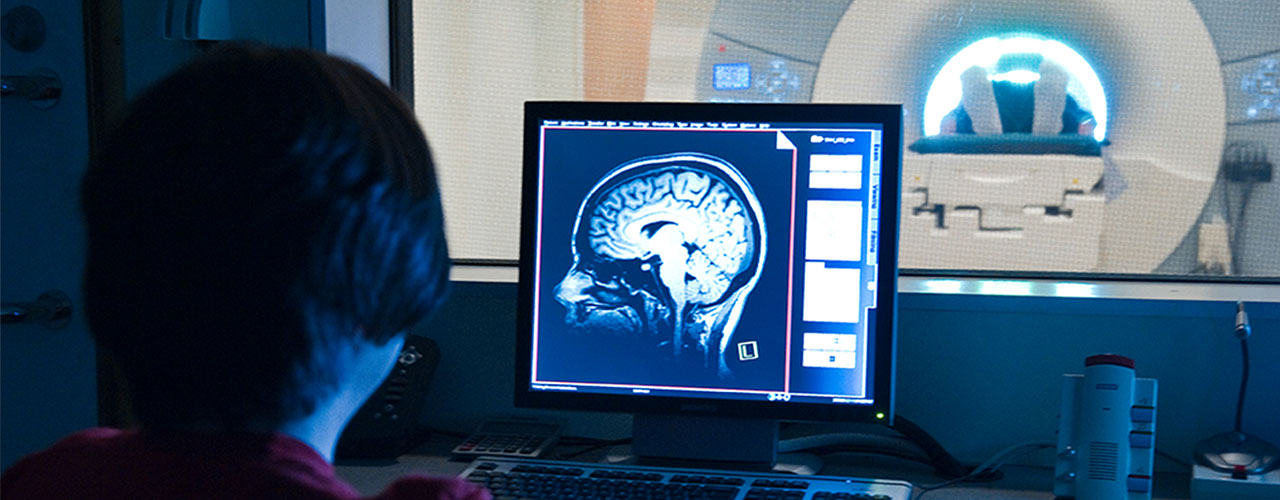
MRI
Magnetic resonance imaging (MRI), the most widely used form of brain imaging, uses magnetic fields and radio waves to produce detailed images of the structure and function of the brain. Functional MRI (fMRI) is a technique that measures the activity of different brain regions as subjects lie inside the MRI scanner. It works by measuring changes in blood flow and blood oxygenation that follow changes in neuronal activity. By comparing the signals obtained as subjects perform different tasks within the scanner, researchers can generate maps that show which parts of the brain are involved in different cognitive processes.
EEG
Electroencephalography (EEG) is a method to record electrical activity in the brain. In this method, electrodes are mounted on a flexible cap that is fitted to the surface of the scalp, allowing researchers to record electrical signals from the brain, which are conducted through the skull. Although EEG lacks the spatial resolution of fMRI, it provides much more detailed information about the timing of brain activity.
NIRS
Near-infrared spectroscopy (NIRS) is a technology that uses the absorption of near-infrared light to reveal changes in blood flow related to brain activity. Unlike fMRI, which requires participants to remain motionless inside the scanner, NIRS involves placing a cap with light emitters and detectors on the participant’s head. This technology is particularly useful for researchers studying brain activity in infants because these young study volunteers can sit on their caregiver’s lap, with some freedom of motion while they participate in research.
Our Staff
The staff at the Martinos Imaging Center direct the research and administrative activities of the center.
MEET OUR STAFF