Artificial “muscles” achieve powerful pulling force

As a cucumber plant grows, it sprouts tightly coiled tendrils that seek out supports in order to pull the plant upward. This ensures the plant receives as much sunlight exposure as possible. Now, researchers at MIT have found a way to imitate this coiling-and-pulling mechanism to produce contracting fibers that could be used as artificial muscles for robots, prosthetic limbs, or other mechanical and biomedical applications.

While many different approaches have been used for creating artificial muscles, including hydraulic systems, servo motors, shape-memory metals, and polymers that respond to stimuli, they all have limitations, including high weight or slow response times. The new fiber-based system, by contrast, is extremely lightweight and can respond very quickly, the researchers say. The findings are being reported today in the journal Science.

The new fibers were developed by MIT postdoc Mehmet Kanik and MIT graduate student Sirma Örgüç, working with professors Polina Anikeeva, Yoel Fink, Anantha Chandrakasan, and C. Cem Taşan, and five others, using a fiber-drawing technique to combine two dissimilar polymers into a single strand of fiber.

The key to the process is mating together two materials that have very different thermal expansion coefficients — meaning they have different rates of expansion when they are heated. This is the same principle used in many thermostats, for example, using a bimetallic strip as a way of measuring temperature. As the joined material heats up, the side that wants to expand faster is held back by the other material. As a result, the bonded material curls up, bending toward the side that is expanding more slowly.
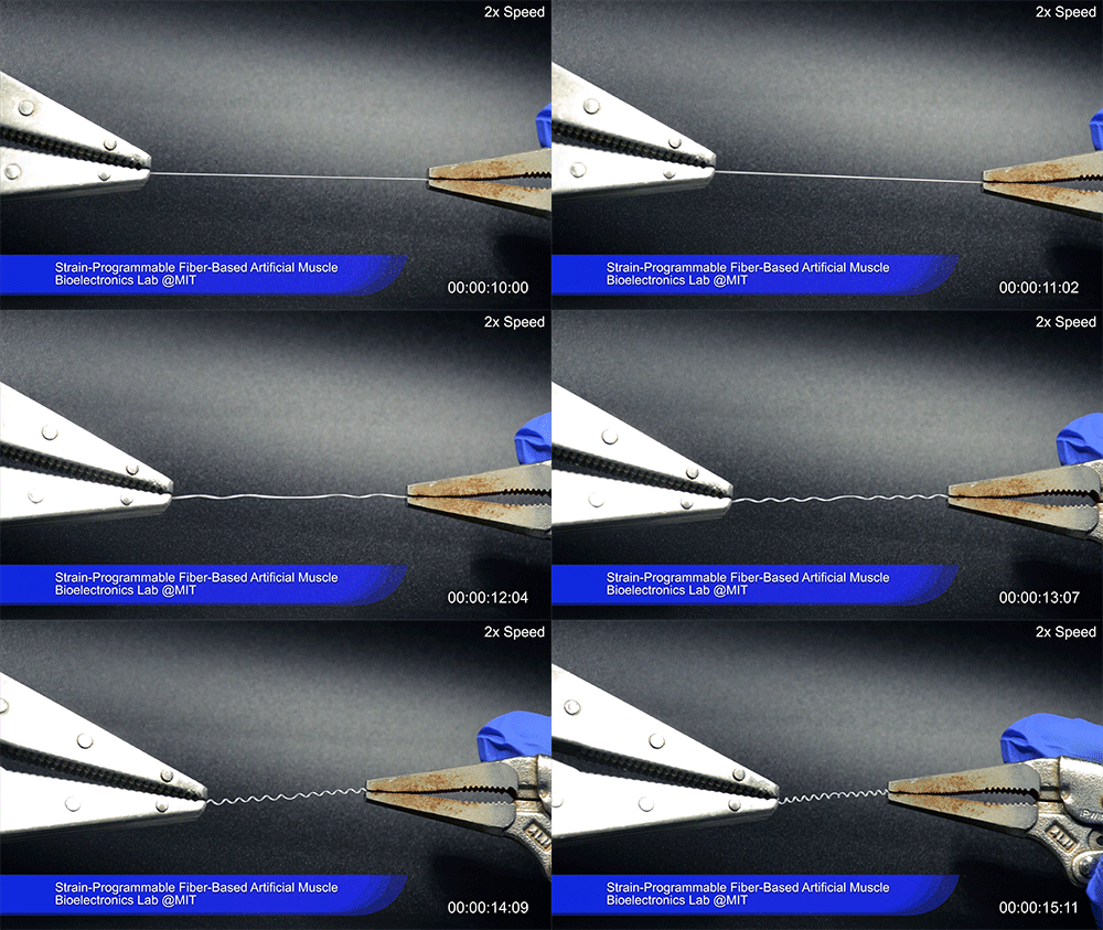
Credit: Courtesy of the researchers

Using two different polymers bonded together, a very stretchable cyclic copolymer elastomer and a much stiffer thermoplastic polyethylene, Kanik, Örgüç and colleagues produced a fiber that, when stretched out to several times its original length, naturally forms itself into a tight coil, very similar to the tendrils that cucumbers produce. But what happened next actually came as a surprise when the researchers first experienced it. “There was a lot of serendipity in this,” Anikeeva recalls.

As soon as Kanik picked up the coiled fiber for the first time, the warmth of his hand alone caused the fiber to curl up more tightly. Following up on that observation, he found that even a small increase in temperature could make the coil tighten up, producing a surprisingly strong pulling force. Then, as soon as the temperature went back down, the fiber returned to its original length. In later testing, the team showed that this process of contracting and expanding could be repeated 10,000 times “and it was still going strong,” Anikeeva says.
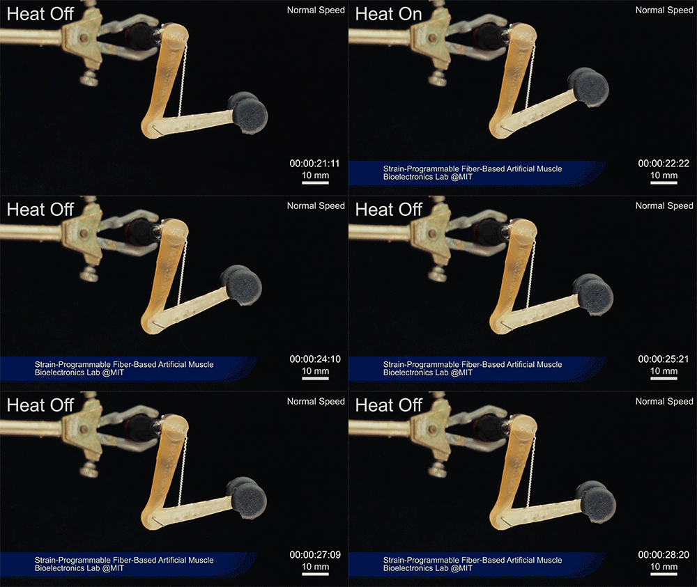
Credit: Courtesy of the researchers

One of the reasons for that longevity, she says, is that “everything is operating under very moderate conditions,” including low activation temperatures. Just a 1-degree Celsius increase can be enough to start the fiber contraction.

The fibers can span a wide range of sizes, from a few micrometers (millionths of a meter) to a few millimeters (thousandths of a meter) in width, and can easily be manufactured in batches up to hundreds of meters long. Tests have shown that a single fiber is capable of lifting loads of up to 650 times its own weight. For these experiments on individual fibers, Örgüç and Kanik have developed dedicated, miniaturized testing setups.
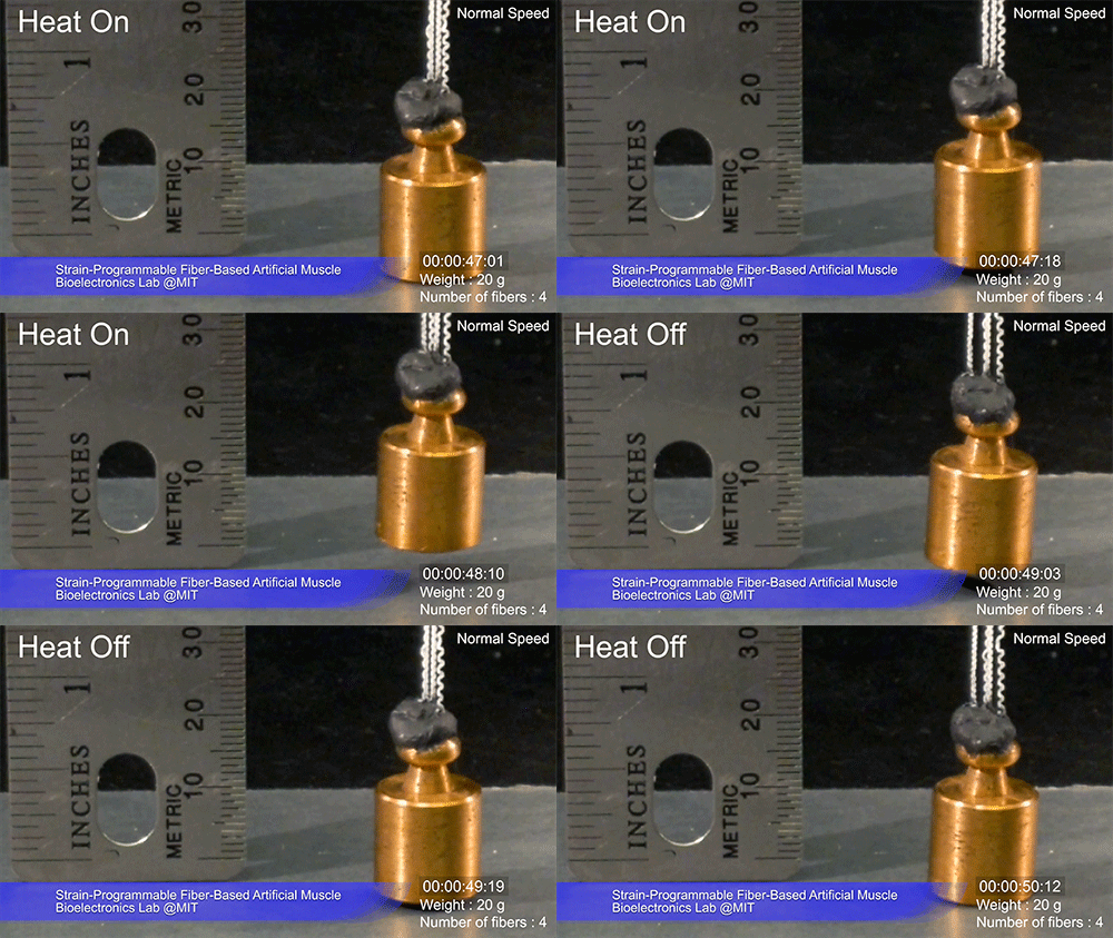
Credit: Courtesy of the researchers

The degree of tightening that occurs when the fiber is heated can be “programmed” by determining how much of an initial stretch to give the fiber. This allows the material to be tuned to exactly the amount of force needed and the amount of temperature change needed to trigger that force.

The fibers are made using a fiber-drawing system, which makes it possible to incorporate other components into the fiber itself. Fiber drawing is done by creating an oversized version of the material, called a preform, which is then heated to a specific temperature at which the material becomes viscous. It can then be pulled, much like pulling taffy, to create a fiber that retains its internal structure but is a small fraction of the width of the preform.

For testing purposes, the researchers coated the fibers with meshes of conductive nanowires. These meshes can be used as sensors to reveal the exact tension experienced or exerted by the fiber. In the future, these fibers could also include heating elements such as optical fibers or electrodes, providing a way of heating it internally without having to rely on any outside heat source to activate the contraction of the “muscle.”

Such fibers could find uses as actuators in robotic arms, legs, or grippers, and in prosthetic limbs, where their slight weight and fast response times could provide a significant advantage.

Some prosthetic limbs today can weigh as much as 30 pounds, with much of the weight coming from actuators, which are often pneumatic or hydraulic; lighter-weight actuators could thus make life much easier for those who use prosthetics. Such fibers might also find uses in tiny biomedical devices, such as a medical robot that works by going into an artery and then being activated,” Anikeeva suggests. “We have activation times on the order of tens of milliseconds to seconds,” depending on the dimensions, she says.

To provide greater strength for lifting heavier loads, the fibers can be bundled together, much as muscle fibers are bundled in the body. The team successfully tested bundles of 100 fibers. Through the fiber drawing process, sensors could also be incorporated in the fibers to provide feedback on conditions they encounter, such as in a prosthetic limb. Örgüç says bundled muscle fibers with a closed-loop feedback mechanism could find applications in robotic systems where automated and precise control are required.

Kanik says that the possibilities for materials of this type are virtually limitless, because almost any combination of two materials with different thermal expansion rates could work, leaving a vast realm of possible combinations to explore. He adds that this new finding was like opening a new window, only to see “a bunch of other windows” waiting to be opened.

“The strength of this work is coming from its simplicity,” he says.

The team also included MIT graduate student Georgios Varnavides, postdoc Jinwoo Kim, and undergraduate students Thomas Benavides, Dani Gonzalez, and Timothy Akintlio. The work was supported by the National Institute of Neurological Disorders and Stroke and the National Science Foundation.

Plugging into the brain

Driven by curiosity and therapeutic goals, Anikeeva leaves no scientific stone unturned in her drive to invent neurotechnology.

The audience sits utterly riveted as Polina Anikeeva highlights the gaps she sees in the landscape of neural tools. With a background in optoelectronics, she has a decidedly unique take on the brain.

“In neuroscience,” says Anikeeva, “we are currently applying silicon-based neural probes with the elastic properties of a knife to a delicate material with the consistency of chocolate pudding—the brain.”

A key problem, summarized by Anikeeva, is that these sharp probes damage tissue, making such interfaces unreliable and thwarting long term brain studies of processes including development and aging. The state of the art is even grimmer in the clinic. An avid climber, Anikeeva recalls a friend sustaining a spinal cord injury. “She made a remarkable recovery,” explains Anikeeva, “but seeing the technology being used to help her was shocking. Not even the simplest electronic tools were used, it was basically lots of screws and physical therapy.” This crude approach, compared to the elegant optoelectronic tools familiar to Anikeeva, sparked a drive to bring advanced materials technology to biological systems.

Outside the box

As the group breaks up after the seminar, the chatter includes boxes, more precisely, thinking outside of them. An associate professor in material sciences and engineering at MIT, Anikeeva’s interest in neuroscience recently led to a McGovern Institute appointment. She sees her journey to neurobiology as serendipitous, having earned her doctorate designing light-emitting devices at MIT.

“I wanted to work on tools that don’t exist, and neuroscience seemed like an obvious choice. Neurons communicate in part through membrane voltage changes and as an electronics designer, I felt that I should be able to use voltage.”

Comfort at the intersection of sciences requires, according to Anikeeva, clarity and focus, also important in her chief athletic pursuits, running and climbing. Through long distant running, Anikeeva finds solitary time (“assuming that no one can chase me”) and the clarity to consider complicated technical questions. Climbing hones something different, absolute focus in the face of the often-tangled information that comes with working at scientific intersections.

“When climbing, you can only think about one thing, your next move. Only the most important thoughts float up.”

This became particularly important when, in Yosemite National Park, she made the decision to go up, instead of down, during an impending thunderstorm. Getting out depended on clear focus, despite imminent hypothermia and being exposed “on one of the tallest features in the area, holding large quantities of metal.” Polina and her climbing partner made it out, but her summary of events echoes her research philosophy: “What you learn and develop is a strong mindset where you don’t do the comfortable thing, the easy thing. Instead you always find, and execute, the most logical strategy.”

In this vein, Anikeeva’s research pursues two very novel, but exceptionally logical, paths to brain research and therapeutics: fiber development and magnetic nanomaterials.

Drawing new fibers

Walking into Anikeeva’s lab, the eye is immediately drawn to a robust metal frame containing, upon closer scrutiny, recognizable parts: a large drill bit, a motor, a heating element. This custom-built machine applies principles from telecommunications to draw multifunctional fibers using more “brain-friendly” materials.

“We start out with a macroscopic model, a preform, of the device that we ultimately want,” explains Anikeeva.

This “preform” is a transparent block of polymers, composites, and soft low-melting temperature metals with optical and electrical properties needed in the final fiber. “So, this could include
electrodes for recording, optical channels for optogenetics, microfluidics for drug delivery, and one day even components that allow chemical or mechanical sensing.” After sitting in a vacuum to remove gases and impurities, the two-inch by one-inch preform arrives at the fiber-drawing tower.

“Then we heat it and pull it, and the macroscopic model becomes a kilometer-long fiber with a lateral dimension of microns, even nanometers,” explains Anikeeva. “Take one of your hairs, and imagine that inside there are electrodes for recording, there are microfluidic channels to infuse drugs, optical channels for stimulation. All of this is combined in a single miniature form
factor, and it can be quite flexible and even stretchable.”

Construction crew

Anikeeva’s lab comprises an eclectic mix of 21 researchers from over 13 different countries, and a range of expertises, including materials science, chemistry, electrical and mechanical engineering, and neuroscience. In 2011, Andres Canales, a materials scientist from Mexico, was the second person to join Anikeeva’s lab.

“There was only an idea, a diagram,” explains Canales. “I didn’t want to work on biology when I arrived at MIT, but talking to Polina, seeing the pictures, thinking about what it would entail, I became very excited by the methods and the potential applications she was thinking of.”

Despite the lack of preliminary models, Anikeeva’s ideas were compelling. Elegant as the fibers are, the road involved painstaking, iterative refinement. From a materials perspective, drawing a fiber containing a continuous conductive element was challenging, as was validation of its properties. But the resulting fiber can deliver optogenetics vectors, monitor expression, and then stimulate neuronal activity in a single surgery, removing the spatial and temporal guesswork usually involved in such an experiment.

Seongjun Park, an electrical engineering graduate student in the lab, explains one biological challenge. “For long term recording in the spinal cord, there was even an additional challenge as the fiber needed to be stretchable to respond to the spine’s movement. For this we developed a drawing process compatible with an elastomer.”

The resulting fibers can be deployed chronically without the scar tissue accumulation that usually prevents long-term optical manipulation and drug delivery, making them good candidates for the treatment of brain disorders. The lab’s current papers find that these implanted fibers are useful for three months, and material innovations make them confident that longer time periods are possible.

Magnetic moments

Another wing of Anikeeva’s research aims to develop entirely non-invasive modalities, and use magnetic nanoparticles to stimulate the brain and deliver therapeutics.

“Magnetic fields are probably the best modality for getting any kind of stimulus to deep tissues,” explains Anikeeva, “because biological systems, except for very specialized systems, do not perceive magnetic fields. They go through us unattenuated, and they don’t couple to our physiology.”

In other words, magnetic fields can safely reach deep tissues, including the brain. Upon reaching their tissue targets these fields can be used to stimulate magnetic nanoparticles, which might one day, for example, be used to deliver dopamine to the brains of Parkinson’s disease patients. The alternating magnetic fields being used in these experiments are tiny, 100-1000 times smaller than fields clinically approved for MRI-based brain imaging.

Tiny fields, but they can be used to powerful effect. By manipulating magnetic moments in these nanoparticles, the magnetic field can cause heat dissipation by the particle that can stimulate thermal receptors in the nervous system. These receptors naturally detect heat, chili peppers and vanilla, but Anikeeva’s magnetic nanoparticles act as tiny heaters that activate these receptors, and, in turn, local neurons. This principle has already been used to activate the brain’s reward center in freely moving mice.

Siyuan Rao, a postdoc who works on the magnetic nanoparticles in collaboration with McGovern Investigator Guoping Feng, is unhesitating when asked what most inspires her.

“As a materials scientist, it is really rewarding to see my materials at work. We can remotely modulate mouse behavior, even turn hopeless behavior into motivation.”

Pushing the boundaries

Such collaborations are valued by Anikeeva. Early on she worked with McGovern Investigator Emilio Bizzi to use the above fiber technology in the spinal cord. “It is important to us to not just make these devices,” explains Anikeeva, “but to use them and show ourselves, and our colleagues, the types of experiments that they can enable.”

Far from an assembly line, the researchers in Anikeeva’s lab follow projects from ideation to deployment. “The student that designs a fiber, performs their own behavioral experiments, and data analysis,” says Anikeeva. “Biology is unforgiving. You can trivially design the most brilliant electrophysiological recording probe, but unless you are directly working in the system, it is easy to miss important design considerations.”

Inspired by this, Anikeeva’s students even started a project with Gloria Choi’s group on their own initiative. This collaborative, can-do ethos spreads beyond the walls of the lab, inspiring people around MIT.

“We often work with a teaching instructor, David Bono, who is an expert on electronics and magnetic instruments,” explains Alex Senko, a senior graduate student in the lab. “In his spare time, he helps those of us who work on electrical engineering flavored projects to hunt down components needed to build our devices.”

These components extend to whatever is needed. When a low frequency source was needed, the Anikeeva lab drafted a guitar amplifier.

Queried about difficulties that she faces having chosen to navigate such a broad swath of fields, Anikeeva is focused, as ever, on the unknown, the boundaries of knowledge.

“Honestly, I really, really enjoy it. It keeps me engaged and not bored. Even when thinking about complicated physics and chemistry, I always have eyes on the prize, that this will allow us to address really interesting neuroscience questions.”

With such thinking, and by relentlessly seeking the tools needed to accomplish scientific goals, Anikeeva and her lab continue to avoid the comfortable route, instead using logical routes toward new technologies.

Polina Anikeeva

Probing the Mind-Body Connection

Polina Anikeeva develops cutting-edge neurotechnologies to probe the flow of information between the brain and peripheral organs in the body.

The brain and the digestive tract are in constant communication, relaying signals that influence our behavior and mental state. Anikeeva’s lab has developed ultrathin, flexible fibers that probe this extensive communication network. The multifunctional fibers are compatible the body’s soft tissue and are equipped with light emitters for activating subsets of cells and tiny channels for delivering genetic cargo or drugs. By deploying these probes within the gastrointestinal tract, Anikeeva’s team has explored how gut-brain circuits contribute to complex behaviors like decision-making and mood.

Anikeeva’s group is also developing magnetic nanoparticles to modulate neural activity wirelessly. Working in conjunction with biological receptors, these non-invasive nanoscale transducers could replace wires in deep-brain stimulation for Parkinson’s disease, or control stress hormones released by adrenal glands. Ultimately, Anikeeva hopes these novel technologies will improve therapies and predictive diagnostics for achieving healthy minds in healthy bodies.

Polina Anikeeva and Feng Zhang awarded 2018 Vilcek Prize

Polina Anikeeva, the Class of 1942 Associate Professor in the Department of Materials Science and Engineering and associate director of the Research Laboratory of Electronics, and Feng Zhang, the James and Patricia Poitras ’63 Professor in Neuroscience at the McGovern Institute, have each been awarded a 2018 Vilcek Prize for Creative Promise in Biomedical Science. Awarded annually by the Vilcek Foundation, the $50,000 prizes recognize younger immigrants who have demonstrated exceptional promise early in their careers.

“The Vilcek Prizes were established in appreciation of the immigrants who chose to dedicate their vision and talent to bettering American society,” says Rick Kinsel, president of the Vilcek Foundation. “This year’s prizewinners honor and continue that legacy with works of astounding, revolutionary importance.”

Polina Anikeeva, who was born in the former Soviet Union, earned her PhD in materials science and engineering at MIT in 2009 and now runs her own bioelectronics lab in the same department focused on the development of materials and devices that enable recording and manipulation of signaling processes within the nervous system. The Vilcek Foundation recognizes Anikeeva for “fashioning ingenious solutions to long-standing challenges in biomedical engineering” including the design of therapeutic devices for conditions such as Parkinson’s disease and spinal cord injury.

Feng Zhang, who is also a core member of the Broad Institute and an associate professor in the departments of Brain and Cognitive Sciences and Biological Engineering, is being recognized for his role in advancing optogenetics (a method for controlling brain activity with light) and developing molecular tools to edit the genome. Thanks to his leadership in inventing precise and efficient gene-editing technologies using CRISPR, Zhang’s work has resulted in a “growing array of applications, such as uncovering the genetic underpinnings of diseases, ushering in gene therapies to cure heritable diseases, and improving agriculture.” Zhang’s family immigrated to the United States from China when he was 11 years of age.

Anikeeva and Zhang will be among eight Vilcek prizewinners honored at an awards gala in New York City in April 2018.

The Vilcek Foundation was established in 2000 by Jan and Marica Vilcek, immigrants from the former Czechoslovakia. The mission of the foundation, to honor the contributions of immigrants to the United States and to foster appreciation of the arts and sciences, was inspired by the couple’s respective careers in biomedical science and art history, as well as their personal experiences and appreciation of the opportunities they received as newcomers to this country.