Searching for self

This story also appears in the Fall 2025 issue of BrainScan

___

The question of how we know ourselves might seem the subject of philosophers, but it is just as much a matter of biology. As modern neuroscientists obtain an increasingly sophisticated understanding of how the brain generates emotions, responds to the external world, and learns from experience, some researchers are returning to a central question: How do we know our experiences, emotions, and physical sensations belong to us?

Curiosity about how the brain generates our sense of self has been a driving force for the research of McGovern Investigator Fan Wang. Following that curiosity has drawn Wang into diverse studies, exploring the origins of pain and the mechanisms we use to control our movements.

“We cannot pinpoint a set of active neurons and say that’s the sense of self. That still remains a mystery,” says Wang, who is also a professor of brain and cognitive sciences and co-director of the K. Lisa Yang and Hock E. Tan Center for Molecular Therapeutics at MIT. But she and other neuroscientists are drilling down into different functions of the brain that together might generate our awareness of ourselves.
Woman wearing blue blazer smiles and gestures off camera with man in white lab coat seated next to her.
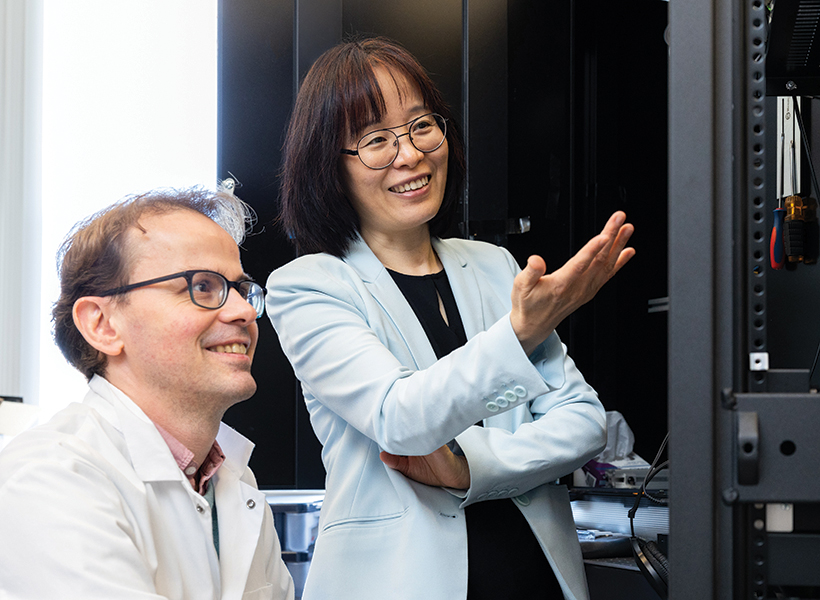
McGovern Investigator Fan Wang (right) with research scientist Vincent Prevosto, who studies brain regions implicated in whisker movement. Photo: Steph Stevens

Wang, who teaches the undergraduate course, “Neurobiology of Self,” explains that there are lots of ways to think about our sense of self, which are probably deeply integrated in the brain. Some are mostly about our physical bodies: How do we experience touch? How do we understand
where we are in space, or recognize the boundary between ourselves and rest of the world? Some consider more internal sensations, like how we experience pain or hunger. Emotion is also key to our sense of self: How do we know that anger or joy are our own, and why do these states change the way our bodies feel?

Wang can trace her initial interest in the brain’s sense of self to work she did as a graduate student in Richard Axel’s lab at Columbia University. The lab had identified receptors expressed by sensory neurons in the nose that detect odorous substances. Wang and others discovered the pathways that information about these smells takes to the brain, and how the brain distinguishes one smell from another.

Who is the “knower” of this information? “The answer,” Wang says, “is ‘I’ or ‘me.’ But understanding where I get the sense of self and how that is constructed, is what drives me to do neuroscience.”

Mechanisms of movement

In her lab at the McGovern Institute, Wang is studying how the brain controls the body’s movements, which she sees as closely tied to the awareness of our physical selves. “The reason I think I am in my body is because I can control my movement. I generate the movement. I cannot control your movement,” says Wang. “Volitional movement gives us a sense of agency, and this sense of agency resembles the sense of self.” For the mice that the group studies, one crucial type of movement comes from the whiskers, which the animals depend on as they explore their environments. Wang’s group has traced the neural circuity that controls whiskers’ rhythmic back-and-forth, which is initiated in the brainstem, where many of the body’s most vital functions are controlled. Wang describes the simple circuit as an oscillator, or a self-generated loop.
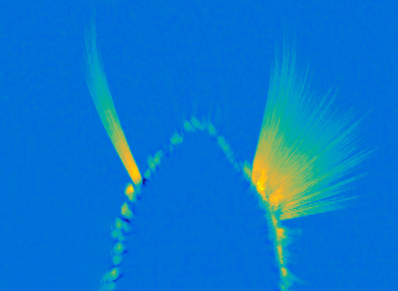
A maximum projection image showing tracked whiskers on the mouse muzzle. The right (control) side shows the back-and-forth rhythmic sweeping of the whiskers, while the experimental side where the whisking oscillator neurons are silenced, the whiskers move very little. Image: Wang Lab

Once it’s started, “the movement can go on unless some other signals stop it,” she says. The movement the circuit generates is simple but voluntary, and can be fine-tuned based on the sensory feedback the whiskers relay back to the brain. They’ve also been investigating how mice move the larynx to generate the squeaks and calls they use to communicate. These intentional movements must be coordinated with the ongoing cycles of respiration since we produce normal sounds only during expiration. Wang’s team has found neurons in the brainstem that generate vocalization-specific movements, and also discovered how respiration-controlling neural circuits can override them, ensuring that breathing is prioritized.

Wang says understanding the circuitry that controls these simple movements sets the stage for figuring out how the brain modifies activity in those circuits to create more complex, intentional movements. “That brings me closer to understanding where this volition is generated — and closer to this sense of self,” she says.

Emotional pain

Still, she knows that volitional movements — even those generated in response to perceptions of the environment — do not, on their own, define a sense of self. As a counterexample, she looks to self-driving cars: “There’s sensory information coming into the central computer, which then generates a motor output — where to drive, where to turn, where to stop. But none of us think a Waymo taxi has a sense of self.”

Wang says when she pondered the ways in which AI-powered cars lack a sense of self, she began thinking about emotions and pain. “If the self-driving Waymo crashes, it will not feel pain,” she says. “But if we hurt ourselves, we will feel pain. And we will hate that, and then we’ll learn.” So her lab is also exploring how the nervous system generates pain perception, including the emotional response that it evokes.
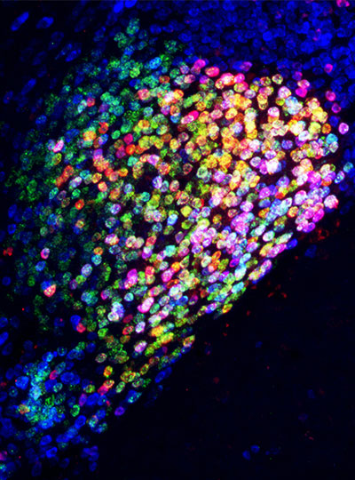
Ensembles of neurons in the amygdala activated by general anesthesia. Image: Fan Wang

In both humans and mice, pain causes emotional suffering that can be recognized and measured through changes in body functions like heart rate and blood pressure. With funding from the K. Lisa Yang Brain-Body Center at MIT, Wang’s lab is carefully tracking these involuntary, or autonomic, functions to gain a more complete understanding of pain’s emotional impact. This approach has helped clarify the role of pain-suppressing neurons in the brain’s amygdala — an important emotion-processing center — that Wang’s team discovered in 2020. When researchers selectively activate those cells in mice, the animals’ behavior makes it clear that the neurons are suppressing pain. Now, the group has learned that activating these neurons suppresses the autonomic response to pain.

Wang says there’s hope that modulating pain’s emotional response might be a way to treat chronic pain in patients. She explains that some patients with damage to another one of the brain’s emotional centers, the cingulate cortex, feel painful stimuli, but experience them as merely intense sensations. That suggests that it might be possible to modulate the emotional response to pain to eliminate patients’ suffering, without blocking the protective information that pain can provide.

The team has also been focusing on another set of anesthesia-activated neurons, which they have found suppress anxiety. When anxiety-suppressing neurons are activated in mice, the animals’ heart rates slow and they become more willing to explore bright, open spaces. Another anxiety-associated measure — heart rate variability — increases. Wang explains that this change is particularly significant: “If you have persistent low heart rate variability, especially in veterans, that is a very good predictor for anxiety developing into depression in the future,” she says.

The team’s findings, which suggest that changes in autonomic functions may themselves relieve anxiety, point toward potential new targets for anti-anxiety therapies. And by highlighting the connection between emotion and bodily responses, they offer more clues about our sense of self. “These neurons are now changing some high-level concept about anxiety,” Wang points out.

That link between emotion and body seems to Wang to be key to the sense of self. The big questions remain unanswered, but that simply stokes her curiosity. “I can be aware of my bodily responses: I am aware of ‘I am anxious’ or ‘I am in pain.’ I can see the pathways from which stimuli go into these nervous systems and come back down to the body and control the response. But I still don’t know who is the person — the knower,” she says. “I haven’t found it, so I’m going to keep looking.”

Harnessing the power of placebo for pain relief

Placebos are inert treatments, generally not expected to impact biological pathways or improve a person’s physical health. But time and again, some patients report that they feel better after taking a placebo. Increasingly, doctors and scientists are recognizing that rather than dismissing placebos as mere trickery, they may be able to help patients by harnessing their power.

To maximize the impact of the placebo effect and design reliable therapeutic strategies, researchers need a better understanding of how it works. Now, with a new animal model developed by scientists at the McGovern Institute, they will be able to investigate the neural circuits that underlie placebos’ ability to elicit pain relief.

“The brain and body interaction has a lot of potential, in a way that we don’t fully understand,” says McGovern investigator Fan Wang. “I really think there needs to be more of a push to understand placebo effect, in pain and probably in many other conditions. Now we have a strong model to probe the circuit mechanism.”

Context-dependent placebo effect

McGovern Investigator Fan Wang. Photo: Caitliin Cunningham

In the September 5, 2024, issue of the journal Current Biology, Wang and her team report that they have elicited strong placebo pain relief in mice by activating pain-suppressing neurons in the brain while the mice are in a specific environment—thereby teaching the animals that they feel better when they are in that context. Following their training, placing the mice in that environment alone is enough to suppress pain. The team’s experiments, which were funded by the National Institutes of Health, the K. Lisa Yang Brain-Body Center and the K. Lisa Yang and Hock E. Tan Center for Molecular Therapeutics within MIT’s Yang Tan Collective show that this context-dependent placebo effect relieves both acute and chronic pain.

Context is critical for the placebo effect. While a pill can help a patient feel better when they expect it to, even if it is made only of sugar or starch, it seems to be not just the pill that sets up those expectations, but the entire scenario in which the pill is taken. For example, being in a hospital and interacting with doctors can contribute to a patient’s perception of care, and these social and environmental factors can make a placebo effect more probable.

Postdoctoral fellows Bin Chen and Nitsan Goldstein used visual and textural cues to define a specific place. Then they activated pain-suppressing neurons in the brain while the animals were in this “pain-relief box.” Those pain-suppressing neurons, which Wang’s lab discovered a few years ago, are located in an emotion-processing center of the brain called the central amygdala. By expressing light-sensitive channels in these neurons, the researchers were able to suppress pain with light in the pain-relief box and leave the neurons inactive when mice were in a control box.

Animals learned to prefer the pain-relief box to other environments. And when the researchers tested their response to potentially painful stimuli after they had made that association, they found the mice were less sensitive while they were there. “Just by being in the context that they had associated with pain suppression, we saw that reduced pain—even though we weren’t actually activating those [pain-suppressing] neurons,” Goldstein explains.

Acute and chronic pain relief

Some scientists have been able to elicit placebo pain relief in rodents by treating the animals with morphine, linking environmental cues to the pain suppression caused by the drugs similar to the way Wang’s team did by directly activating pain-suppressing neurons. This drug-based approach works best for setting up expectations of relief for acute pain; its placebo effect is short-lived and mostly ineffective against chronic pain. So Wang, Chen, and Goldstein were particularly pleased to find that their engineered placebo effect was effective for relieving both acute and chronic pain.

In their experiments, animals experiencing a chemotherapy-induced hypersensitivity to touch exhibited a preference for the pain relief box as much as animals who were exposed to a chemical that induces acute pain, days after their initial conditioning. Once there, their chemotherapy-induced pain sensitivity was eliminated; they exhibited no more sensitivity to painful stimuli than they had prior to receiving chemotherapy.

One of the biggest surprises came when the researchers turned their attention back to the pain-suppressing neurons in the central amygdala that they had used to trigger pain relief. They suspected that those neurons might be reactivated when mice returned to the pain-relief box. Instead, they found that after the initial conditioning period, those neurons remained quiet. “These neurons are not reactivated, yet the mice appear to be no longer in pain,” Wang says. “So it suggests this memory of feeling well is transferred somewhere else.”

Goldstein adds that there must be a pain-suppressing neural circuit somewhere that is activated by pain-relief-associated contexts—and the team’s new placebo model sets researchers up to investigate those pathways. A deeper understanding of that circuitry could enable clinicians to deploy the placebo effect—alone or in combination with active treatments—to better manage patients’ pain in the future.

How touch dampens the brain’s response to painful stimuli

McGovern Investigator Fan Wang. Photo: Caitliin Cunningham

When we press our temples to soothe an aching head or rub an elbow after an unexpected blow, it often brings some relief. It is believed that pain-responsive cells in the brain quiet down when these neurons also receive touch inputs, say scientists at MIT’s McGovern Institute, who for the first time have watched this phenomenon play out in the brains of mice.

The team’s discovery, reported November 16, 2022, in the journal Science Advances, offers researchers a deeper understanding of the complicated relationship between pain and touch and could offer some insights into chronic pain in humans. “We’re interested in this because it’s a common human experience,” says McGovern Investigator Fan Wang. “When some part of your body hurts, you rub it, right? We know touch can alleviate pain in this way.” But, she says, the phenomenon has been very difficult for neuroscientists to study.

Modeling pain relief

Touch-mediated pain relief may begin in the spinal cord, where prior studies have found pain-responsive neurons whose signals are dampened in response to touch. But there have been hints that the brain was involved too. Wang says this aspect of the response has been largely unexplored, because it can be hard to monitor the brain’s response to painful stimuli amidst all the other neural activity happening there—particularly when an animal moves.

So while her team knew that mice respond to a potentially painful stimulus on the cheek by wiping their faces with their paws, they couldn’t follow the specific pain response in the animals’ brains to see if that rubbing helped settle it down. “If you look at the brain when an animal is rubbing the face, movement and touch signals completely overwhelm any possible pain signal,” Wang explains.

She and her colleagues have found a way around this obstacle. Instead of studying the effects of face-rubbing, they have focused their attention on a subtler form of touch: the gentle vibrations produced by the movement of the animals’ whiskers. Mice use their whiskers to explore, moving them back and forth in a rhythmic motion known as whisking to feel out their environment. This motion activates touch receptors in the face and sends information to the brain in the form of vibrotactile signals. The human brain receives the same kind of touch signals when a person shakes their hand as they pull it back from a painfully hot pan—another way we seek touch-mediate pain relief.

If you look at the brain when an animal is rubbing the face, movement and touch signals completely overwhelm any possible pain signal, says Wang.

Wang and her colleagues found that this whisker movement alters the way mice respond to bothersome heat or a poke on the face—both of which usually lead to face rubbing. “When the unpleasant stimuli were applied in the presence of their self-generated vibrotactile whisking…they respond much less,” she says. Sometimes, she says, whisking animals entirely ignore these painful stimuli.

In the brain’s somatosensory cortex, where touch and pain signals are processed, the team found signaling changes that seem to underlie this effect. “The cells that preferentially respond to heat and poking are less frequently activated when the mice are whisking,” Wang says. “They’re less likely to show responses to painful stimuli.” Even when whisking animals did rub their faces in response to painful stimuli, the team found that neurons in the brain took more time to adopt the firing patterns associated with that rubbing movement. “When there is a pain stimulation, usually the trajectory the population dynamics quickly moved to wiping. But if you already have whisking, that takes much longer,” Wang says.

Wang notes that even in the fraction of a second before provoked mice begin rubbing their faces, when the animals are relatively still, it can be difficult to sort out which brain signals are related to perceiving heat and poking and which are involved in whisker movement. Her team developed computational tools to disentangle these, and are hoping other neuroscientists will use the new algorithms to make sense of their own data.

Whisking’s effects on pain signaling seem to depend on dedicated touch-processing circuitry that sends tactile information to the somatosensory cortex from a brain region called the ventral posterior thalamus. When the researchers blocked that pathway, whisking no longer dampened the animals’ response to painful stimuli. Now, Wang says, she and her team are eager to learn how this circuitry works with other parts of the brain to modulate the perception and response to painful stimuli.

Wang says the new findings might shed light on a condition called thalamic pain syndrome, a chronic pain disorder that can develop in patients after a stroke that affects the brain’s thalamus. “Such strokes may impair the functions of thalamic circuits that normally relay pure touch signals and dampen painful signals to the cortex,” she says.

New research center focused on brain-body relationship established at MIT

The inextricable link between our brains and our bodies has been gaining increasing recognition among researchers and clinicians over recent years. Studies have shown that the brain-body pathway is bidirectional — meaning that our mental state can influence our physical health and vice versa. But exactly how the two interact is less clear.

A new research center at MIT, funded by a $38 million gift to the McGovern Institute for Brain Research from philanthropist K. Lisa Yang, aims to unlock this mystery by creating and applying novel tools to explore the multidirectional, multilevel interplay between the brain and other body organ systems. This gift expands Yang’s exceptional philanthropic support of human health and basic science research at MIT over the past five years.

“Lisa Yang’s visionary gift enables MIT scientists and engineers to pioneer revolutionary technologies and undertake rigorous investigations into the brain’s complex relationship with other organ systems,” says MIT President L. Rafael Reif.  “Lisa’s tremendous generosity empowers MIT scientists to make pivotal breakthroughs in brain and biomedical research and, collectively, improve human health on a grand scale.”

The K. Lisa Yang Brain-Body Center will be directed by Polina Anikeeva, professor of materials science and engineering and brain and cognitive sciences at MIT and an associate investigator at the McGovern Institute. The center will harness the power of MIT’s collaborative, interdisciplinary life sciences research and engineering community to focus on complex conditions and diseases affecting both the body and brain, with a goal of unearthing knowledge of biological mechanisms that will lead to promising therapeutic options.

“Under Professor Anikeeva’s brilliant leadership, this wellspring of resources will encourage the very best work of MIT faculty, graduate fellows, and research — and ultimately make a real impact on the lives of many,” Reif adds.

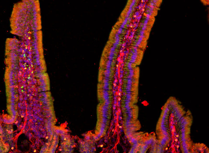
microscope image of gut
Mouse small intestine stained to reveal cell nucleii (blue) and peripheral nerve fibers (red).
Image: Polina Anikeeva, Marie Manthey, Kareena Villalobos

Center goals  

Initial projects in the center will focus on four major lines of research:

  • Gut-Brain: Anikeeva’s group will expand a toolbox of new technologies and apply these tools to examine major neurobiological questions about gut-brain pathways and connections in the context of autism spectrum disorders, Parkinson’s disease, and affective disorders.
  • Aging: CRISPR pioneer Feng Zhang, the James and Patricia Poitras Professor of Neuroscience at MIT and investigator at the McGovern Institute, will lead a group in developing molecular tools for precision epigenomic editing and erasing accumulated “errors” of time, injury, or disease in various types of cells and tissues.
  • Pain: The lab of Fan Wang, investigator at the McGovern Institute and professor of brain and cognitive sciences, will design new tools and imaging methods to study autonomic responses, sympathetic-parasympathetic system balance, and brain-autonomic nervous system interactions, including how pain influences these interactions.
  • Acupuncture: Wang will also collaborate with Hilda (“Scooter”) Holcombe, a veterinarian in MIT’s Division of Comparative Medicine, to advance techniques for documenting changes in brain and peripheral tissues induced by acupuncture in mouse models. If successful, these techniques could lay the groundwork for deeper understandings of the mechanisms of acupuncture, specifically how the treatment stimulates the nervous system and restores function.

A key component of the K. Lisa Yang Brain-Body Center will be a focus on educating and training the brightest young minds who aspire to make true breakthroughs for individuals living with complex and often devastating diseases. A portion of center funding will endow the new K. Lisa Yang Brain-Body Fellows Program, which will support four annual fellowships for MIT graduate students and postdocs working to advance understanding of conditions that affect both the body and brain.

Mens sana in corpore sano

“A phrase I remember reading in secondary school has always stuck with me: ‘mens sana in corpore sano’ ‘a healthy mind in a healthy body,’” says Lisa Yang, a former investment banker committed to advocacy for individuals with visible and invisible disabilities. “When we look at how stress, nutrition, pain, immunity, and other complex factors impact our health, we truly see how inextricably linked our brains and bodies are. I am eager to help MIT scientists and engineers decode these links and make real headway in creating therapeutic strategies that result in longer, healthier lives.”

“This center marks a once-in-a-lifetime opportunity for labs like mine to conduct bold and risky studies into the complexities of brain-body connections,” says Anikeeva, who works at the intersection of materials science, electronics, and neurobiology. “The K. Lisa Yang Brain-Body Center will offer a pathbreaking, holistic approach that bridges multiple fields of study. I have no doubt that the center will result in revolutionary strides in our understanding of the inextricable bonds between the brain and the body’s peripheral organ systems, and a bold new way of thinking in how we approach human health overall.”

On a mission to alleviate chronic pain

About 50 million Americans suffer from chronic pain, which interferes with their daily life, social interactions, and ability to work. MIT Professor Fan Wang wants to develop new ways to help relieve that pain, by studying and potentially modifying the brain’s own pain control mechanisms.

Her recent work has identified an “off switch” for pain, located in the brain’s amygdala. She hopes that finding ways to control this switch could lead to new treatments for chronic pain.

“Chronic pain is a major societal issue,” Wang says. “By studying pain-suppression neurons in the brain’s central amygdala, I hope to create a new therapeutic approach for alleviating pain.”

Wang, who joined the MIT faculty in January 2021, is also the leader of a new initiative at the McGovern Institute for Brain Research that is studying drug addiction, with the goal of developing more effective treatments for addiction.

“Opioid prescription for chronic pain is a major contributor to the opioid epidemic. With the Covid pandemic, I think addiction and overdose are becoming worse. People are more anxious, and they seek drugs to alleviate such mental pain,” Wang says. “As scientists, it’s our duty to tackle this problem.”

Sensory circuits

Wang, who grew up in Beijing, describes herself as “a nerdy child” who loved books and math. In high school, she took part in science competitions, then went on to study biology at Tsinghua University. She arrived in the United States in 1993 to begin her PhD at Columbia University. There, she worked on tracing the connection patterns of olfactory receptor neurons in the lab of Richard Axel, who later won the Nobel Prize for his discoveries of odorant receptors and how the olfactory system is organized.

After finishing her PhD, Wang decided to switch gears. As a postdoc at the University of California at San Francisco and then Stanford University, she began studying how the brain perceives touch.

In 2003, Wang joined the faculty at Duke University School of Medicine. There, she began developing techniques to study the brain circuits that underlie the sense of touch, tracing circuits that carry sensory information from the whiskers of mice to the brain. She also studied how the brain integrates movements of touch organs with signals of sensory stimuli to generate perception (such as using stretching movements to sense elasticity).

As she pursued her sensory perception studies, Wang became interested in studying pain perception, but she felt she needed to develop new techniques to tackle it. While at Duke, she invented a technique called CANE (capturing activated neural ensembles), which can identify networks of neurons that are activated by a particular stimulus.

Using this approach in mice, she identified neurons that become active in response to pain, but so many neurons across the brain were activated that it didn’t offer much useful information. As a way to indirectly get at how the brain controls pain, she decided to use CANE to explore the effects of drugs used for general anesthesia. During general anesthesia, drugs render a patient unconscious, but Wang hypothesized that the drugs might also shut off pain perception.

“At that time, it was just a wild idea,” Wang recalls. “I thought there may be other mechanisms — that instead of just a loss of consciousness, anesthetics may do something to the brain that actually turns pain off.”

Support for the existence of an “off switch” for pain came from the observation that wounded soldiers on a battlefield can continue to fight, essentially blocking out pain despite their injuries.

In a study of mice treated with anesthesia drugs, Wang discovered that the brain does have this kind of switch, in an unexpected location: the amygdala, which is involved in regulating emotion. She showed that this cluster of neurons can turn off pain when activated, and when it is suppressed, mice become highly sensitive to ordinary gentle touch.

“There’s a baseline level of activity that makes the animals feel normal, and when you activate these neurons, they’ll feel less pain. When you silence them, they’ll feel more pain,” Wang says.

Turning off pain

That finding, which Wang reported in 2020, raised the possibility of somehow modulating that switch in humans to try to treat chronic pain. This is a long-term goal of Wang’s, but more work is required to achieve it, she says. Currently her lab is working on analyzing the RNA expression patterns of the neurons in the cluster she identified. They also are measuring the neurons’ electrical activity and how they interact with other neurons in the brain, in hopes of identifying circuits that could be targeted to tamp down the perception of pain.

One way of modulating these circuits could be to use deep brain stimulation, which involves implanting electrodes in certain areas of the brain. Focused ultrasound, which is still in early stages of development and does not require surgery, could be a less invasive alternative.

Another approach Wang is interested in exploring is pairing brain stimulation with a context such as looking at a smartphone app. This kind of pairing could help train the brain to shut off pain using the app, without the need for the original stimulation (deep brain stimulation or ultrasound).

“Maybe you don’t need to constantly stimulate the brain. You may just need to reactivate it with a context,” Wang says. “After a while you would probably need to be restimulated, or reconditioned, but at least you have a longer window where you don’t need to go to the hospital for stimulation, and you just need to use a context.”

Wang, who was drawn to MIT in part by its focus on fostering interdisciplinary collaborations, is now working with several other McGovern Institute members who are taking different angles to try to figure out how the brain generates the state of craving that occurs in drug addiction, including opioid addiction.

“We’re going to focus on trying to understand this craving state: how it’s created in the brain and how can we sort of erase that trace in the brain, or at least control it. And then you can neuromodulate it in real time, for example, and give people a chance to get back their control,” she says.

Fan Wang

Sensing the World

Why do we feel pain? What causes us to have intense cravings? How do we manage move so effortlessly through the world?

Fan Wang’s research focuses on the neural circuits governing the bidirectional interactions between the brain and body. She is specifically interested in the circuits that control the sensory and emotional aspects of pain and addiction, as well as the sensory and motor circuits that work together to execute behaviors such as eating, drinking, and moving. She has explored how anesthesia suppresses pain, how brain circuits generate rhythmic behaviors, how the brain coordinates speaking and breathing, and how drugs of abuse influence brain circuits that drive addiction. Wang’s lab deploys a range of techniques to gain traction in these studies, including genetic and viral methods, in vivo electrophysiology, in vivo imaging, and behavioral and autonomic response tracking. Her research has profound implications for real-world problems, including chronic pain and addiction.