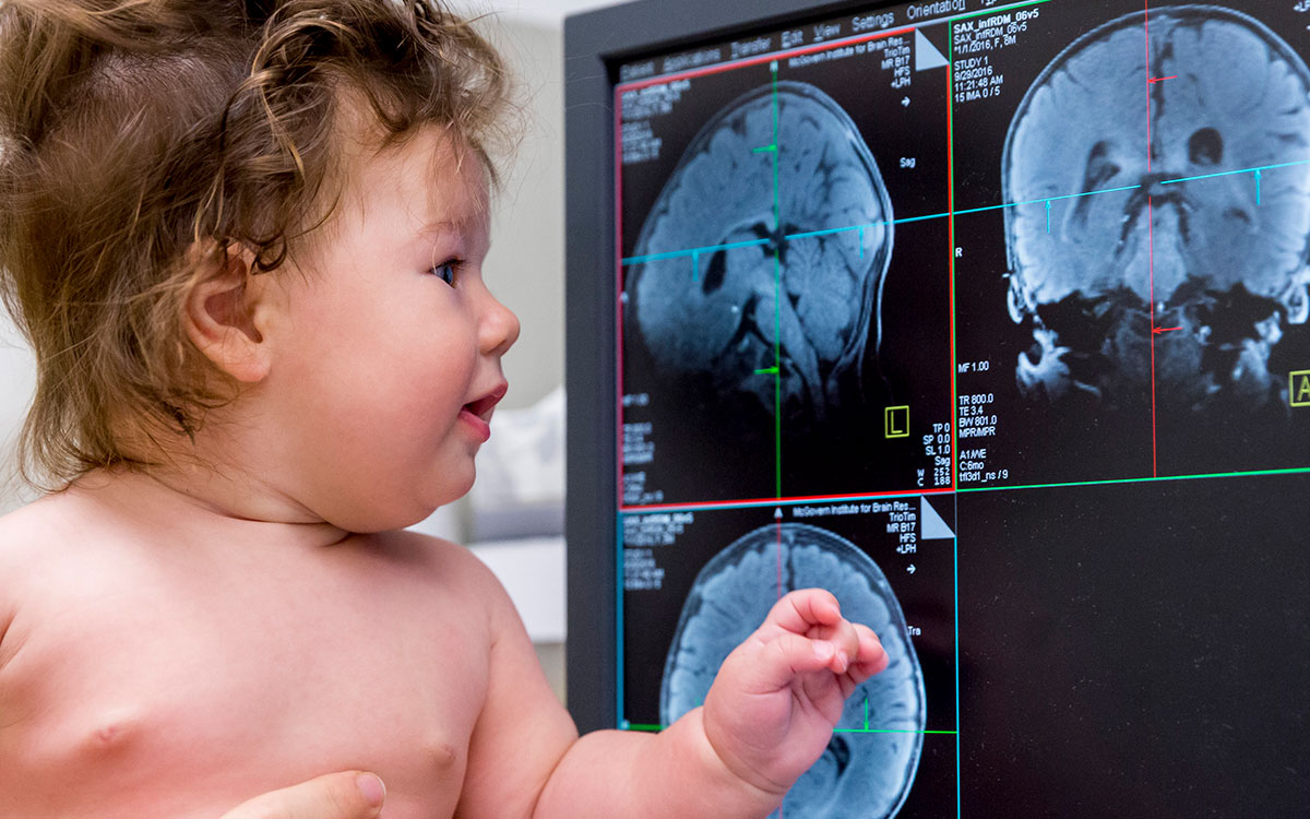
Powerful imaging methods like MRI and MEG provide neuroscientists with a detailed view of the human brain – including the spatial and temporal changes that occur as we interact with the world. Our researchers are using these tools to study how the brain develops from infancy, which regions underlie different aspects of our mental lives, and whether imaging can be used to predict the onset of disease.
To view this video please enable JavaScript, and consider upgrading to a web browser that supports HTML5 video

Brain Imaging
brain mapping · functional connectivity · fMRI · EEG · MEG · predictive imaging · precision interventions · contrast agents · theory of mind · the developing brain · learning

Featured Researcher
Satrajit Ghosh
Satrajit Ghosh uses neuroimaging, speech communication, and machine learning to improve assessments and treatments for mental health.
224
The typical number of images our scanners take for a 3D brain reconstruction.
3.5-6 minutes
The time typically taken to generate a structural scan of the adult brain using MRI.
Recent Publications
- Rutledge, O, Goyette, RB, Wang, KL, Park, MS, Gabrieli, JDE. Functional neuroimaging of Cannabidiol in stress and anxiety: a systematic review. Front Neuroimaging. 2026;5 :1860919. doi: 10.3389/fnimg.2026.1860919. PubMed PMID:42494831 PubMed Central PMC13391337.
- Ahn, G, Li, CE, Liang, A, Choi, W, Ahn, S, Roberts, C et al.. Large Language Model Few-Shot Learning for Predicting Individual Treatment Response to Smartphone-Based Mindfulness in Autistic Adults With Anxiety: Secondary Analysis of a Randomized Controlled Trial. JMIR AI. 2026;5 :e89054. doi: 10.2196/89054. PubMed PMID:42490583 PubMed Central PMC13394852.
- Radkani, S, Saxe, R. Delegation to legitimate authority as a resource rational mechanism. Behav Brain Sci. 2026;49 :e152. doi: 10.1017/S0140525X25101581. PubMed PMID:42342633 .