Inside the adult ADHD brain
Brain scans differentiate adults who have recovered from childhood ADHD and those whose difficulties linger.
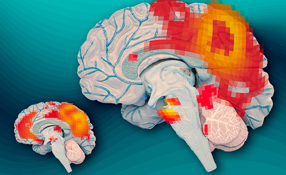
In the first study to compare patterns of brain activity in adults who recovered from childhood ADHD and those who did not, McGovern Institute neuroscientists have discovered key differences in a brain communication network that is active when the brain is at wakeful rest and not focused on a particular task.
The findings offer evidence of a biological basis for adult ADHD and should help to validate the criteria used to diagnose the disorder, according to the researchers. Read more >>