Detecting the brain’s magnetic signals with MEG
What MEG reveals about brain activity that other neuroimaging methods don't.
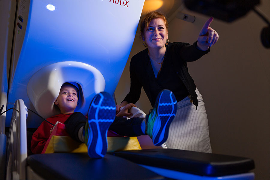
Magnetoencephalography (MEG) is a noninvasive technique for measuring neuronal activity in the human brain. Electrical currents flowing through neurons generate weak magnetic fields that can be recorded at the surface of the head using very sensitive magnetic detectors known as superconducting quantum interference devices (SQUIDs).
MEG is a purely passive method that relies on detection of signals that are produced naturally by the brain. It does not involve exposure to radiation or strong magnetic fields, and there are no known hazards associated with MEG.

Magnetic signals from the brain are very small compared to the magnetic fluctuations that are produced by interfering sources such as nearby electrical equipment or moving metal objects. Therefore MEG scans are typically performed within a special magnetically shielded room that blocks this external interference.
It is fitting that MIT should have a state-of-the-art MEG scanner, since the MEG technology was pioneered by David Cohen in the early 1970s while he was a member of MIT’s Francis Bitter Magnet Laboratory.
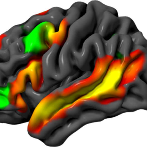
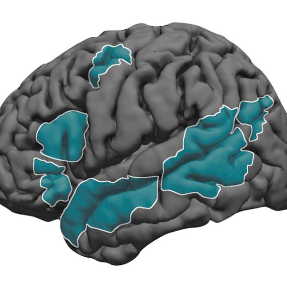
MEG can detect the timing of magnetic signals with millisecond precision. This is the timescale on which neurons communicate, and MEG is thus well suited to measuring the rapid signals that reflect communication between different parts of the human brain.
MEG is complementary to other brain imaging modalities such as functional magnetic resonance imaging (fMRI) and positron emission tomography (PET), which depend on changes in blood flow, and which have higher spatial resolution but much lower temporal resolution than MEG.
Our MEG scanner, an Elekta Neuromag Triux with 306 channels plus 128 channels for EEG, was installed in 2011 and is the first of its kind in North America. It is housed within a magnetically shielded room to reduce background noise.
The MEG lab is part of the Martinos Imaging Center at MIT, operating as a core facility, and accessible to all members of the local research community. Potential users should contact Dimitrios Pantazis for more information.
The MEG Lab was made possible through a grant from the National Science Foundation and through the generous support of the following donors: Thomas F. Peterson, Jr. ’57; Edward and Kay Poitras; The Simons Foundation; and an anonymous donor.