Making invisible therapy targets visible
A powerful new technology reveals hidden molecular structures in cells and tissues.
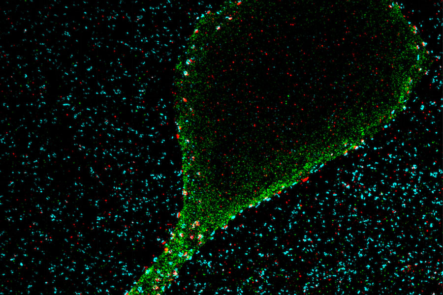
The lab of Edward Boyden, the Y. Eva Tan Professor in Neurotechnology, has developed a powerful technology called Expansion Revealing (ExR) that makes visible molecular structures that were previously too hidden to be seen with even the most powerful microscopes. It “reveals” the nanoscale alterations in synapses, neural wiring, and other molecular assemblies using ordinary lab microscopes. It does so this way: Inside a cell, proteins and other molecules are often tightly packed together. These dense clusters can be difficult to image because the fluorescent labels used to make them visible can’t wedge themselves between the molecules. ExR “de-crowds” the molecules by expanding the cell using a chemical process, making the molecules accessible to fluorescent tags.

“This technology can be used to answer a lot of biological questions about dysfunction in synaptic proteins, which are involved in neurodegenerative diseases,” says Jinyoung Kang, a J. Douglas Tan Postdoctoral Fellow in the labs of Boyden and Guoping Feng, the James W. (1963) and Patricia T. Poitras Professor of Brain and Cognitive Sciences. “Until now, there has been no tool to visualize synapses very well at nanoscale.”
Over the past year, the Boyden team has been using ExR to explore the underlying mechanisms of brain disorders, including autism spectrum disorder (ASD) and Alzheimer’s disease. Since the method can be applied iteratively, Boyden imagines it may one day succeed in creating a 100-fold magnification of molecular structures.
“Using earlier technology, researchers may be missing entire categories of molecular phenomena, both functional and dysfunctional,” says Boyden. “It’s critical to bring these nanostructures into view so that we can identify potential targets for new therapeutics that can restore functional molecular arrangements.”
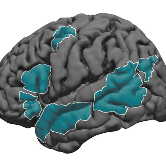
The team is applying ExR to the study of mutant-animal-model brain slices to expose complex synapse 3D nanoarchitecture and configuration. Among their questions: How do synapses differ when mutations that cause autism and other neurological conditions are present?
Using the new technology, Kang and her collaborator Menglong Zeng characterized the molecular architecture of excitatory synapses on parvalbumin interneurons, cells that drastically influence the downstream effects of neuronal signaling and ultimately change cognitive behaviors. They discovered condensed AMPAR clustering in parvalbumin interneurons is essential for normal brain function. The next step is to explore their role in the function of parvalbumin interneurons, which are vulnerable to stressors and have been implicated in brain disorders including autism and Alzheimer’s disease.
The researchers are now investigating whether ExR can reveal abnormal protein nanostructures in SHANK3 knockout mice and marmosets. Mutations in the SHANK3 gene lead to one of the most severe types of ASD, Phelan-McDermid syndrome, which accounts for about 2 percent of all ASD patients with intellectual disability.