Musicians’ enhanced attention

In a world full of competing sounds, we often have to filter out a lot of noise to hear what’s most important. This critical skill may come more easily for people with musical training, according to scientists at MIT’s McGovern Institute who used brain imaging to follow what happens when people try to focus their attention on certain sounds.

When Cassia Low Manting, a postdoctoral researcher working in the labs of McGovern Institute Investigators John Gabrieli and Dimitrios Pantazis, asked people to focus on a particular melody while another melody played at the same time, individuals with musical backgrounds were, unsurprisingly, better able to follow the target tune. An analysis of study participants’ brain activity suggests this advantage arises because musical training sharpens neural mechanisms that amplify the sounds they want to listen to while turning down distractions. “This points to the idea that we can train this selective attention ability,” Manting says.

The research team, including senior author Daniel Lundqvist at the Karolinska Institute in Sweden, reported their findings September 17, 2025, in the journal Science Advances. Manting, who is now at the Karolinska Institute, notes that the research is part of an ongoing collaboration between the two institutions.

Overcoming challenges

Participants in the study had vastly difference backgrounds when it came to music. Some were professional musicians with deep training and experience, while others struggled to differentiate between the two tunes they were played, despite each one’s distinct pitch. This disparity allowed the researchers to explore how the brain’s capacity for attention might change with experience. “Musicians are very fun to study because their brains have been morphed in ways based on their training,” Manting says. “It’s a nice model to study these training effects.”

Still, the researchers had significant challenges to overcome. It has been hard to study how the brain manages auditory attention, because when researchers use neuroimaging to monitor brain activity, they see the brain’s response to all sounds: those that the listener cares most about, as well as those the listener is trying to ignore. It is usually difficult to figure out which brain signals were triggered by which sounds.

Manting and her colleagues overcame this challenge with a method called frequency tagging. Rather than playing the melodies in their experiments at a constant volume, the volume of each melody oscillated, rising and falling with a particular frequency. Each melody had its own frequency, creating detectable patterns in the brain signals that responded to it. “When you play these two sounds simultaneously to the subject and you record the brain signal, you can say, this 39-Hertz activity corresponds to the lower pitch sound and the 43-Hertz activity corresponds specifically to the higher pitch sound,” Manting explains. “It is very clean and very clear.”

When they paired frequency tagging with magnetoencephalography, a noninvasive method of monitoring brain activity, the team was able to track how their study participants’ brains responded to each of two melodies during their experiments. While the two tunes played, subjects were instructed to follow either the higher pitched or the lower pitched melody. When the music stopped, they were asked about the final notes of the target tune: did they rise or did they fall? The researchers could make this task harder by making the two tunes closer together in pitch, as well as by altering the timing of the notes.

Manting used a survey that asked about musical experience to score each participant’s musicality, and this measure had an obvious effect on task performance: The more musical a person was, the more successful they were at following the tune they had been asked to track.

To look for differences in brain activity that might explain this, the research team developed a new machine-learning approach to analyze their data. They used it to tease apart what was happening in the brain as participants focused on the target tune—even, in some cases, when the notes of the distracting tune played at the exact same time.

Top-down vs bottom-up attention

What they found was a clear separation of brain activity associated with two kinds of attention, known as top-down and bottom-up attention. Manting explains that top-down attention is goal-oriented, involving a conscious focus—the kind of attention listeners called on as they followed the target tune. Bottom-up attention, on the other hand, is triggered by the nature of the sound itself. A fire alarm would be expected to trigger this kind of attention, both with its volume and its suddenness. The distracting tune in the team’s experiments triggered activity associated with bottom-up attention—but more so in some people than in others.

“The more musical someone is, the better they are at focusing their top-down selective attention, and the less the effect of bottom-up attention is,” Manting explains.

Manting expects that musicians use their heightened capacity for top-down attention in other situations, as well. For example, they might be better than others at following a conversation in a room filled with background chatter. “I would put my bet on it that there is a high chance that they will be great at zooming into sounds,” she says.

She wonders, however, if one kind of distraction might actually be harder for a musician to filter out: the sound of their own instrument. Manting herself plays both the piano and the Chinese harp, and she says hearing those instruments is “like someone calling my name.” It’s one of many questions about how musical training affects cognition that she plans to explore in her future work.

3 Questions: On humanizing scientists

Alan Lightman has spent much of his authorial career writing about scientific discovery, the boundaries of knowledge, and remarkable findings from the world of research. His latest book “The Shape of Wonder,” co-authored with the lauded English astrophysicist Martin Rees and published this month by Penguin Random House, offers both profiles of scientists and an examination of scientific methods, humanizing researchers and making an affirmative case for the value of their work. Lightman is a professor of the practice of the humanities in MIT’s Comparative Media Studies/Writing Program; Rees is a fellow of Trinity College at Cambridge University and the UK’s Astronomer Royal. Lightman talked with MIT News about the new volume.

Q: What is your new book about?

A: The book tries to show who scientists are and how they think. Martin and I wrote it to address several problems. One is mistrust in scientists and their institutions, which is a worldwide problem. We saw this problem illustrated during the pandemic. That mistrust I think is associated with a belief by some people that scientists and their institutions are part of the elite establishment, a belief that is one feature of the populist movement worldwide. In recent years there’s been considerable misinformation about science. And, many people don’t know who scientists are.

Another thing, which is very important, is a lack of understanding about evidence-based critical thinking. When scientists get new data and information, their theories and recommendations change. But this process, part of the scientific method, is not well-understood outside of science. Those are issues we address in the book. We have profiles of a number of scientists and show them as real people, most of whom work for the benefit of society or out of intellectual curiosity, rather than being driven by political or financial interests. We try to humanize scientists while showing how they think.

Q: You profile some well-known figures in the book, as well as some lesser-known scientists. Who are some of the people you feature in it?

A: One person is a young neuroscientist, Lace Riggs, who works at the McGovern Institute for Brain Research at MIT. She grew up in difficult circumstances in southern California, decided to go into science, got a PhD in neuroscience, and works as a postdoc researching the effect of different compounds on the brain and how that might lead to drugs to combat certain mental illnesses. Another very interesting person is Magdalena Lenda, an ecologist in Poland. When she was growing up, her father sold fish for a living, and took her out in the countryside and would identify plants, which got her interested in ecology. She works on stopping invasive species. The intention is to talk about people’s lives and interests, and show them as full people.

While humanizing scientists in the book, we show how critical thinking works in science. By the way, critical thinking is not owned by scientists. Accountants, doctors, and many others use critical thinking. I’ve talked to my car mechanic about what kinds of problems come into the shop. People don’t know what causes the check engine light to go on — the catalytic converter, corroded spark plugs, etc. — so mechanics often start from the simplest and cheapest possibilities and go to the next potential problem, down the list. That’s a perfect example of critical thinking. In science, it is checking your ideas and hypotheses against data, then updating them if needed.

Q: Are there common threads linking together the many scientists you feature in the book?

A: There are common threads, but also no single scientific stereotype. There’s a wide range of personalities in the sciences. But one common thread is that all the scientists I know are passionate about what they’re doing. They’re working for the benefit of society, and out of sheer intellectual curiosity. That links all the people in the book, as well as other scientists I’ve known. I wish more people in America would realize this: Scientists are working for their overall benefit. Science is a great success story. Thanks to scientific advances, since 1900 the expected lifespan in the U.S, has increased from a little more than 45 years to almost 80 years, in just a century, largely due to our ability to combat diseases. What’s more vital than your lifespan?

This book is just a drop in the bucket in terms of what needs to be done. But we all do what we can.

International neuroscience collaboration unveils comprehensive cellular-resolution map of brain activity

The first comprehensive map of mouse brain activity has been unveiled by a large international collaboration of neuroscientists. Researchers from the International Brain Laboratory (IBL), including McGovern Investigator Ila Fiete, published their findings today in two papers in Nature, revealing insights into how decision-making unfolds across the entire brain in mice at single-cell resolution. This brain-wide activity map challenges the traditional hierarchical view of information processing in the brain and shows that decision-making is distributed across many regions in a highly coordinated way.

“This is the first time anyone has produced a full, brain-wide map of the activity of single neurons during decision-making,” explains Co-Founder of IBL Alexandre Pouget. “The scale is unprecedented as we recorded from over half a million neurons across mice in 12 labs, covering 279 brain areas, which together represent 95% of the mouse brain volume. The decision-making activity, and particularly reward, lit up the brain like a Christmas tree,” adds Pouget, who is also a Group Leader at the University of Geneva.

Brain-wide map showing 75,000 analyzed neurons lighting up during different stages of decision-making. At the beginning of the trial, the activity is quiet. Then it builds up in the visual areas at the back of the brain, followed by a rise in activity spreading across the brain as evidence accumulates towards a decision. Next, motor areas light up as there is movement onset and finally there is a spike in activity everywhere in the brain as the animal is rewarded.

Modeling decision-making

The brain map was made possible by a major international collaboration of neuroscientists from multiple universities, including MIT. Researchers across 12 labs used state-of-the-art silicon electrodes, called Neuropixels probes,  for simultaneous neural recordings to measure brain activity while mice were carrying out a decision-making task.

McGovern Associate Investigator Ila Fiete. Photo: Caitlin Cunningham

“Participating in the International Brain Laboratory has added new ways for our group to contribute to science,” says Fiete, who is also a professor of brain and cognitive sciences director of the K. Lisa Yang ICoN Center at MIT. “Our lab has helped standardize methods to analyze and generate robust conclusions from data. As computational neuroscientists interested in building models of how the brain works, access to brainwide recordings is incredible: the traditional approach of recording from one or a few brain areas limited our ability to build and test theories, resulting in fragmented models. Now we have the delightful but formidable task to make sense of how all parts of the brain coordinate to perform a behavior. Surprisingly, having a full view of the brain leads to simplifications in the models of decision making.”

The labs collected data from mice performing a decision-making task with sensory, motor, and cognitive components. In the task, a mouse sits in front of a screen and a light appears on the left or right side. If the mouse then responds by moving a small wheel in the correct direction, it receives a reward.

In some trials, the light is so faint that the animal must guess which way to turn the wheel, for which it can use prior knowledge: the light tends to appear more frequently on one side for a number of trials, before the high-frequency side switches. Well-trained mice learn to use this information to help them make correct guesses. These challenging trials therefore allowed the researchers to study how prior expectations influence perception and decision-making.

Brain-wide results

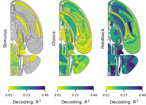
The first paper, “A brain-wide map of neural activity during complex behaviour,” showed that decision-making signals are surprisingly distributed across the brain, not localized to specific regions. This adds brain-wide evidence to a growing number of studies that challenge the traditional hierarchical model of brain function and emphasizes that there is constant communication across brain areas during decision-making, movement onset, and even reward. This means that neuroscientists will need to take a more holistic, brain-wide approach when studying complex behaviors in future.

Flat maps of the mouse brain showing which areas have significant changes in activity during each of three task intervals. Credit: Michael Schartner & International Brain Laboratory

“The unprecedented breadth of our recordings pulls back the curtain on how the entire brain performs the whole arc of sensory processing, cognitive decision-making, and movement generation,” says Fiete. “Structuring a collaboration that collects a large standardized dataset which single labs could not assemble is a revolutionary new direction for systems neuroscience, initiating the field into the hyper-collaborative mode that has contributed to leaps forward in particle physics and human genetics. Beyond our own conclusions, the dataset and associated technologies, which were released much earlier as part of the IBL mission, have already become a massively used resource for the entire neuroscience community.”

The second paper, “Brain-wide representations of prior information,” showed that prior expectations, our beliefs about what is likely to happen based on our recent experience, are encoded throughout the brain. Surprisingly, these expectations are not only found in cognitive areas, but also brain areas that process sensory information and control actions. For example, expectations are even encoded in early sensory areas such as the thalamus, the brain’s first relay for visual input from the eye. This supports the view that the brain acts as a prediction machine, but with expectations encoded across multiple brain structures playing a central role in guiding behavior responses. These findings could have implications for understanding conditions such as schizophrenia and autism, which are thought to be caused by differences in the way expectations are updated in the brain.

“Much remains to be unpacked: if it is possible to find a signal in a brain area, does it mean that this area is generating the signal, or simply reflecting a signal generated somewhere else? How strongly is our perception of the world is shaped by our expectations? Now we can generate some quantitative answers and begin the next phase experiments to learn about the origins of the expectation signals by intervening to modulate their activity,” says Fiete.

Looking ahead, the team at IBL plan to expand beyond their initial focus on decision-making to explore a broader range of neuroscience questions. With renewed funding in hand, IBL aims to expand its research scope and continue to support large-scale, standardized experiments.

New model of collaborative neuroscience

Officially launched in 2017, IBL introduced a new model of collaboration in neuroscience that uses a standardized set of tools and data processing pipelines shared across multiple labs, enabling the collection of massive datasets while ensuring data alignment and reproducibility. This approach to democratize and accelerate science draws inspiration from large-scale collaborations in physics and biology, such as CERN and the Human Genome Project.

All data from these studies, along with detailed specifications of the tools and protocols used for data collection, are openly accessible to the global scientific community for further analysis and research. Summaries of these resources can be viewed and downloaded on the IBL website under the sections: Data, Tools, Protocols.

This research was supported by grants from Wellcome (209558 and 216324), the Simons Foundation, The National Institutes of Health (NIH U19NS12371601), the National Science Foundation (NSF 1707398), the Gatsby Charitable Foundation (GAT3708), andby the Max Planck Society and the Humboldt Foundation.


Searching for self

This story also appears in the Fall 2025 issue of BrainScan

___

The question of how we know ourselves might seem the subject of philosophers, but it is just as much a matter of biology. As modern neuroscientists obtain an increasingly sophisticated understanding of how the brain generates emotions, responds to the external world, and learns from experience, some researchers are returning to a central question: How do we know our experiences, emotions, and physical sensations belong to us?

Curiosity about how the brain generates our sense of self has been a driving force for the research of McGovern Investigator Fan Wang. Following that curiosity has drawn Wang into diverse studies, exploring the origins of pain and the mechanisms we use to control our movements.

“We cannot pinpoint a set of active neurons and say that’s the sense of self. That still remains a mystery,” says Wang, who is also a professor of brain and cognitive sciences and co-director of the K. Lisa Yang and Hock E. Tan Center for Molecular Therapeutics at MIT. But she and other neuroscientists are drilling down into different functions of the brain that together might generate our awareness of ourselves.

Woman wearing blue blazer smiles and gestures off camera with man in white lab coat seated next to her.
McGovern Investigator Fan Wang (right) with research scientist Vincent Prevosto, who studies brain regions implicated in whisker movement. Photo: Steph Stevens

Wang, who teaches the undergraduate course, “Neurobiology of Self,” explains that there are lots of ways to think about our sense of self, which are probably deeply integrated in the brain. Some are mostly about our physical bodies: How do we experience touch? How do we understand
where we are in space, or recognize the boundary between ourselves and rest of the world? Some consider more internal sensations, like how we experience pain or hunger. Emotion is also key to our sense of self: How do we know that anger or joy are our own, and why do these states change the way our bodies feel?

Wang can trace her initial interest in the brain’s sense of self to work she did as a graduate student in Richard Axel’s lab at Columbia University. The lab had identified receptors expressed by sensory neurons in the nose that detect odorous substances. Wang and others discovered the pathways that information about these smells takes to the brain, and how the brain distinguishes one smell from another.

Who is the “knower” of this information? “The answer,” Wang says, “is ‘I’ or ‘me.’ But understanding where I get the sense of self and how that is constructed, is what drives me to do neuroscience.”

Mechanisms of movement

In her lab at the McGovern Institute, Wang is studying how the brain controls the body’s movements, which she sees as closely tied to the awareness of our physical selves. “The reason I think I am in my body is because I can control my movement. I generate the movement. I cannot control your movement,” says Wang. “Volitional movement gives us a sense of agency, and this sense of agency resembles the sense of self.” For the mice that the group studies, one crucial type of movement comes from the whiskers, which the animals depend on as they explore their environments. Wang’s group has traced the neural circuity that controls whiskers’ rhythmic back-and-forth, which is initiated in the brainstem, where many of the body’s most vital functions are controlled. Wang describes the simple circuit as an oscillator, or a self-generated loop.

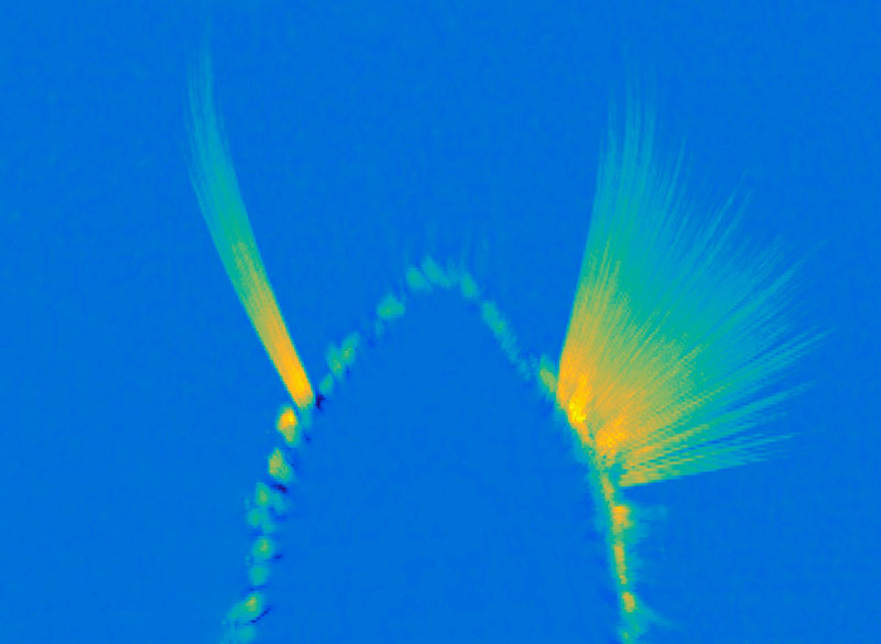
A maximum projection image showing tracked whiskers on the mouse muzzle. The right (control) side shows the back-and-forth rhythmic sweeping of the whiskers, while the experimental side where the whisking oscillator neurons are silenced, the whiskers move very little. Image: Wang Lab

Once it’s started, “the movement can go on unless some other signals stop it,” she says. The movement the circuit generates is simple but voluntary, and can be fine-tuned based on the sensory feedback the whiskers relay back to the brain. They’ve also been investigating how mice move the larynx to generate the squeaks and calls they use to communicate. These intentional movements must be coordinated with the ongoing cycles of respiration since we produce normal sounds only during expiration. Wang’s team has found neurons in the brainstem that generate vocalization-specific movements, and also discovered how respiration-controlling neural circuits can override them, ensuring that breathing is prioritized.

Wang says understanding the circuitry that controls these simple movements sets the stage for figuring out how the brain modifies activity in those circuits to create more complex, intentional movements. “That brings me closer to understanding where this volition is generated — and closer to this sense of self,” she says.

Emotional pain

Still, she knows that volitional movements — even those generated in response to perceptions of the environment — do not, on their own, define a sense of self. As a counterexample, she looks to self-driving cars: “There’s sensory information coming into the central computer, which then generates a motor output — where to drive, where to turn, where to stop. But none of us think a Waymo taxi has a sense of self.”

Wang says when she pondered the ways in which AI-powered cars lack a sense of self, she began thinking about emotions and pain. “If the self-driving Waymo crashes, it will not feel pain,” she says. “But if we hurt ourselves, we will feel pain. And we will hate that, and then we’ll learn.” So her lab is also exploring how the nervous system generates pain perception, including the emotional response that it evokes.

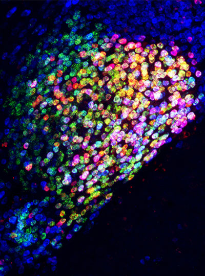
Ensembles of neurons in the amygdala activated by general anesthesia. Image: Fan Wang

In both humans and mice, pain causes emotional suffering that can be recognized and measured through changes in body functions like heart rate and blood pressure. With funding from the K. Lisa Yang Brain-Body Center at MIT, Wang’s lab is carefully tracking these involuntary, or autonomic, functions to gain a more complete understanding of pain’s emotional impact. This approach has helped clarify the role of pain-suppressing neurons in the brain’s amygdala — an important emotion-processing center — that Wang’s team discovered in 2020. When researchers selectively activate those cells in mice, the animals’ behavior makes it clear that the neurons are suppressing pain. Now, the group has learned that activating these neurons suppresses the autonomic response to pain.

Wang says there’s hope that modulating pain’s emotional response might be a way to treat chronic pain in patients. She explains that some patients with damage to another one of the brain’s emotional centers, the cingulate cortex, feel painful stimuli, but experience them as merely intense sensations. That suggests that it might be possible to modulate the emotional response to pain to eliminate patients’ suffering, without blocking the protective information that pain can provide.

The team has also been focusing on another set of anesthesia-activated neurons, which they have found suppress anxiety. When anxiety-suppressing neurons are activated in mice, the animals’ heart rates slow and they become more willing to explore bright, open spaces. Another anxiety-associated measure — heart rate variability — increases. Wang explains that this change is particularly significant: “If you have persistent low heart rate variability, especially in veterans, that is a very good predictor for anxiety developing into depression in the future,” she says.

The team’s findings, which suggest that changes in autonomic functions may themselves relieve anxiety, point toward potential new targets for anti-anxiety therapies. And by highlighting the connection between emotion and bodily responses, they offer more clues about our sense of self. “These neurons are now changing some high-level concept about anxiety,” Wang points out.

That link between emotion and body seems to Wang to be key to the sense of self. The big questions remain unanswered, but that simply stokes her curiosity. “I can be aware of my bodily responses: I am aware of ‘I am anxious’ or ‘I am in pain.’ I can see the pathways from which stimuli go into these nervous systems and come back down to the body and control the response. But I still don’t know who is the person — the knower,” she says. “I haven’t found it, so I’m going to keep looking.”