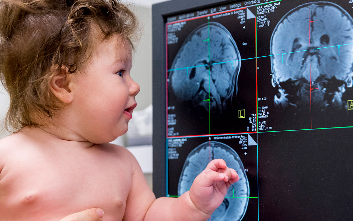
Powerful imaging methods like MRI and MEG provide neuroscientists with a detailed view of the human brain – including the spatial and temporal changes that occur as we interact with the world. Our researchers are using these tools to study how the brain develops from infancy, which regions underlie different aspects of our mental lives, and whether imaging can be used to predict the onset of disease.
To view this video please enable JavaScript, and consider upgrading to a web browser that supports HTML5 video

Brain Imaging
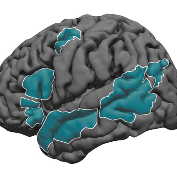
brain mapping · functional connectivity · fMRI · EEG · MEG · predictive imaging · precision interventions · contrast agents · theory of mind · the developing brain · learning
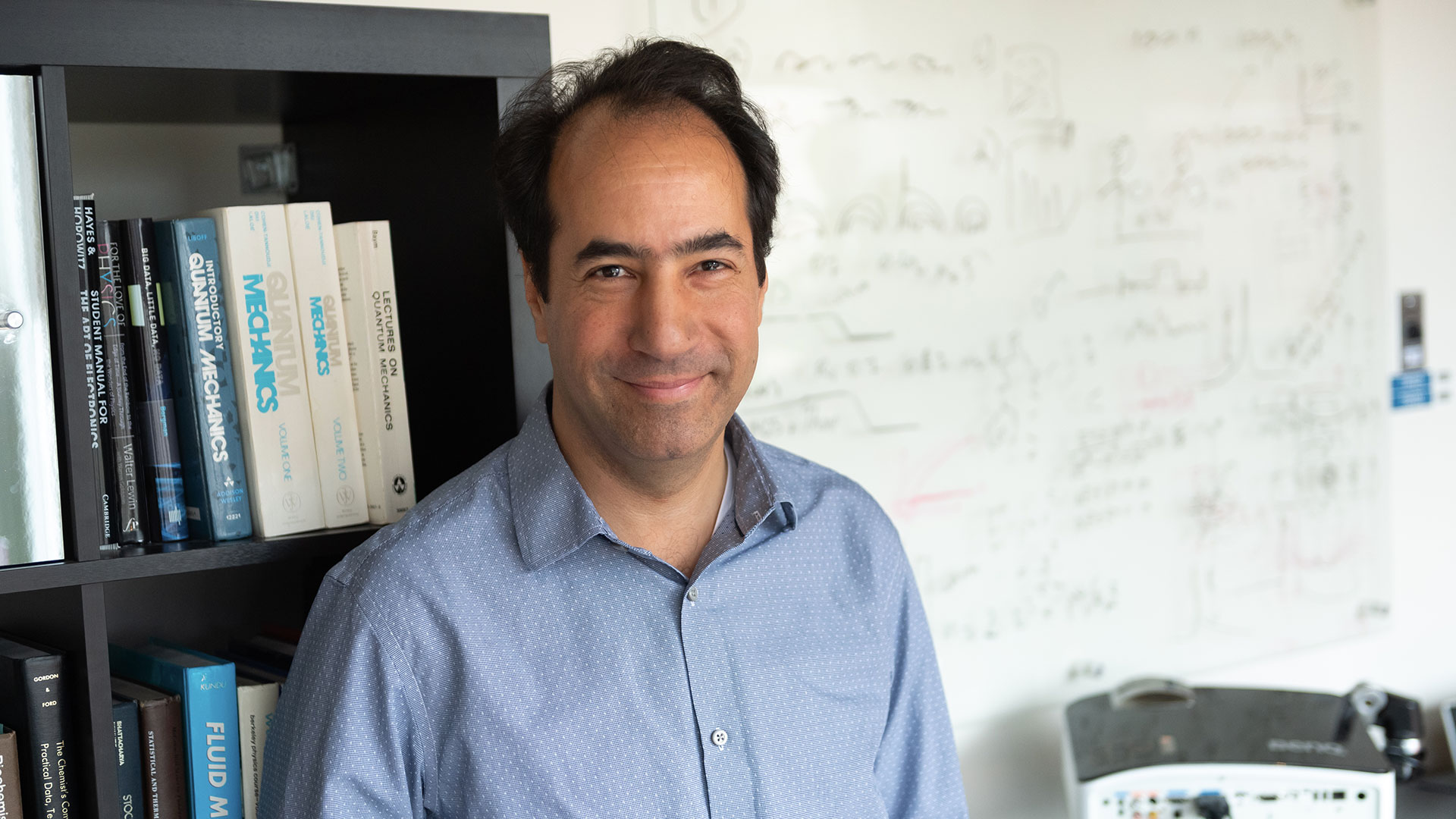
Featured Researcher
Alan Jasanoff
Alan Jasanoff develops and applies next generation imaging methods for studying the biology underlying behavior and cognition.
224
The typical number of images our scanners take for a 3D brain reconstruction.
3.5-6 minutes
The time typically taken to generate a structural scan of the adult brain using MRI.

Recent Publications
- Chandra, K, Saxe, RR, Ragan-Kelley, J, Tenenbaum, JB. Conniving With Continuations: Representing Goals in a Domain-Specific Language of Thought. Top Cogn Sci. 2026; :e70054. doi: 10.1111/tops.70054. PubMed PMID:42165328 .
- Ray, PL, Miller, JA, Jarecka, D, Smith, KA, Baker, PM, Ng, L et al.. A layered standards framework for integrating single-cell and spatial omics data into brain cell atlases. bioRxiv. 2026; :. doi: 10.64898/2026.04.30.722039. PubMed PMID:42146500 PubMed Central PMC13174332.
- Ozernov-Palchik, O, O'Brien, AM, Lee, EJ, Richardson, H, Romeo, R, Poliak, M et al.. Precision fMRI reveals that the language network exhibits adult-like left-hemispheric lateralization by 4 years of age. Nat Commun. 2026; :. doi: 10.1038/s41467-026-72916-5. PubMed PMID:42143030 .