Author: Julie Pryor
Colloquium: Joshua D. Berke | Oct 30 | 4PM | 46-3002
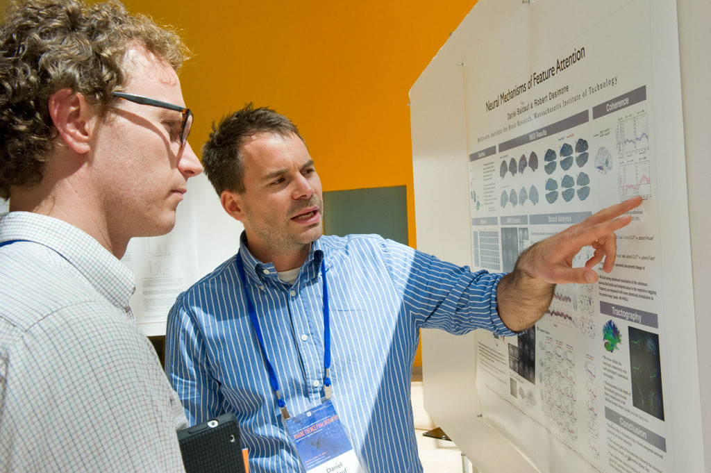
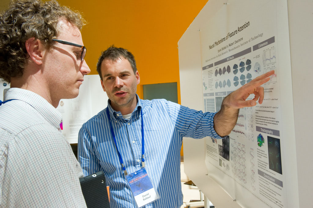
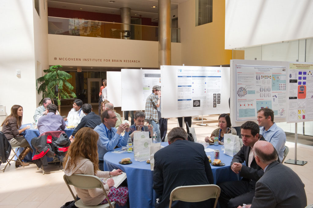
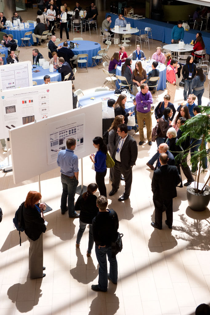
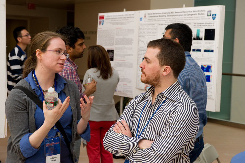
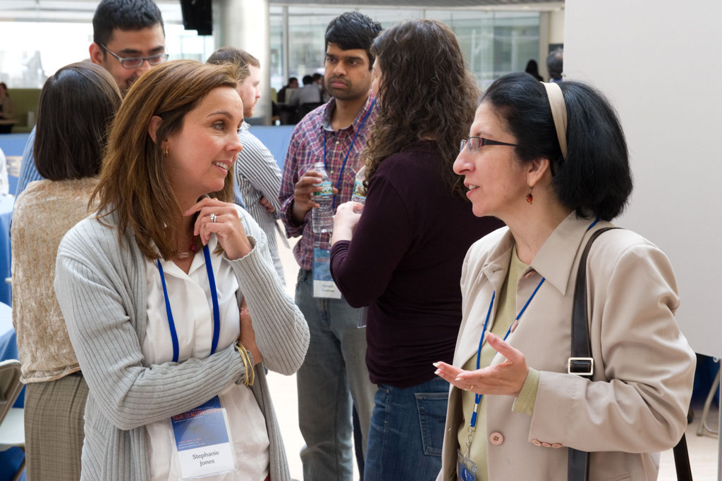
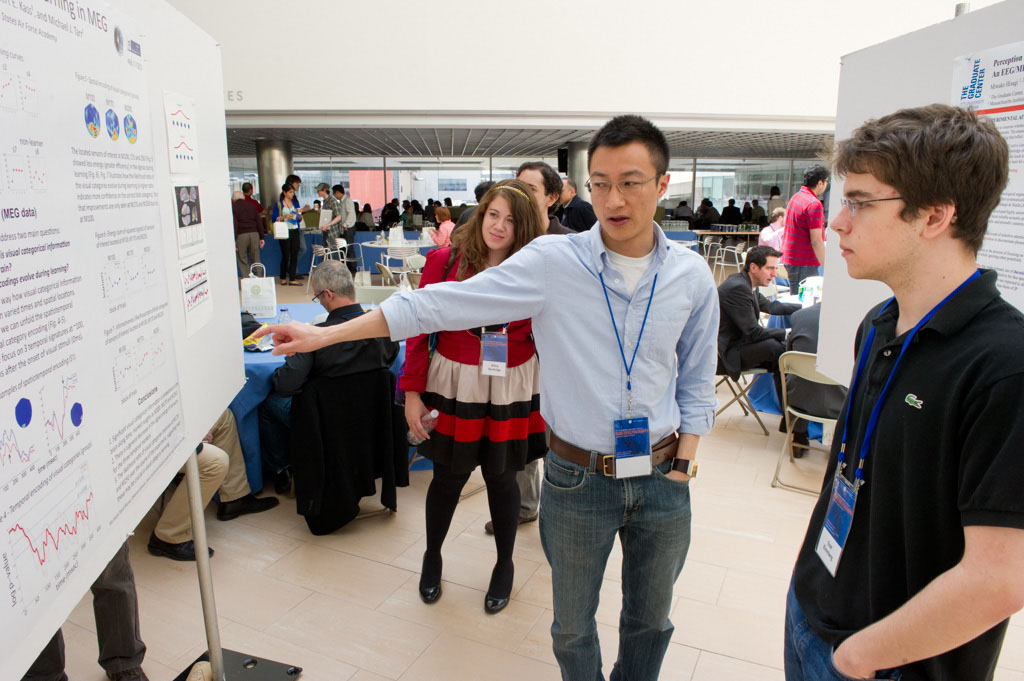
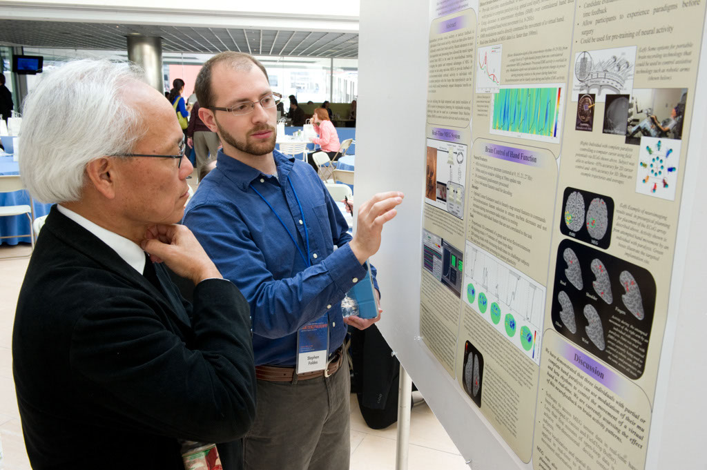
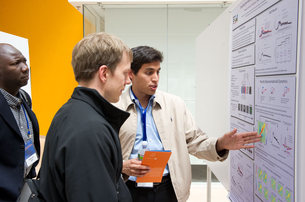
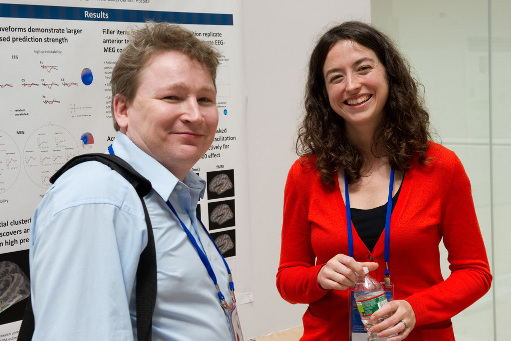
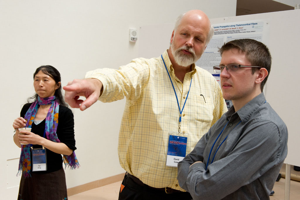
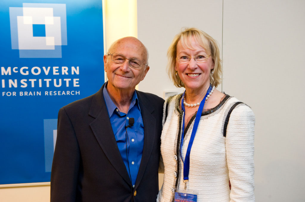
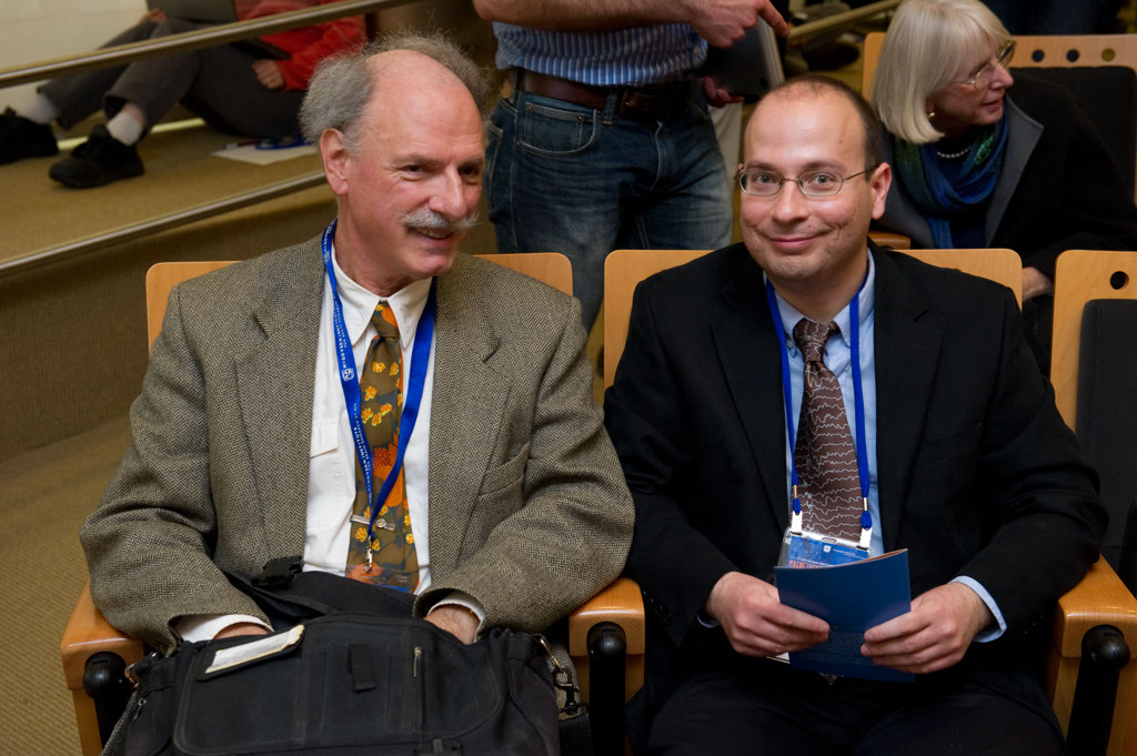
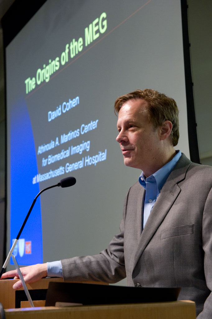
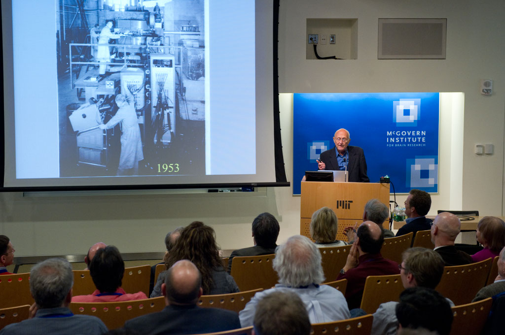
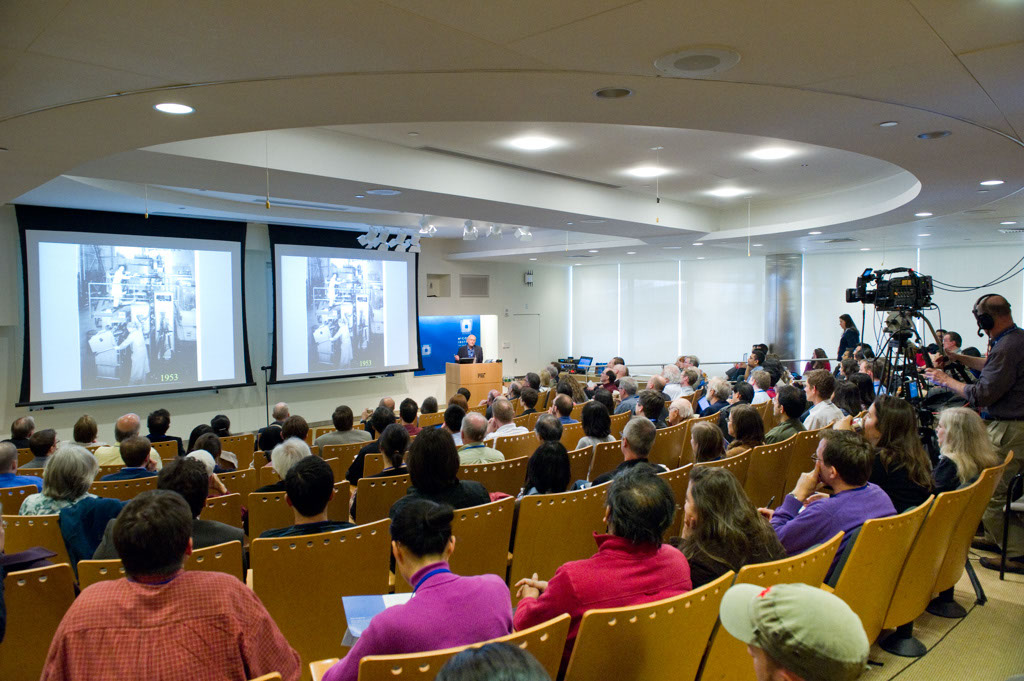
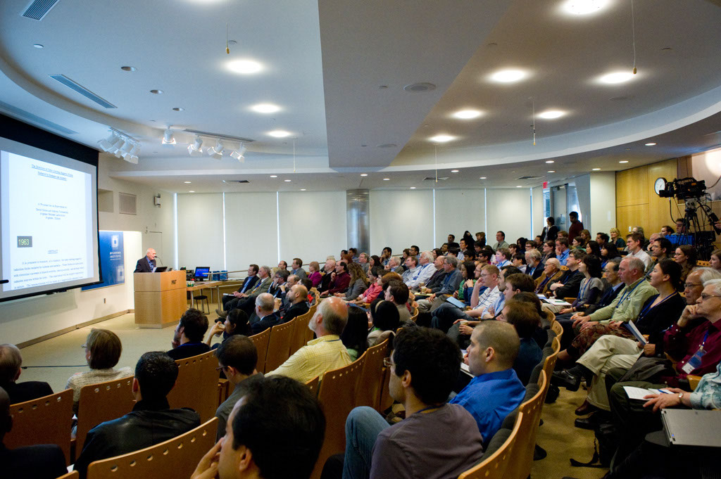
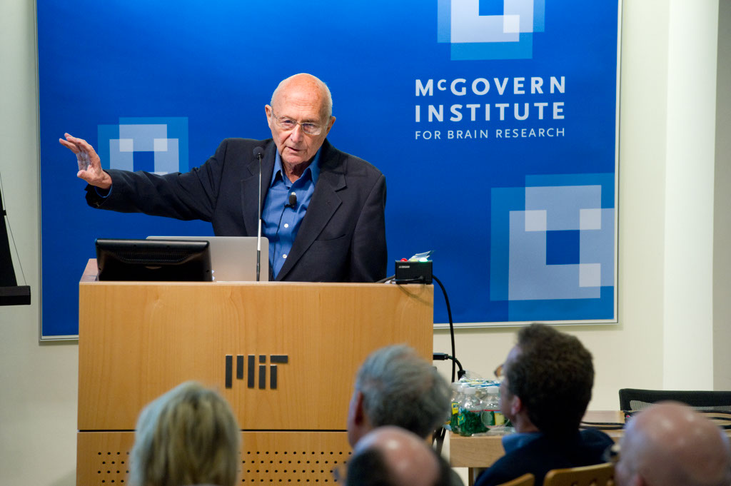
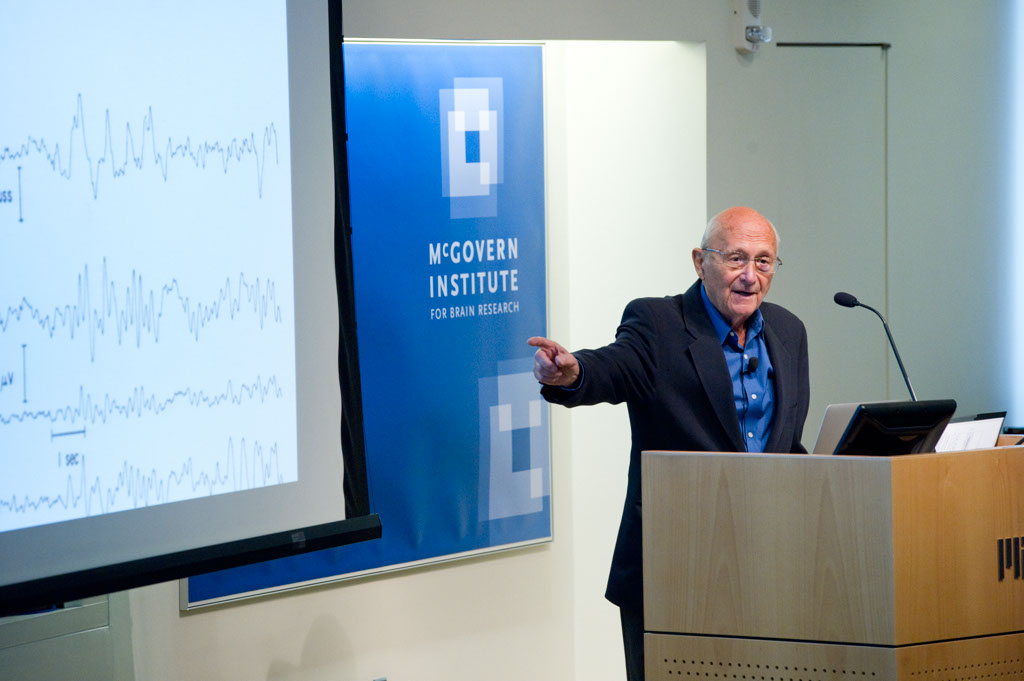
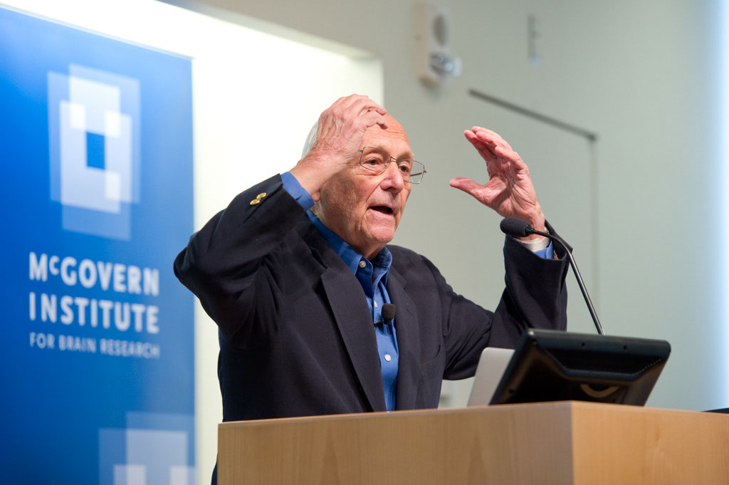
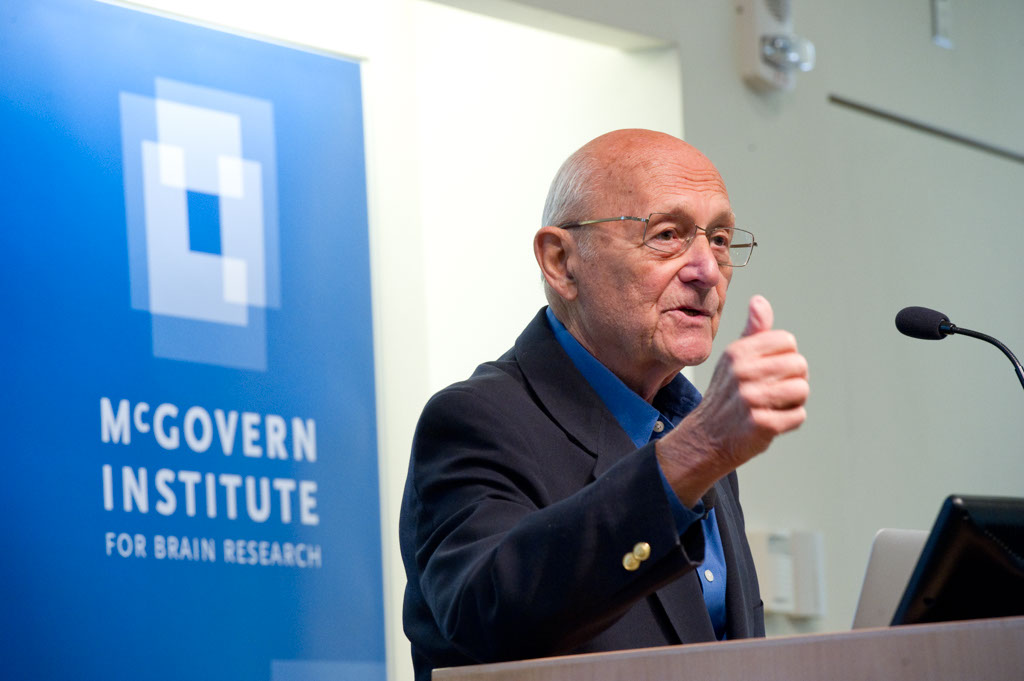
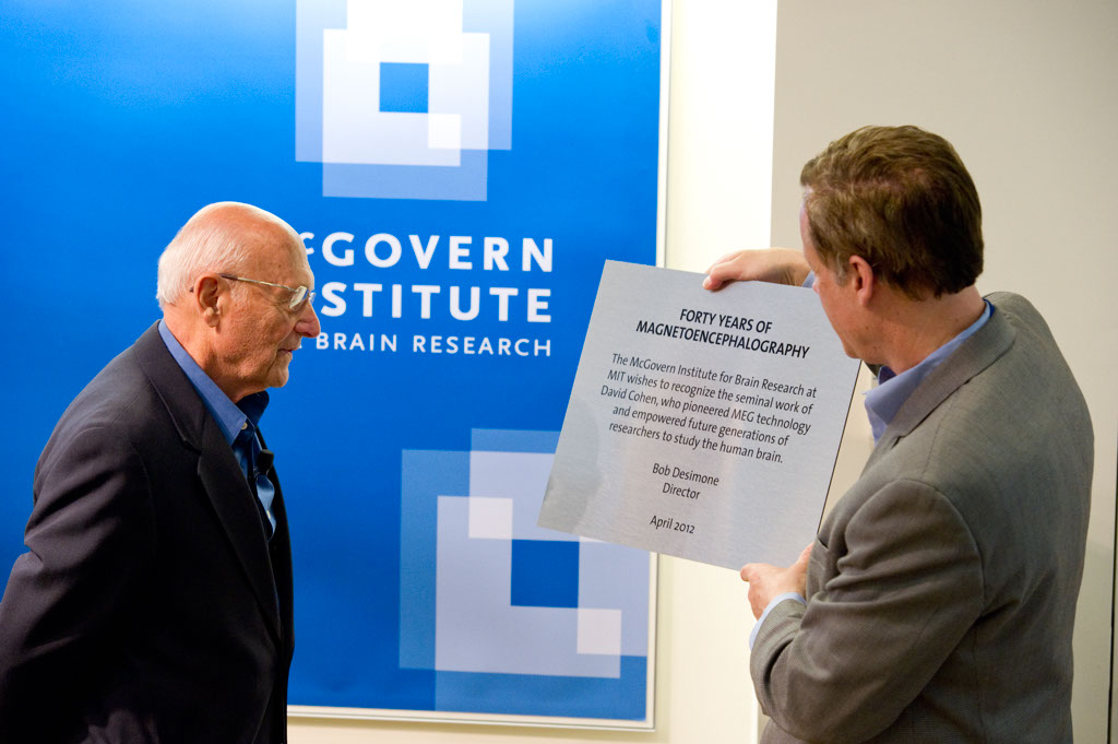
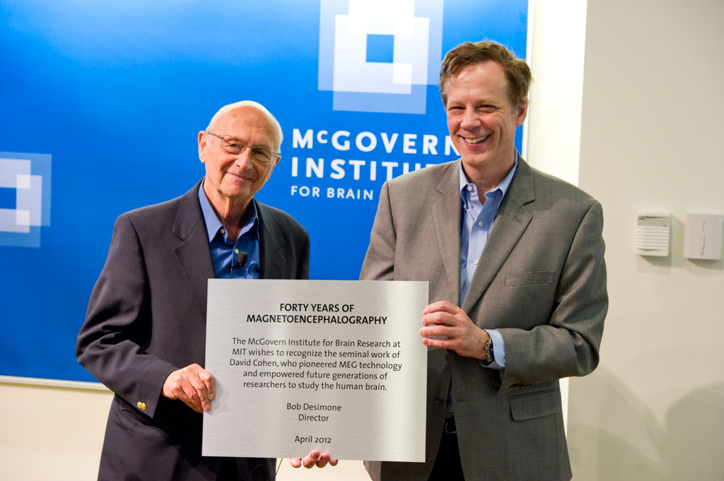
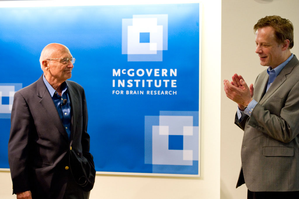
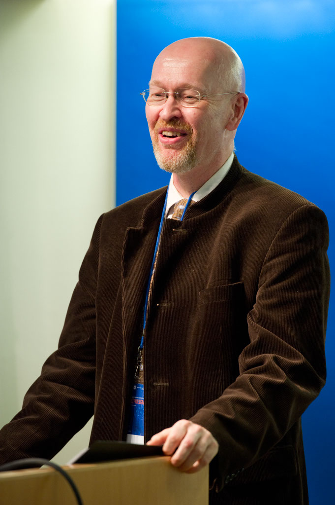
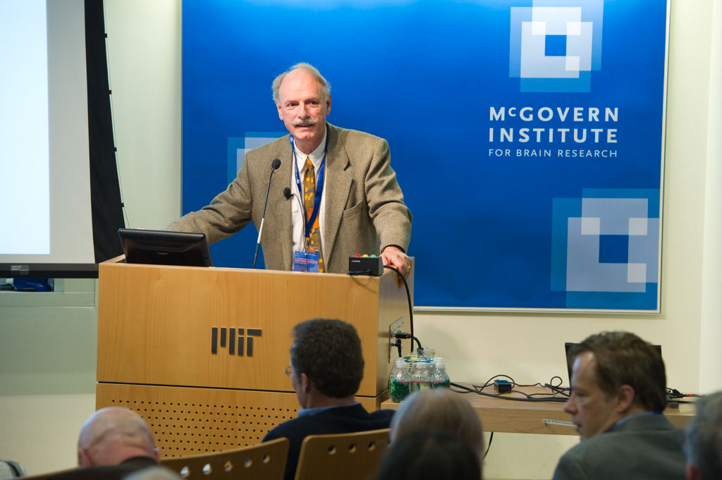
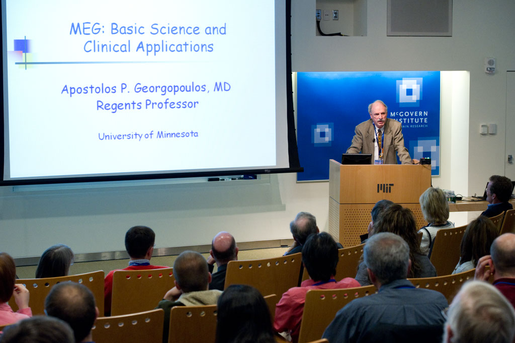
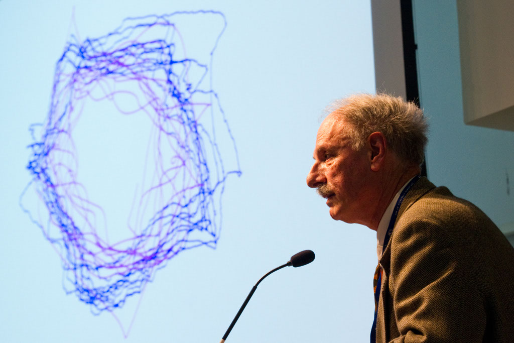
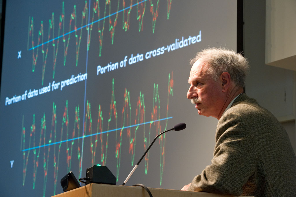
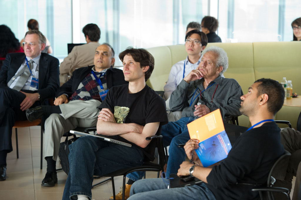
2012 MEG Symposium: Applications to Cognitive Neuroscience
To view the 2012 MEG Symposium gallery, please click on one of the thumbnail images below.
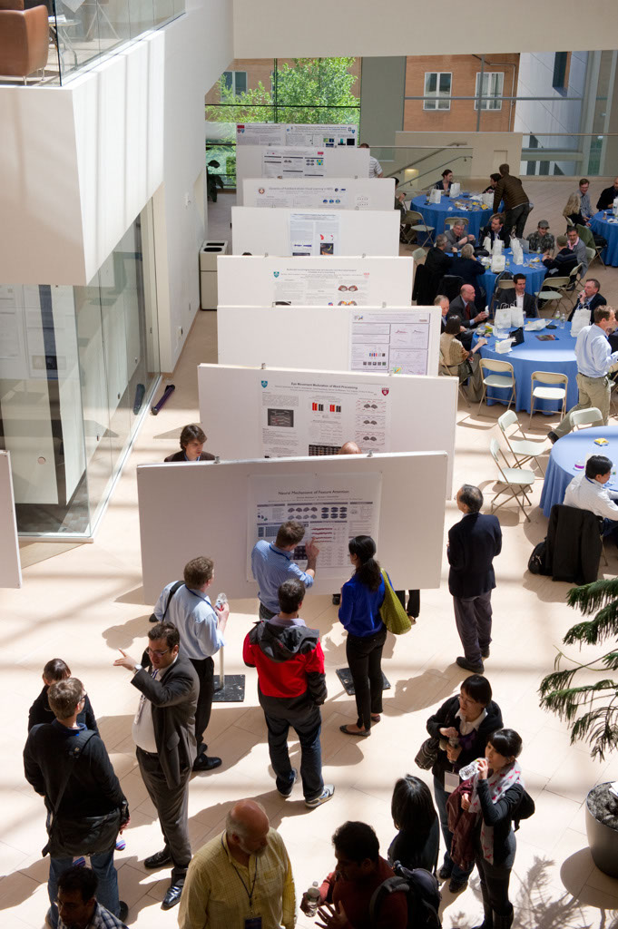


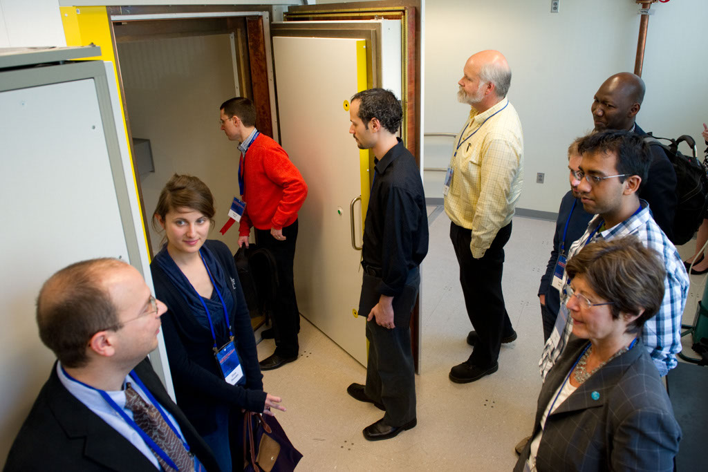

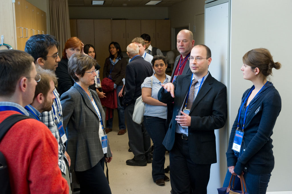
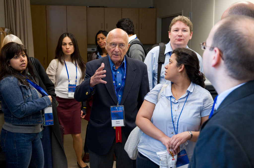


Magnetoencephalography (MEG) Lab at the McGovern Institute
Postdoc Daniel Baldauf and visiting student Yasaman Bagherzadeh are using MEG to study attention. Photo: Justin Knight
The brain is a Swiss Army Knife: Nancy Kanwisher at TED2014
Brain imaging pioneer Nancy Kanwisher, who uses fMRI scans to see activity in brain regions (often her own), shares what she and her colleagues have learned: The brain is made up of both highly specialized components and general-purpose “machinery.” Another surprise: There’s so much left to learn.
Learn more about Nancy’s research by visiting her website, which contains short talks on the different scientific methods neuroscientists can use to study the human mind and brain.
Fifteen MIT scientists receive NIH BRAIN Initiative grants
Today, the National Institutes of Health (NIH) announced their first round of BRAIN Initiative award recipients. Six teams and 15 researchers from the Massachusetts Institute of Technology were recipients.
Mriganka Sur, principal investigator at the Picower Institute for Learning and Memory and the Paul E. Newton Professor of Neuroscience in MIT’s Department of Brain and Cognitive Sciences (BCS) leads a team studying cortical circuits and information flow during memory-guided perceptual decisions. Co-principal investigators include Emery Brown, BCS professor of computational neuroscience and the Edward Hood Taplin Professor of Medical Engineering; Kwanghun Chung, Picower Institute principal investigator and assistant professor in the Department of Chemical Engineering and the Institute for Medical Engineering and Science (IMES); and Ian Wickersham, research scientist at the McGovern Institute for Brain Research and head of MIT’s Genetic Neuroengineering Group.
Elly Nedivi, Picower Institute principal investigator and professor in BCS and the Department of Biology, leads a team studying new methods for high-speed monitoring of sensory-driven synaptic activity across all inputs to single living neurons in the context of the intact cerebral cortex. Her co-principal investigator is Peter So, professor of mechanical and biological engineering, and director of the MIT Laser Biomedical Research Center.
Ian Wickersham will lead a team looking at novel technologies for nontoxic transsynaptic tracing. His co-principal investigators include Robert Desimone, director of the McGovern Institute and the Doris and Don Berkey Professor of Neuroscience in BCS; Li-Huei Tsai, director of the Picower Institute and the Picower Professor of Neuroscience in BCS; and Kay Tye, Picower Institute principal investigator and assistant professor of neuroscience in BCS.
Robert Desimone will lead a team studying vascular interfaces for brain imaging and stimulation. Co-principal investigators include Ed Boyden, associate professor at the MIT Media Lab, McGovern Institute, and departments of BCS and Biological Engineering; head of MIT’s Synthetic Neurobiology Group, and co-director of MIT’s Center for Neurobiological Engineering; and Elazer Edelman, the Thomas D. and Virginia W. Cabot Professor of Health Sciences and Technology in IMES and director of the Harvard-MIT Biomedical Engineering Center. Collaborators on this project include: Rodolfo Llinas (New York University), George Church (Harvard University), Jan Rabaey (University of California at Berkeley), Pablo Blinder (Tel Aviv University), Eric Leuthardt (Washington University/St. Louis), Michel Maharbiz (Berkeley), Jose Carmena (Berkeley), Elad Alon (Berkeley), Colin Derdeyn (Washington University in St. Louis), Lowell Wood (Bill and Melinda Gates Foundation), Xue Han (Boston University), and Adam Marblestone (MIT).
Ed Boyden will be co-principal investigator with Mark Bathe, associate professor of biological engineering, and Peng Yin of Harvard on a project to study ultra-multiplexed nanoscale in situ proteomics for understanding synapse types.
Alan Jasanoff, associate professor of biological engineering and director of the MIT Center for Neurobiological Engineering, will lead a team looking at calcium sensors for molecular fMRI. Stephen Lippard, the Arthur Amos Noyes Professor of Chemistry, is co-principal investigator.
In addition, Sur and Wickersham also received BRAIN Early Concept Grants for Exploratory Research (EAGER) from the National Science Foundation (NSF). Sur will focus on massive-scale multi-area single neuron recordings to reveal circuits underlying short-term memory. Wickersham, in collaboration with Li-Huei Tsai, Kay Tye, and Robert Desimone, will develop cell-type specific optogenetics in wild-type animals. Additional information about NSF support of the BRAIN initiative can be found at NSF.gov/brain.
The BRAIN Initiative, spearheaded by President Obama in April 2013, challenges the nation’s leading scientists to advance our sophisticated understanding of the human mind and discover new ways to treat, prevent, and cure neurological disorders like Alzheimer’s, schizophrenia, autism, and traumatic brain injury. The scientific community is charged with accelerating the invention of cutting-edge technologies that can produce dynamic images of complex neural circuits and illuminate the interaction of lightning-fast brain cells. The new capabilities are expected to provide greater insights into how brain functionality is linked to behavior, learning, memory, and the underlying mechanisms of debilitating disease. BRAIN was launched with approximately $100 million in initial investments from the NIH, the National Science Foundation, and the Defense Advanced Research Projects Agency (DARPA).
BRAIN Initiative scientists are engaged in a challenging and transformative endeavor to explore how our minds instantaneously processes, store, and retrieve vast quantities of information. Their discoveries will unlock many of the remaining mysteries inherent in the brain’s billions of neurons and trillions of connections, leading to a deeper understanding of the underlying causes of many neurological and psychiatric conditions. Their findings will enable scientists and doctors to develop the groundbreaking arsenal of tools and technologies required to more effectively treat those suffering from these devastating disorders.