Biologists discover a trigger for cell extrusion

For all animals, eliminating some cells is a necessary part of embryonic development. Living cells are also naturally sloughed off in mature tissues; for example, the lining of the intestine turns over every few days.

One way that organisms get rid of unneeded cells is through a process called extrusion, which allows cells to be squeezed out of a layer of tissue without disrupting the layer of cells left behind. MIT biologists have now discovered that this process is triggered when cells are unable to replicate their DNA during cell division.

The researchers discovered this mechanism in the worm C. elegans, and they showed that the same process can be driven by mammalian cells; they believe extrusion may serve as a way for the body to eliminate cancerous or precancerous cells.

“Cell extrusion is a mechanism of cell elimination used by organisms as diverse as sponges, insects, and humans,” says H. Robert Horvitz, the David H. Koch Professor of Biology at MIT, a member of the McGovern Institute for Brain Research and the Koch Institute for Integrative Cancer Research, a Howard Hughes Medical Institute investigator, and the senior author of the study. “The discovery that extrusion is driven by a failure in DNA replication was unexpected and offers a new way to think about and possibly intervene in certain diseases, particularly cancer.”

MIT postdoc Vivek Dwivedi is the lead author of the paper, which appears today in Nature. Other authors of the paper are King’s College London research fellow Carlos Pardo-Pastor, MIT research specialist Rita Droste, MIT postdoc Ji Na Kong, MIT graduate student Nolan Tucker, Novartis scientist and former MIT postdoc Daniel Denning, and King’s College London professor of biology Jody Rosenblatt.

Stuck in the cell cycle

In the 1980s, Horvitz was one of the first scientists to analyze a type of programmed cell suicide called apoptosis, which organisms use to eliminate cells that are no longer needed. He made his discoveries using C. elegans, a tiny nematode that contains exactly 959 cells. The developmental lineage of each cell is known, and embryonic development follows the same pattern every time. Throughout this developmental process, 1,090 cells are generated, and 131 cells undergo programmed cell suicide by apoptosis.

Horvitz’s lab later showed that if the worms were genetically mutated so that they could not eliminate cells by apoptosis, a few of those 131 cells would instead be eliminated by cell extrusion, which appears to be able to serve as a backup mechanism to apoptosis. How this extrusion process gets triggered, however, remained a mystery.

To unravel this mystery, Dwivedi performed a large-scale screen of more than 11,000 C. elegans genes. One by one, he and his colleagues knocked down the expression of each gene in worms that could not perform apoptosis. This screen allowed them to identify genes that are critical for turning on cell extrusion during development.

To the researchers’ surprise, many of the genes that turned up as necessary for extrusion were involved in the cell division cycle. These genes were primarily active during first steps of the cell cycle, which involve initiating the cell division cycle and copying the cell’s DNA.

Further experiments revealed that cells that are eventually extruded do initially enter the cell cycle and begin to replicate their DNA. However, they appear to get stuck in this phase, leading them to be extruded.

Most of the cells that end up getting extruded are unusually small, and are produced from an unequal cell division that results in one large daughter cell and one much smaller one. The researchers showed that if they interfered with the genes that control this process, so that the two daughter cells were closer to the same size, the cells that normally would have been extruded were able to successfully complete the cell cycle and were not extruded.

The researchers also showed that the failure of the very small cells to complete the cell cycle stems from a shortage of the proteins and DNA building blocks needed to copy DNA. Among other key proteins, the cells likely don’t have enough of an enzyme called LRR-1, which is critical for DNA replication. When DNA replication stalls, proteins that are responsible for detecting replication stress quickly halt cell division by inactivating a protein called CDK1. CDK1 also controls cell adhesion, so the researchers hypothesize that when CDK1 is turned off, cells lose their stickiness and detach, leading to extrusion.

Cancer protection

Horvitz’s lab then teamed up with researchers at King’s College London, led by Rosenblatt, to investigate whether the same mechanism might be used by mammalian cells. In mammals, cell extrusion plays an important role in replacing the lining of the intestines, lungs, and other organs.

The researchers used a chemical called hydroxyurea to induce DNA replication stress in canine kidney cells grown in cell culture. The treatment quadrupled the rate of extrusion, and the researchers found that the extruded cells made it into the phase of the cell cycle where DNA is replicated before being extruded. They also showed that in mammalian cells, the well-known cancer suppressor p53 is involved in initiating extrusion of cells experiencing replication stress.

That suggests that in addition to its other cancer-protective roles, p53 may help to eliminate cancerous or precancerous cells by forcing them to extrude, Dwivedi says.

“Replication stress is one of the characteristic features of cells that are precancerous or cancerous. And what this finding suggests is that the extrusion of cells that are experiencing replication stress is potentially a tumor suppressor mechanism,” he says.

The fact that cell extrusion is seen in so many animals, from sponges to mammals, led the researchers to hypothesize that it may have evolved as a very early form of cell elimination that was later supplanted by programmed cell suicide involving apoptosis.

“This cell elimination mechanism depends only on the cell cycle,” Dwivedi says. “It doesn’t require any specialized machinery like that needed for apoptosis to eliminate these cells, so what we’ve proposed is that this could be a primordial form of cell elimination. This means it may have been one of the first ways of cell elimination to come into existence, because it depends on the same process that an organism uses to generate many more cells.”

Dwivedi, who earned his PhD at MIT, was a Khorana scholar before entering MIT for graduate school. This research was supported by the Howard Hughes Medical Institute and the National Institutes of Health.

Josh McDermott seeks to replicate the human auditory system

The human auditory system is a marvel of biology. It can follow a conversation in a noisy restaurant, learn to recognize words from languages we’ve never heard before, and identify a familiar colleague by their footsteps as they walk by our office.

So far, even the most sophisticated computational models cannot perform such tasks as well as the human auditory system, but MIT neuroscientist Josh McDermott hopes to change that. Achieving this goal would be a major step toward developing new ways to help people with hearing loss, says McDermott, who recently earned tenure in MIT’s Department of Brain and Cognitive Sciences.

“Our long-term goal is to build good predictive models of the auditory system,” McDermott says.

“If we were successful in that goal, then it would really transform our ability to make people hear better, because we could design a computer program to figure out what to do to incoming sound to make it easier to recognize what somebody said or where a sound is coming from.”

McDermott’s lab also explores how exposure to different types of music affects people’s music preferences and even how they perceive music. Such studies can help to reveal elements of sound perception that are “hardwired” into our brains, and other elements that are influenced by exposure to different kinds of sounds.

“We have found that there is cross-cultural variation in things that people had widely supposed were universal and possibly even innate,” McDermott says.

Sound perception

As an undergraduate at Harvard University, McDermott originally planned to study math and physics, but “I was very quickly seduced by the brain,” he says. At the time, Harvard did not offer a major in neuroscience, so McDermott created his own, with a focus on vision.

After earning a master’s degree from University College London, he came to MIT to do a PhD in brain and cognitive sciences. His focus was still on vision, which he studied with Ted Adelson, the John and Dorothy Wilson Professor of Vision Science, but he found himself increasingly interested in audition. He had always loved music, and around this time, he started working as a radio and club DJ. “I was spending a lot of time thinking about sound and why things sound the way they do,” he recalls.

To pursue his new interest, he served as a postdoc at the University of Minnesota, where he worked in a lab devoted to psychoacoustics — the study of how humans perceive sound. There, he studied auditory phenomena such as the “cocktail party effect,” or the ability to focus on a particular person’s voice while tuning out background noise. During another postdoc at New York University, he started working on computational models of the auditory system. That interest in computation is part of what drew him back to MIT as a faculty member, in 2013.

“The culture here surrounding brain and cognitive science really prioritizes and values computation, and that was a perspective that was important to me,” says McDermott, who is also a member of MIT’s McGovern Institute for Brain Research and the Center for Brains, Minds and Machines. “I knew that was the kind of work I really wanted to do in my lab, so it just felt like a natural environment for doing that work.”

One aspect of audition that McDermott’s lab focuses on is “auditory scene analysis,” which includes tasks such as inferring what events in the environment caused a particular sound, and determining where a particular sound came from. This requires the ability to disentangle sounds produced by different events or objects, and the ability to tease out the effects of the environment. For instance, a basketball bouncing on a hardwood floor in a gym makes a different sound than a basketball bouncing on an outdoor paved court.

“Sounds in the world have very particular properties, due to physics and the way that the world works,” McDermott says. “We believe that the brain internalizes those regularities, and you have models in your head of the way that sound is generated. When you hear something, you are performing an inference in that model to figure out what is likely to have happened that caused the sound.”

A better understanding of how the brain does this may eventually lead to new strategies to enhance human hearing, McDermott says.

“Hearing impairment is the most common sensory disorder. It affects almost everybody as they get older, and the treatments are OK, but they’re not great,” he says. “We’re eventually going to all have personalized hearing aids that we walk around with, and we just need to develop the right algorithms in order to tell them what to do. That’s something we’re actively working on.”

Music in the brain

About 10 years ago, when McDermott was a postdoc, he started working on cross-cultural studies of how the human brain perceives music. Richard Godoy, an anthropologist at Brandeis University, asked McDermott to join him for some studies of the Tsimane’ people, who live in the Amazon rainforest. Since then, McDermott and some of his students have gone to Bolivia most summers to study sound perception among the Tsimane’. The Tsimane’ have had very little exposure to Western music, making them ideal subjects to study how listening to certain kinds of music influences human sound perception.

These studies have revealed both differences and similarities between Westerners and the Tsimane’ people. McDermott, who counts soul, disco, and jazz-funk among his favorite types of music, has found that Westerners and the Tsimane’ differ in their perceptions of dissonance. To Western ears, for example, the chord of C and F# sounds very unpleasant, but not to the Tsimane’.

He has also shown that that people in Western society perceive sounds that are separated by an octave to be similar, but the Tsimane’ do not. However, there are also some similarities between the two groups. For example, the upper limit of frequencies that can be perceived appears to be the same regardless of music exposure.

“We’re finding both striking variation in some perceptual traits that many people presumed were common across cultures and listeners, and striking similarities in others,” McDermott says. “The similarities and differences across cultures dissociate aspects of perception that are tightly coupled in Westerners, helping us to parcellate perceptual systems into their underlying components.”

Investigating the embattled brain

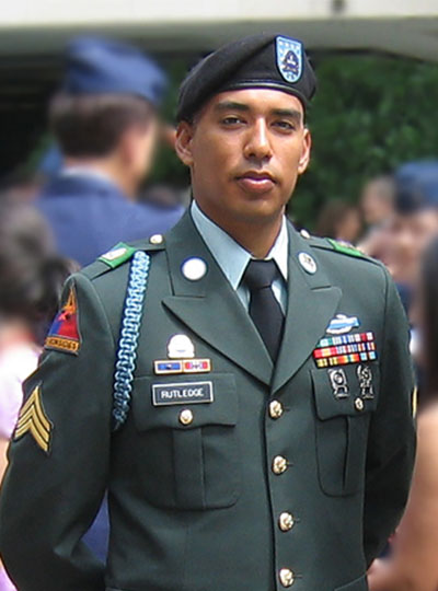
Omar Rutledge served as a US Army infantryman in the 1st Armored and 25th Infantry Divisions. He was deployed in support of Operation Iraqi Freedom from March 2003 to July 2004. Photo: Omar Rutledge

As an Iraq war veteran, Omar Rutledge is deeply familiar with post-traumatic stress – recurring thoughts and memories that persist long after a danger has passed – and he knows that a brain altered by trauma is not easily fixed. But as a graduate student in the Department of Brain and Cognitive Sciences, Rutledge is determined to change that. He wants to understand exactly how trauma alters the brain – and whether the tools of neuroscience can be used to help fellow veterans with post-traumatic stress disorder (PTSD) heal from their experiences.

“In the world of PTSD research, I look to my left and to my right, and I don’t see other veterans, certainly not former infantrymen,” says Rutledge, who served in the US Army and was deployed to Iraq from March 2003 to July 2004. “If there are so few of us in this space, I feel like I have an obligation to make a difference for all who suffer from the traumatic experiences of war.”

Rutledge is uniquely positioned to make such a difference in the lab of McGovern Investigator John Gabrieli, where researchers use technologies like magnetic resonance imaging (MRI), electroencephalography (EEG), and magnetoencephalography (MEG) to peer into the human brain and explore how it powers our thoughts, memories, and emotions. Rutledge is studying how PTSD weakens the connection between the amygdala, which is responsible for emotions like fear, and the prefrontal cortex, which regulates or controls these emotional responses. He hopes these studies will eventually lead to the development of wearable technologies that can retrain the brain to be less responsive to triggering events.

“I feel like it has been a mission of mine to do this kind of work.”

Though Covid-19 has unexpectedly paused some aspects of his research, Rutledge is pursuing another line of research inspired both by the mandatory social distancing protocols imposed during the lockdown and his own experiences with social isolation. Does chronic social isolation cause physical or chemical changes in the brain similar to those seen in PTSD? And does loneliness exacerbate symptoms of PTSD?

“There’s this hypervigilance that occurs in loneliness, and there’s also something very similar that occurs in PTSD — a heightened awareness of potential threats,” says Rutledge, who is the recipient of Michael Ferrara Graduate Fellowship provided by the Poitras Center, a fellowship made possible by the many friends and family of Michael Ferrara. “The combination of the two may lead to more adverse reactions in people with PTSD.”

In the future, Rutledge hopes to explore whether chronic loneliness impairs reasoning and logic skills and has a deeper impact on veterans who have PTSD.

Although his research tends to resurface painful memories of his own combat experiences, Rutledge says if it can help other veterans heal, it’s worth it.  “In the process, it makes me a little bit stronger as well,” he adds.

Nine MIT students awarded 2021 Paul and Daisy Soros Fellowships for New Americans

An MIT senior and eight MIT graduate students are among the 30 recipients of this year’s P.D. Soros Fellowships for New Americans. In addition to senior Fiona Chen, MIT’s newest Soros winners include graduate students Aziza Almanakly, Alaleh Azhir, Brian Y. Chang PhD ’18, James Diao, Charlie ChangWon Lee, Archana Podury, Ashwin Sah ’20, and Enrique Toloza. Six of the recipients are enrolled at the Harvard-MIT Program in Health Sciences and Technology.

P.D. Soros Fellows receive up to $90,000 to fund their graduate studies and join a lifelong community of new Americans from different backgrounds and fields. The 2021 class was selected from a pool of 2,445 applicants, marking the most competitive year in the fellowship’s history.

The Paul & Daisy Soros Fellowships for New Americans program honors the contributions of immigrants and children of immigrants to the United States. As Fiona Chen says, “Being a new American has required consistent confrontation with the struggles that immigrants and racial minorities face in the U.S. today. It has meant frequent difficulties with finding security and comfort in new contexts. But it has also meant continual growth in learning to love the parts of myself — the way I look; the things that my family and I value — that have marked me as different, or as an outsider.”

Students interested in applying to the P.D. Soros fellowship should contact Kim Benard, assistant dean of distinguished fellowships in Career Advising and Professional Development.

Aziza Almanakly

Aziza Almanakly, a PhD student in electrical engineering and computer science, researches microwave quantum optics with superconducting qubits for quantum communication under Professor William Oliver in the Department of Physics. Almanakly’s career goal is to engineer multi-qubit systems that push boundaries in quantum technology.

Born and raised in northern New Jersey, Almanakly is the daughter of Syrian immigrants who came to the United States in the early 1990s in pursuit of academic opportunities. As the civil war in Syria grew dire, more of her relatives sought asylum in the U.S. Almanakly grew up around extended family who built a new version of their Syrian home in New Jersey.

Following in the footsteps of her mathematically minded father, Almanakly studied electrical engineering at The Cooper Union for the Advancement of Science and Art. She also pursued research opportunities in experimental quantum computing at Princeton University, the City University of New York, New York University, and Caltech.

Almanakly recognizes the importance of strong mentorship in diversifying engineering. She uses her unique experience as a New American and female engineer to encourage students from underrepresented backgrounds to enter STEM fields.

Alaleh Azhir

Alaleh Azhir grew up in Iran, where she pursued her passion for mathematics. She immigrated with her mother to the United States at age 14. Determined to overcome strict gender roles she had witnessed for women, Azhir is dedicated to improving health care for them.

Azhir graduated from Johns Hopkins University in 2019 with a perfect GPA as a triple major in biomedical engineering, computer science, and applied mathematics and statistics. A Rhodes and Barry Goldwater Scholar, she has developed many novel tools for visualization and analysis of genomics data at Johns Hopkins University, Harvard University, MIT, the National Institutes of Health, and laboratories in Switzerland.

After completing a master’s in statistical science at Oxford University, Azhir began her MD studies in the Harvard-MIT Program in Health Sciences and Technology. Her thesis focuses on the role of X and Y sex chromosomes on disease manifestations. Through medical training, she aims to build further computational tools specifically for preventive care for women. She has also founded and directs the nonprofit organization, Frappa, aimed at mentoring women living in Iran and helping them to immigrate abroad through the graduate school application process.

Brian Y. Chang PhD ’18

Born in Johnson City, New York, Brian Y. Chang PhD ’18 is the son of immigrants from the Shanghai municipality and Shandong Province in China. He pursued undergraduate and master’s degrees in mechanical engineering at Carnegie Mellon University, graduating in a combined four years with honors.

In 2018, Chang completed a PhD in medical engineering at MIT. Under the mentorship of Professor Elazer Edelman, Chang developed methods that make advanced cardiac technologies more accessible. The resulting approaches are used in hospitals around the world. Chang has published extensively and holds five patents.

With the goal of harnessing the power of engineering to improve patient care, Chang co-founded X-COR Therapeutics, a seed-funded medical device startup developing a more accessible treatment for lung failure with the potential to support patients with severe Covid-19 and chronic obstructive pulmonary disease.

After spending time in the hospital connecting with patients and teaching cardiovascular pathophysiology to medical students, Chang decided to attend medical school. He is currently a medical student in the Harvard-MIT Program in Health Sciences and Technology. Chang hopes to advance health care through medical device innovation and education as a future physician-scientist, entrepreneur, and educator.

Fiona Chen

MIT senior Fiona Chen was born in Cedar Park, Texas, the daughter of immigrants from China. Witnessing how her own and many other immigrant families faced significant difficulties finding work and financial stability sparked her interest in learning about poverty and economic inequality.

At MIT, Chen has pursued degrees in economics and mathematics. Her economics research projects have examined important policy issues — social isolation among students, global development and poverty, universal health-care systems, and the role of technology in shaping the labor market.

An active member of the MIT community, Chen has served as the officer on governance and officer on policy of the Undergraduate Association, MIT’s student government; the opinion editor of The Tech student newspaper; the undergraduate representative of several Institute-wide committees, including MIT’s Corporation Joint Advisory Committee; and one of the founding members of MIT Students Against War. In each of these roles, she has worked to advocate for policies to support underrepresented groups at MIT.

As a Soros fellow, Chen will pursue a PhD in economics to deepen her understanding of economic policy. Her ultimate goal is to become a professor who researches poverty and economic inequality, and applies her findings to craft policy solutions.

James Diao

James Diao graduated from Yale University with degrees in statistics and biochemistry and is currently a medical student at the Harvard-MIT Program in Health Sciences and Technology. He aspires to give voice to patient perspectives in the development and evaluation of health-care technology.

Diao grew up in Houston’s Chinatown, and spent summers with his extended family in Jiangxian. Diao’s family later moved to Fort Bend, Texas, where he found a pediatric oncologist mentor who introduced him to the wonders of modern molecular biology.

Diao’s interests include the responsible development of technology. At Apple, he led projects to validate wearable health features in diverse populations; at PathAI, he built deep learning models to broaden access to pathologist services; at Yale, where he worked on standardizing analyses of exRNA biomarkers; and at Harvard, he studied the impacts of clinical guidelines on marginalized groups.

Diao’s lead author research in the New England Journal of Medicine and JAMA systematically compared race-based and race-free equations for kidney function, and demonstrated that up to 1 million Black Americans may receive unequal kidney care due to their race. He has also published articles on machine learning and precision medicine.

Charlie ChangWon Lee

Born in Seoul, South Korea, Charlie ChangWon Lee was 10 when his family immigrated to the United States and settled in Palisades Park, New Jersey. The stress of his parents’ lack of health coverage ignited Lee’s determination to study the reasons for the high cost of health care in the U.S. and learn how to care for uninsured families like his own.

Lee graduated summa cum laude in integrative biology from Harvard College, winning the Hoopes Prize for his thesis on the therapeutic potential of human gut microbes. Lee’s research on novel therapies led him to question how newly approved, and expensive, medications could reach more patients.

At the Program on Regulation, Therapeutics, and Law (PORTAL) at Brigham and Women’s Hospital, Lee studied policy issues involving pharmaceutical drug pricing, drug development, and medication use and safety. His articles have appeared in JAMA, Health Affairs, and Mayo Clinic Proceedings.

As a first-year medical student at the Harvard-MIT Health Sciences and Technology program, Lee is investigating policies to incentivize vaccine and biosimilar drug development. He hopes to find avenues to bridge science and policy and translate medical innovations into accessible, affordable therapies.

Archana Podury

The daughter of Indian immigrants, Archana Podury was born in Mountain View, California. As an undergraduate at Cornell University, she studied the neural circuits underlying motor learning. Her growing interest in whole-brain dynamics led her to the Princeton Neuroscience Institute and Neuralink, where she discovered how brain-machine interfaces could be used to understand diffuse networks in the brain.

While studying neural circuits, Podury worked at a syringe exchange in Ithaca, New York, where she witnessed firsthand the mechanics of court-based drug rehabilitation. Now, as an MD student in the Harvard-MIT Health Sciences and Technology program, Podury is interested in combining computational and social approaches to neuropsychiatric disease.

In the Boyden Lab at the MIT McGovern Institute for Brain Research, Podury is developing human brain organoid models to better characterize circuit dysfunction in neurodevelopmental disorders. Concurrently, her work in the Dhand Lab at Brigham and Women’s Hospital applies network science tools to understand how patients’ social environments influence their health outcomes following acute neurological injury.

Podury hopes that focusing on both neural and social networks can lead toward a more comprehensive, and compassionate, approach to health and disease.

Ashwin Sah ’20

Ashwin Sah ’20 was born and raised in Portland, Oregon, the son of Indian immigrants. He developed a passion for mathematics research as an undergraduate at MIT, where he conducted research under Professor Yufei Zhao, as well as at the Duluth and Emory REU (Research Experience for Undergraduates) programs.

Sah has given talks on his work at multiple professional venues. His undergraduate research in varied areas of combinatorics and discrete mathematics culminated in the Barry Goldwater Scholarship and the Frank and Brennie Morgan Prize for Outstanding Research in Mathematics by an Undergraduate Student. Additionally, his work on diagonal Ramsey numbers was recently featured in Quanta Magazine.

Beyond research, Sah has pursued opportunities to give back to the math community, helping to organize or grade competitions such as the Harvard-MIT Mathematics Tournament and the USA Mathematical Olympiad. He has also been a grader at the Mathematical Olympiad Program, a camp for talented high-school students in the United States, and an instructor for the Monsoon Math Camp, a virtual program aimed at teaching higher mathematics to high school students in India.

Sah is currently a PhD student in mathematics at MIT, where he continues to work with Zhao.

Enrique Toloza

Enrique Toloza was born in Los Angeles, California, the child of two immigrants: one from Colombia who came to the United States for a PhD and the other from the Philippines who grew up in California and went on to medical school. Their literal marriage of science and medicine inspired Toloza to become a physician-scientist.

Toloza majored in physics and Spanish literature at the University of North Carolina at Chapel Hill. He eventually settled on an interest in theoretical neuroscience after a summer research internship at MIT and completing an honors thesis on noninvasive brain stimulation.

After college, Toloza joined Professor Mark Harnett’s laboratory at MIT for a year. He went on to enroll in the Harvard-MIT MD/PhD program, studying within the Health Sciences and Technology MD curriculum at Harvard and the PhD program at MIT. For his PhD, Toloza rejoined Harnett to conduct research on the biophysics of dendritic integration and the contribution of dendrites to cortical computations in the brain.

Toloza is passionate about expanding health care access to immigrant populations. In college, he led the interpreting team at the University of North Carolina at Chapel Hill’s student-run health clinic; at Harvard Medical School, he has worked with Spanish-speaking patients as a student clinician.

Method offers inexpensive imaging at the scale of virus particles

Using an ordinary light microscope, MIT engineers have devised a technique for imaging biological samples with accuracy at the scale of 10 nanometers — which should enable them to image viruses and potentially even single biomolecules, the researchers say.

The new technique builds on expansion microscopy, an approach that involves embedding biological samples in a hydrogel and then expanding them before imaging them with a microscope. For the latest version of the technique, the researchers developed a new type of hydrogel that maintains a more uniform configuration, allowing for greater accuracy in imaging tiny structures.

This degree of accuracy could open the door to studying the basic molecular interactions that make life possible, says Edward Boyden, the Y. Eva Tan Professor in Neurotechnology, a professor of biological engineering and brain and cognitive sciences at MIT, and a member of MIT’s McGovern Institute for Brain Research and Koch Institute for Integrative Cancer Research.

“If you could see individual molecules and identify what kind they are, with single-digit-nanometer accuracy, then you might be able to actually look at the structure of life.”

“And structure, as a century of modern biology has told us, governs function,” says Boyden, who is the senior author of the new study.

The lead authors of the paper, which appears today in Nature Nanotechnology, are MIT Research Scientist Ruixuan Gao and Chih-Chieh “Jay” Yu PhD ’20. Other authors include Linyi Gao PhD ’20; former MIT postdoc Kiryl Piatkevich; Rachael Neve, director of the Gene Technology Core at Massachusetts General Hospital; James Munro, an associate professor of microbiology and physiological systems at University of Massachusetts Medical School; and Srigokul Upadhyayula, a former assistant professor of pediatrics at Harvard Medical School and an assistant professor in residence of cell and developmental biology at the University of California at Berkeley.

Low cost, high resolution

Many labs around the world have begun using expansion microscopy since Boyden’s lab first introduced it in 2015. With this technique, researchers physically enlarge their samples about fourfold in linear dimension before imaging them, allowing them to generate high-resolution images without expensive equipment. Boyden’s lab has also developed methods for labeling proteins, RNA, and other molecules in a sample so that they can be imaged after expansion.

“Hundreds of groups are doing expansion microscopy. There’s clearly pent-up demand for an easy, inexpensive method of nanoimaging,” Boyden says. “Now the question is, how good can we get? Can we get down to single-molecule accuracy? Because in the end, you want to reach a resolution that gets down to the fundamental building blocks of life.”

Other techniques such as electron microscopy and super-resolution imaging offer high resolution, but the equipment required is expensive and not widely accessible. Expansion microscopy, however, enables high-resolution imaging with an ordinary light microscope.

In a 2017 paper, Boyden’s lab demonstrated resolution of around 20 nanometers, using a process in which samples were expanded twice before imaging. This approach, as well as the earlier versions of expansion microscopy, relies on an absorbent polymer made from sodium polyacrylate, assembled using a method called free radical synthesis. These gels swell when exposed to water; however, one limitation of these gels is that they are not completely uniform in structure or density. This irregularity leads to small distortions in the shape of the sample when it’s expanded, limiting the accuracy that can be achieved.

To overcome this, the researchers developed a new gel called tetra-gel, which forms a more predictable structure. By combining tetrahedral PEG molecules with tetrahedral sodium polyacrylates, the researchers were able to create a lattice-like structure that is much more uniform than the free-radical synthesized sodium polyacrylate hydrogels they previously used.

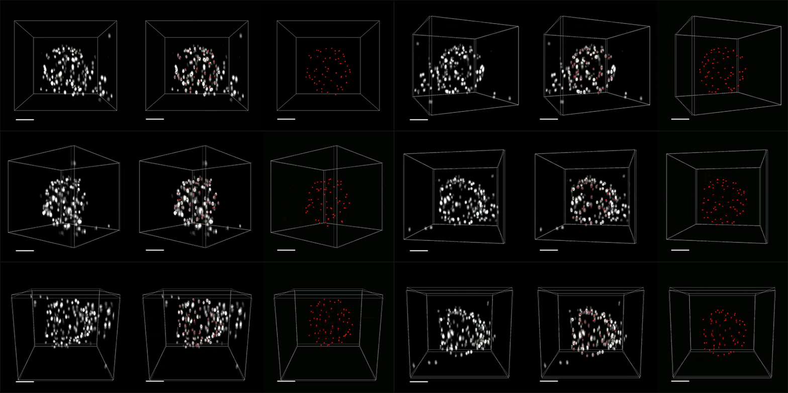
Three-dimensional (3D) rendered movie of envelope proteins of an herpes simplex virus type 1 (HSV-1) virion expanded by tetra-gel (TG)-based three-round iterative expansion. The deconvolved puncta (white), the overlay of the deconvolved puncta (white) and the fitted centroids (red), and the extracted centroids (red) are shown from left to right. Expansion factor, 38.3×. Scale bars, 100 nm.
Credit: Ruixuan Gao and Boyden Lab

The researchers demonstrated the accuracy of this approach by using it to expand particles of herpes simplex virus type 1 (HSV-1), which have a distinctive spherical shape. After expanding the virus particles, the researchers compared the shapes to the shapes obtained by electron microscopy and found that the distortion was lower than that seen with previous versions of expansion microscopy, allowing them to achieve an accuracy of about 10 nanometers.

“We can look at how the arrangements of these proteins change as they are expanded and evaluate how close they are to the spherical shape. That’s how we validated it and determined how faithfully we can preserve the nanostructure of the shapes and the relative spatial arrangements of these molecules,” Ruixuan Gao says.

Single molecules

The researchers also used their new hydrogel to expand cells, including human kidney cells and mouse brain cells. They are now working on ways to improve the accuracy to the point where they can image individual molecules within such cells. One limitation on this degree of accuracy is the size of the antibodies used to label molecules in the cell, which are about 10 to 20 nanometers long. To image individual molecules, the researchers would likely need to create smaller labels or to add the labels after expansion was complete.

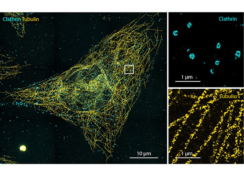
Left, HeLa cell with two-color labeling of clathrin-coated pits/vesicles and microtubules, expanded by TG-based two-round iterative expansion. Expansion factor, 15.6×. Scale bar, 10 μm (156 μm). Right, magnified view of the boxed region for each color channel. Scale bars, 1 μm (15.6 μm). Image: Boyden Lab

They are also exploring whether other types of polymers, or modified versions of the tetra-gel polymer, could help them realize greater accuracy.

If they can achieve accuracy down to single molecules, many new frontiers could be explored, Boyden says. For example, scientists could glimpse how different molecules interact with each other, which could shed light on cell signaling pathways, immune response activation, synaptic communication, drug-target interactions, and many other biological phenomena.

“We’d love to look at regions of a cell, like the synapse between two neurons, or other molecules involved in cell-cell signaling, and to figure out how all the parts talk to each other,” he says. “How do they work together and how do they go wrong in diseases?”

The research was funded by Lisa Yang, John Doerr, Open Philanthropy, the National Institutes of Health, the Howard Hughes Medical Institute Simons Faculty Scholars Program, the Intelligence Advanced Research Projects Activity, the U.S. Army Research Laboratory, the US-Israel Binational Science Foundation, the National Science Foundation, the Friends of the McGovern Fellowship, and the Fellows program of the Image and Data Analysis Core at Harvard Medical School.

Eyeless roundworms sense color

Roundworms don’t have eyes or the light-absorbing molecules required to see. Yet, new research shows they can somehow sense color. The study, published on March 5 in the journal Science, suggests worms use this ability to assess the risk of feasting on potentially dangerous bacteria that secrete blue toxins. The researchers pinpointed two genes that contribute to this spectral sensitivity and are conserved across many organisms, including humans.

“It’s amazing to me that a tiny worm — with neither eyes nor the molecular machinery used by eyes to detect colors — can identify and avoid a toxic bacterium based, in part, on its blue color,” says H. Robert Horvitz, the David H. Koch Professor of Biology at MIT, a member of the McGovern Institute for Brain Research and the Koch Institute for Integrative Cancer Research, Howard Hughes Medical Institute Investigator, and the co-senior author of the study.

“One of the joys of being a biologist is the opportunity to discover things about nature that no one has ever imagined before,” says Horvitz.

A model organism

The roundworm in question, Caenorhabditis elegans, is only about a millimeter long. Despite their minute stature and simple nervous system, these nematodes display a complex repertoire of behaviors. They can smell, taste, sense touch, react to temperature, and even escape or change their feeding patterns in response to bright, blue light. Although researchers once thought that these worms bury themselves deep in soil, it’s becoming increasingly clear that C. elegans prefers compost heaps above ground that offer some sun exposure. As a result, roundworms may have a need for light- and color-sensing capabilities after all.

The decomposing organic matter where C. elegans resides offers an array of scrumptious microbes, including bacteria like Pseudomonas aeruginosa, which secretes a distinctive blue toxin. Previous studies showed that worms in the lab feed on a lawn of P. aeruginosa for a few hours and then begin avoiding their food — perhaps because the bacteria continue to divide and excrete more of the colorful poison. Dipon Ghosh, Horvitz lab postdoc and the study’s first author, wondered whether the worms were using the distinctive color to determine if their meal was too toxic to consume.

A spectrum of behavior

Over the course of his experiments, Ghosh noticed that his worms were more likely to flee the colorful bacterial lawn if it was bathed in white light from a nearby LED bulb. This finding was curious on its own, but Ghosh wanted know if the blue toxin played a role as well.

To test this theory, he first exchanged the blue toxin for a harmless dye of the same color, and then for a clear, colorless toxin. On its own, neither substitute was sufficient to spur avoidance. Only together did they prompt a response — suggesting the worms were assessing both the toxic nature and the color of the P. aeruginosa secretions simultaneously. Once again, this behavioral pattern only emerged in the presence of the LED’s white light.

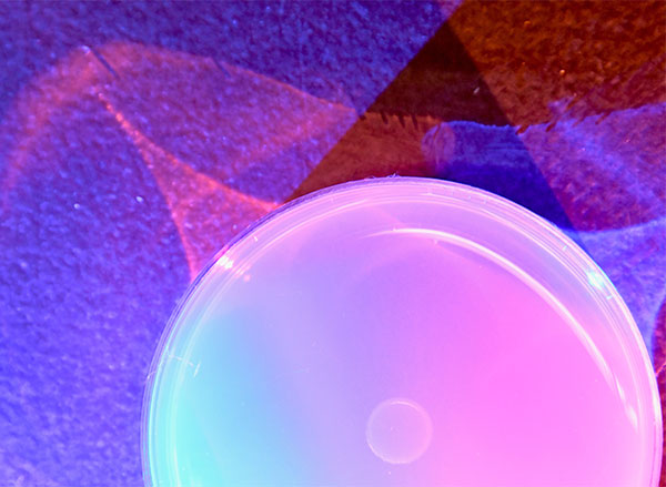
To test how worms sense color, the researchers placed C. elegans on an agar plate under tinted lights. Image: Eugene Lee

Intrigued, Ghosh wanted to examine what it was about the blue color that triggered avoidance. This time, he used two colored LED lights, one blue and one amber, to tint the ambient light. In doing so, he could control the ratio of wavelengths without changing the total energy delivered to the worms. The beam had previously contained the entire visible spectrum, but mixing the amber and blue bulbs allowed Ghosh to tweak the relative amounts of short-wavelength blue light and long-wavelength amber light. Surprisingly, the worms only fled the bacterial lawn when their environment was bathed in light with specific blue:amber ratios.

“We were able to definitively show that worms aren’t sensing the world in grayscale and simply evaluating the levels of brightness and darkness,” Ghosh says. “They’re actually comparing ratios of wavelengths and using that information to make decisions — which was thoroughly unexpected.”

It wasn’t until Ghosh ran his experiments again, this time using various types of wild C. elegans, that he realized the popular laboratory strain he’d been using was actually less color-sensitive compared to its close relatives. After analyzing the genomes of these worms, he was able to identify two genes in particular (called jkk-1 and lec-3) that contributed to these variations in color-dependent foraging.

Although the two genes play many important functions in a variety of organisms, including humans, they are both involved in molecular pathways that help cells respond to stress caused by damaging ultraviolet light.

“We’ve discovered that the color of light in the worm’s environment can influence how the worm navigates the world,” Ghosh says. “But our work suggests that many genes, in addition to the two we’ve already identified, can affect color sensitivity, and we’re now exploring how.”

Nature’s innovation

The notion that worms can sense color is “astounding” and showcases nature’s innovation, according to Leslie Vosshall, Robin Chemers Neustein Professor and Howard Hughes Medical Institute Investigator at The Rockefeller University, who was not involved in the study. “These worms are sliding around in a dim muck with colorful, toxic bacteria. It would be helpful to see and avoid them, so the worms somehow evolved a completely new way to see.”

Vosshall is curious about which cells in C. elegans help discriminate light, as well as the specific roles that the jkk-1 and lec-3 genes play in mediating light perception. “This paper, like all important papers, raises many additional questions,” she says.

Ghosh suspects the lab’s findings could generalize to other critters besides roundworms. If nothing else, it’s clear that light-sensitivity does not always require vision — or eyes. C. elegans are seeing the light, and now so are the biologists.

This research was funded by the Howard Hughes Medical Institute and National Institute of General Medical Sciences.

A high-resolution glimpse of gene expression in cells

Using a novel technique for expanding tissue, MIT and Harvard Medical School researchers have devised a way to label individual molecules of messenger RNA within a tissue sample and then sequence the RNA.

This approach offers a unique snapshot of which genes are being expressed in different parts of a cell, and could allow scientists to learn much more about how gene expression is influenced by a cell’s location or its interactions with nearby cells. The technique could also be useful for mapping cells in the brain or other tissues and classifying them according to their function.

“Gene expression is one of the most fundamental processes in all of biology, and it plays roles in all biological processes, both healthy and disease-related. However, you need to know more than just whether a gene is on or off,” says Ed Boyden, the Y. Eva Tan Professor in Neurotechnology and a professor of biological engineering, media arts and sciences, and brain and cognitive sciences at MIT.

“You want to know where the gene products are located. You care what cell types they’re in, which individual cells they play roles in, and even which parts of cells they work in,” says Boyden.

In a study appearing today in Science, the researchers showed that they could use this technique to locate and then sequence thousands of different messenger RNA molecules within the mouse brain and in human tumor samples.

The senior authors of the study are Boyden, an investigator at the MIT McGovern Institute and the Howard Hughes Medical Institute; George Church, a professor of genetics at Harvard Medical School; and Adam Marblestone, a former MIT research scientist. The paper’s lead authors are Shahar Alon, a former MIT postdoc who is now a senior lecturer at Bar-Ilan University; Daniel Goodwin, an MIT graduate student; Anubhav Sinha ’14 MNG ’15, an MIT graduate student; Asmamaw Wassie ’12, PhD ’19; and Fei Chen PhD ’17, who is an assistant professor of stem cell and regenerative biology at Harvard University and a member of the Broad Institute of MIT and Harvard.

Tissue expansion

The new sequencing technique builds on a method that Boyden’s group devised in 2015 for expanding tissue samples and then imaging them. By embedding water-absorbent polymers into a tissue sample, researchers can swell the tissue sample while keeping its overall organization intact. Using this approach, tissues can be expanded by a factor of 100 or more, allowing scientists to obtain very high-resolution images of the brain or other tissues using a regular light microscope.

In 2014, Church’s lab developed an RNA sequencing technique known as FISSEQ (fluorescent in situ sequencing), which allows thousands of mRNA molecules to be located and sequenced within cells grown in a lab dish. The Boyden and Church labs decided to join forces to combine tissue expansion and in situ RNA sequencing, creating a new technique they call expansion sequencing (ExSeq).

Expanding the tissue before performing RNA sequencing has two main benefits: It offers a higher-resolution look at the RNA in cells, and it makes it easier to sequence those RNA molecules. “When you separate these molecules in the expanding sample, and move them away from each other, that gives you more room to actually perform the chemical reactions of in situ sequencing,” Marblestone says.

Once the tissue is expanded, the researchers can label and sequence thousands of RNA molecules in a sample, at a resolution that allows them to pinpoint the molecules’ locations not only within cells but within specific compartments such as dendrites — the tiny extensions of neurons that receive communications from other neurons.

“We know that the location of RNA in these small regions is important for learning and memory, but until now, we didn’t have any way to measure these locations because they are very small, on the order of nanometers,” Alon says.

Using an “untargeted” version of this technique, meaning that they are not looking for specific RNA sequences, the researchers can turn up thousands of different sequences. They estimate that in a given sample, they can sequence between 20 and 50 percent of all of the genes present.

In the mouse hippocampus, this technique yielded some surprising results. For one, the researchers found mRNA containing introns, which are sections of RNA that are normally edited out of mRNA in the nucleus, in dendrites. They also discovered mRNA molecules encoding transcription factors in the dendrites, which may help with novel forms of dendrite-to-nucleus communication.

“These are just examples of things that we never would have gone looking for intentionally, but now that we can sequence RNA exactly where it is in the neuron, we’re able to explore a lot more biology,” Goodwin says.

Cellular interactions

The researchers also showed that they could explore gene expression in a more targeted way, looking for a specific set of RNA sequences that correspond to genes of interest. In the visual cortex of the mouse, the researchers used this approach to classify neurons into different types based on an analysis of 42 different genes that they express.

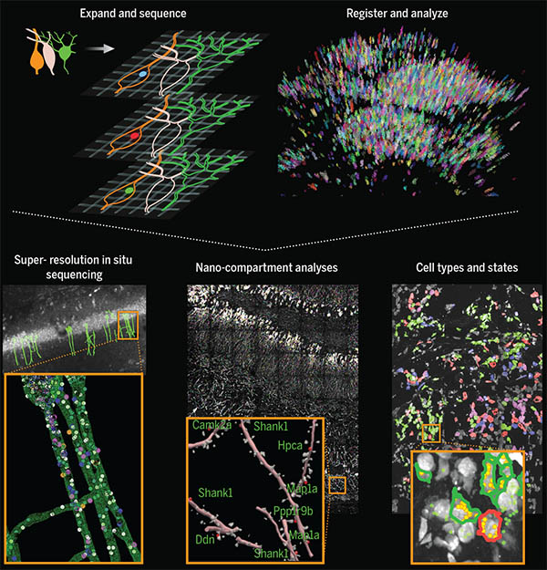
In situ sequencing of physically expanded specimens enables multiplexed mapping of RNAs at nanoscale, subcellular resolution throughout intact tissues. Top: schematics of physical expansion and in situ sequencing (left), and image analysis (right). Bottom: characterization of nanoscale transcriptomic compartmentalization in mouse hippocampal neuron dendrites and spines (left, middle), and maps of cell types and states in a metastatic human breast cancer biopsy (right). Image courtesy of the researchers.

This technology could also be useful to analyze many other kinds of tissues, such as tumor biopsies. In this paper, the researchers studied breast cancer metastases, which contain many different cell types, including cancer cells and immune cells. The study revealed that these cell types can behave differently depending on their location within a tumor. For example, the researchers found that B cells that were near tumor cells expressed certain inflammatory genes at a higher level than B cells that were farther from tumor cells.

“The tumor microenvironment has been studied in many different contexts for a long time, but it’s been difficult to study it with any depth,” Sinha says. “A cancer biologist can give you a list of 20 or 30 marker genes that will identify most of the cell types in the tissue. Here, since we interrogated 297 different RNA transcripts in the sample, we can ask and answer more detailed questions about gene expression.”

The researchers now plan to further study the interactions between cancer cells and immune cells, as well as gene expression in the brain in healthy and disease states. They also plan to extend their techniques to allow them to map additional types of biomolecules, such as proteins, alongside RNA.

The research was funded, in part, by the National Institutes of Health and the National Science Foundation, as well as by Lisa Yang, John Doerr, the Open Philanthropy Project, Cancer Research UK, the Chan Zuckerberg Initiative Human Cell Atlas pilot program, and HHMI.

Four MIT scientists honored with 2021 National Academy of Sciences awards

Four MIT scientists are among the 20 recipients of the 2021 Academy Honors for major contributions to science, the National Academy of Sciences (NAS) announced at its annual meeting. The individuals are recognized for their “extraordinary scientific achievements in a wide range of fields spanning the physical, biological, social, and medical sciences.”

The awards recognize: Pablo Jarillo-Herrero, for contributions to the fields of nanoscience and nanotechnology through his discovery of correlated insulator behavior and unconventional superconductivity in magic-angle graphene superlattices; Aviv Regev, for using interdisciplinary information or techniques to solve a contemporary challenge; Susan Solomon, for contributions to understanding and communicating the causes of ozone depletion and climate change; and Feng Zhang, for pioneering achievements developing CRISPR tools with the potential to diagnose and treat disease.

Pablo Jarillo-Herrero: Award for Scientific Discovery

Pablo Jarillo-Herrero, a Cecil and Ida Green Professor of Physics, is the recipient of the NAS Award for Scientific Discovery for his pioneering developments in nanoscience and nanotechnology, which is presented to scientists in the fields of astronomy, materials science, or physics. His findings expand nanoscience by demonstrating for the first time that orientation can be used to dramatically control nanomaterial properties and to design new nanomaterials. This work lays the groundwork for developing a whole new family of 2D materials and has had a transformative impact on the field and on condensed-matter physics.

The biannual award recognizes “an accomplishment or discovery in basic research, achieved within the previous five years, that is expected to have a significant impact on one or more of the following fields: astronomy, biochemistry, biophysics, chemistry, materials science, or physics.”

In 2018, his research group discovered that by rotating two layers of graphene relative to each other by a magic angle, the bilayer material can be turned from a metal into an electrical insulator or even a superconductor. This discovery has fostered new theoretical and experimental research, inspiring the interest of technologists in nanoelectronics. The result is a new field in condensed-matter physics that has the potential to result in materials that conduct electricity without resistance at room temperature.

Aviv Regev: James Prize in Science and Technology Integration

Aviv Regev, who is a professor of biology, a core member of the Broad Institute of Harvard and MIT, a member of the Koch Institute, and a Howard Hughes Medical Institute investigator has been selected for the inaugural James Prize in Science and Technology Integration, along with Harvard Medical School Professor Allon Kelin, for “their concurrent development of now widely adopted massively parallel single-cell genomics to interrogate the gene expression profiles that define, at the level of individual cells, the distinct cell types in metazoan tissues, their developmental trajectories, and disease states, which integrated tools from molecular biology, engineering, statistics, and computer science.”

The prize recognizes individuals “who are able to adopt or adapt information or techniques from outside their fields” to “solve a major contemporary challenge not addressable from a single disciplinary perspective.”

Regev is credited with forging new ways to unite the disciplines of biology, computational science, and engineering as a pioneer in the field of single-cell biology, including developing some of its core experimental and analysis tools, and their application to discover cell types, states, programs, environmental responses, development, tissue locations, and regulatory circuits, and deploying these to assemble cellular atlases of the human body that illuminate mechanisms of disease with remarkable fidelity.

Susan Solomon: Award for Chemistry in Service to Society

Susan Solomon, the Lee and Geraldine Martin Professor of Environmental Studies in the Department of Earth, Atmospheric and Planetary Sciences who holds a secondary appointment in the Department of Chemistry, is the recipient of the Award for Chemistry in Service to Society for “influential and incisive application of atmospheric chemistry to understand our most critical environmental issues — ozone layer depletion and climate change — and for her effective communication of environmental science to leaders to facilitate policy changes.”

The award is given biannually for “contributions to chemistry, either in fundamental science or its application, that clearly satisfy a societal need.”

Solomon is globally recognized as a leader in atmospheric science, notably for her insights in explaining the cause of the Antarctic ozone “hole.” She and her colleagues have made important contributions to understanding chemistry-climate coupling, including pioneering research on the irreversibility of global warming linked to anthropogenic carbon dioxide emissions, and on the influence of the ozone hole on the climate of the southern hemisphere.

Her work has had an enormous effect on policy and society, including the transition away from ozone-depleting substances and to environmentally benign chemicals. The work set the stage for the Paris Agreement on climate, and she continues to educate policymakers, the public, and the next generation of scientists.

Feng Zhang: Richard Lounsbery Award

Feng Zhang, who is the James and Patricia Poitras Professor of Neuroscience at MIT, an investigator at the McGovern Institute for Brain Research and the Howard Hughes Medical Institute, a professor of brain and cognitive sciences and biological engineering at MIT, and a core member of the Broad Institute of MIT and Harvard, is the recipient of the Richard Lounsbery Award for pioneering CRISPR-mediated genome editing.

The award recognizes “extraordinary scientific achievement in biology and medicine” as well as stimulating research and encouraging reciprocal scientific exchanges between the United States and France.

Zhang continues to lead the field through the discovery of novel CRISPR systems and their development as molecular tools with the potential to diagnose and treat disease, such as disorders affecting the nervous system. His contributions in genome engineering, as well as his earlier work developing optogenetics, are enabling a deeper understanding of behavioral neural circuits and advances in gene therapy for treating disease.

In addition, Zhang has championed the open sharing of the technologies he has developed through extensive resource sharing. The tools from his lab are being used by thousands of scientists around the world to accelerate research in nearly every field of the life sciences. Even as biomedical researchers around the world adopt Zhang’s discoveries and his tools enter the clinic to treat genetic diseases, he continues to innovate and develop new technologies to advance science.

The National Academy of Sciences is a private, nonprofit society of distinguished scholars, established in 1863 by the U.S. Congress. The NAS is charged with providing independent, objective advice to the nation on matters related to science and technology as well as encouraging education and research, recognize outstanding contributions to knowledge, and increasing public understanding in matters of science, engineering, and medicine. Winners received their awards, which include a monetary prize, during a virtual ceremony at the 158th NAS Annual Meeting.

This story is a modified compilation from several National Academy of Sciences press releases.

James DiCarlo named director of the MIT Quest for Intelligence

James DiCarlo, the Peter de Florez Professor of Neuroscience, has been appointed to the role of director of the MIT Quest for Intelligence. MIT Quest was launched in 2018 to discover the basis of natural intelligence, create new foundations for machine intelligence, and deliver new tools and technologies for humanity.

As director, DiCarlo will forge new collaborations with researchers within MIT and beyond to accelerate progress in understanding intelligence and developing the next generation of intelligence tools.

“We have discovered and developed surprising new connections between natural and artificial intelligence,” says DiCarlo, currently head of the Department of Brain and Cognitive Sciences (BCS). “The scientific understanding of natural intelligence, and advances in building artificial intelligence with positive real-world impact, are interlocked aspects of a unified, collaborative grand challenge, and MIT must continue to lead the way.”

Aude Oliva, senior research scientist at the Computer Science and Artificial Intelligence Laboratory (CSAIL) and the MIT director of the MIT-IBM Watson AI Lab, will lead industry engagements as director of MIT Quest Corporate. Nicholas Roy, professor of aeronautics and astronautics and a member of CSAIL, will lead the development of systems to deliver on the mission as director of MIT Quest Systems Engineering. Daniel Huttenlocher, dean of the MIT Schwarzman College of Computing, will serve as chair of MIT Quest.

“The MIT Quest’s leadership team has positioned this initiative to spearhead our understanding of natural and artificial intelligence, and I am delighted that Jim is taking on this role,” says Huttenlocher, the Henry Ellis Warren (1894) Professor of Electrical Engineering and Computer Science.

DiCarlo will step down from his current role as head of BCS, a position he has held for nearly nine years, and will continue as faculty in BCS and as an investigator in the McGovern Institute for Brain Research.

“Jim has been a highly productive leader for his department, the School of Science, and the Institute at large. I’m excited to see the impact he will make in this new role,” says Nergis Mavalvala, dean of the School of Science and the Curtis and Kathleen Marble Professor of Astrophysics.

As department head, DiCarlo oversaw significant progress in the department’s scientific and educational endeavors. Roughly a quarter of current BCS faculty were hired on his watch, strengthening the department’s foundations in cognitive, systems, and cellular and molecular brain science. In addition, DiCarlo developed a new departmental emphasis in computation, deepening BCS’s ties with the MIT Schwarzman College of Computing and other MIT units such as the Center for Brains, Minds and Machines. He also developed and leads an NIH-funded graduate training program in computationally-enabled integrative neuroscience. As a result, BCS is one of the few departments in the world that is attempting to decipher, in engineering terms, how the human mind emerges from the biological components of the brain.

To prepare students for this future, DiCarlo collaborated with BCS Associate Department Head Michale Fee to design and execute a total overhaul of the Course 9 curriculum. In addition, partnering with the Department of Electrical Engineering and Computer Science, BCS developed a new major, Course 6-9 (Computation and Cognition), to fill the rapidly growing interest in this interdisciplinary topic. In only its second year, Course 6-9 already has more than 100 undergraduate majors.

DiCarlo has also worked tirelessly to build a more open, connected, and supportive culture across the entire BCS community in Building 46. In this work, as in everything, DiCarlo sought to bring people together to address challenges collaboratively. He attributes progress to strong partnerships with Li-Huei Tsai, the Picower Professor of Neuroscience in BCS and director of the Picower Institute for Learning and Memory; Robert Desimone, the Doris and Don Berkey Professor in BCS and director of the McGovern Institute for Brain Research; and to the work of dozens of faculty and staff. For example, in collaboration with associate department head Professor Rebecca Saxe, the department has focused on faculty mentorship of graduate students, and, in collaboration with postdoc officer Professor Mark Bear, the department developed postdoc salary and benefit standards. Both initiatives have become models for the Institute. In recent months, DiCarlo partnered with new associate department head Professor Laura Schulz to constructively focus renewed energy and resources on initiatives to address systemic racism and promote diversity, equity, inclusion, and social justice.

“Looking ahead, I share Jim’s vision for the research and educational programs of the department, and for enhancing its cohesiveness as a community, especially with regard to issues of diversity, equity, inclusion, and justice,” says Mavalvala. “I am deeply committed to supporting his successor in furthering these goals while maintaining the great intellectual strength of BCS.”

In his own research, DiCarlo uses a combination of large-scale neurophysiology, brain imaging, optogenetic methods, and high-throughput computational simulations to understand the neuronal mechanisms and cortical computations that underlie human visual intelligence. Working in animal models, he and his research collaborators have established precise connections between the internal workings of the visual system and the internal workings of particular computer vision systems. And they have demonstrated that these science-to-engineering connections lead to new ways to modulate neurons deep in the brain as well as to improved machine vision systems. His lab’s goals are to help develop more human-like machine vision, new neural prosthetics to restore or augment lost senses, new learning strategies, and an understanding of how visual cognition is impaired in agnosia, autism, and dyslexia.

DiCarlo earned both a PhD in biomedical engineering and an MD from The Johns Hopkins University in 1998, and completed his postdoc training in primate visual neurophysiology at Baylor College of Medicine. He joined the MIT faculty in 2002.

A search committee will convene early this year to recommend candidates for the next department head of BCS. DiCarlo will continue to lead the department until that new head is selected.

Stars, brains, and enzymes: a celebration of MIT science

“Our topic tonight, science and discovery, lives at the heart of MIT.” In his welcoming remarks for the first virtual MIT Better World gathering, W. Eric L. Grimson, MIT chancellor for academic advancement, detailed some of the ways MIT excels as a hub of scientific research and innovation. “Institute researchers are plumbing the secrets of the universe; modeling climate at a local, regional, and global scale; striving to understand how brains and bodies give rise to cognition and mind; and racing to find treatments and cures for diseases ranging from the acute, like Covid-19, to the chronic, like cancers and maladies of the aging brain,” said Grimson, who is also the Bernard M. Gordon Professor of Medical Engineering.

Members of the MIT community from around the globe were invited to attend the MIT Better World (Science) event, held online in November, to hear from Institute leaders, faculty, students, and alumni about the pursuit of scientific knowledge. Alumni in more than 80 countries registered to attend, and the evening put a special emphasis on Canada, which is home to a group of alumni and friends who served as virtual hosts, and to which Grimson and all of the opening session speakers captured in the video above have personal ties.

Grimson’s remarks were followed by presentations from the new dean of the MIT School of Science, Nergis Mavalvala; as well as Rebecca Saxe, the John W. Jarve (1978) Professor in Brain and Cognitive Sciences and associate investigator at the McGovern Institute for Brain Research; and microbiology PhD student Linda Zhong-Johnson.

Mavalvala, the Curtis (1963) and Kathleen Marble Professor of Astrophysics, described how she and colleagues have worked to improve the sensitivity of instruments used to detect gravitational waves through LIGO—the landmark research endeavor that has revealed, among other recent discoveries, that colliding neutron stars are the “factories” in which heavy elements like gold and platinum are manufactured. Having begun the role of School of Science dean this fall, Mavalvala now takes joy in enabling discoveries across the MIT community, including those focused on our own corner of the universe. “It’s a vast world out there, and for us to make a better world, we must first understand that world. At MIT, that’s just what we do.”

Saxe, who uses brain imaging to study human social cognition, described prescient experiments on social isolation conducted by her lab between 2017 and 2019. “Sometimes we do science just out of curiosity,” said Saxe as she explained why she, former postdoc Livia Tomova, and fellow researchers pursued a project with uncertain applications — only to find themselves writing what Saxe now calls “the most timely and relevant paper in my life” in March, just as the Covid-19 pandemic triggered widespread isolation measures.

The third speaker, Linda Zhong-Johnson, discussed her PhD research in the labs of Anthony J. Sinskey, professor of biology, and Christopher A. Voigt, the Daniel I.C. Wang Professor of Advanced Biotechnology. Her goal is to reduce the amount of plastic in landfills and oceans by studying enzymes that could digest polyethylene terephthalate, or PET, the plastic used to make most water bottles. “We’re getting closer to the answer,” she said. “I’m grateful to be at MIT, where we have the mandate and resources to keep exploring.”

More virtual MIT Better World events on the topics of health and sustainability are planned for this coming February and March. Meanwhile, watch the full session (above) and a range of breakout sessions on topics such as the politics of molecular medicine and the Mars 2020 mission, and learn more about the MIT Campaign for a Better World at betterworld.mit.edu.