Jacqueline Lees and Rebecca Saxe named associate deans of science

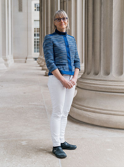
Jaqueline Lees and Rebecca Saxe have been named associate deans serving in the MIT School of Science. Lees is the Virginia and D.K. Ludwig Professor for Cancer Research and is currently the associate director of the Koch Institute for Integrative Cancer Research, as well as an associate department head and professor in the Department of Biology at MIT. Saxe is the John W. Jarve (1978) Professor in Brain and Cognitive Sciences and the associate head of the Department of Brain and Cognitive Sciences (BCS); she is also an associate investigator in the McGovern Institute for Brain Research.

Lees and Saxe will both contribute to the school’s diversity, equity, inclusion, and justice (DEIJ) activities, as well as develop and implement mentoring and other career-development programs to support the community. From their home departments, Saxe and Lees bring years of DEIJ and mentorship experience to bear on the expansion of school-level initiatives.

Lees currently serves on the dean’s science council in her capacity as associate director of the Koch Institute. In this new role as associate dean for the School of Science, she will bring her broad administrative and programmatic experiences to bear on the next phase for DEIJ and mentoring activities.

Lees joined MIT in 1994 as a faculty member in MIT’s Koch Institute (then the Center for Cancer Research) and Department of Biology. Her research focuses on regulators that control cellular proliferation, terminal differentiation, and stemness — functions that are frequently deregulated in tumor cells. She dissects the role of these proteins in normal cell biology and development, and establish how their deregulation contributes to tumor development and metastasis.

Since 2000, she has served on the Department of Biology’s graduate program committee, and played a major role in expanding the diversity of the graduate student population. Lees also serves on DEIJ committees in her home department, as well as at the Koch Institute.

With co-chair with Boleslaw Wyslouch, director of the Laboratory for Nuclear Science, Lees led the ReseArch Scientist CAreer LadderS (RASCALS) committee tasked to evaluate career trajectories for research staff in the School of Science and make recommendations to recruit and retain talented staff, rewarding them for their contributions to the school’s research enterprise.

“Jackie is a powerhouse in translational research, demonstrating how fundamental work at the lab bench is critical for making progress at the patient bedside,” says Nergis Mavalvala, dean of the School of Science. “With Jackie’s dedicated and thoughtful partnership, we can continue to lead in basic research and develop the recruitment, retention, and mentoring and necessary to support our community.”

Saxe will join Lees in supporting and developing programming across the school that could also provide direction more broadly at the Institute.

“Rebecca is an outstanding researcher in social cognition and a dedicated educator — someone who wants our students not only to learn, but to thrive,” says Mavalvala. “I am grateful that Rebecca will join the dean’s leadership team and bring her mentorship and leadership skills to enhance the school.”

For example, in collaboration with former department head James DiCarlo, the BCS department has focused on faculty mentorship of graduate students; and, in collaboration with Professor Mark Bear, the department developed postdoc salary and benefit standards. Both initiatives have become models at MIT.

With colleague Laura Schulz, Saxe also served as co-chair of the Committee on Medical Leave and Hospitalizations (CMLH), which outlined ways to enhance MIT’s current leave and hospitalization procedures and policies for undergraduate and graduate students. Saxe was also awarded MIT’s Committed to Caring award for excellence in graduate student mentorship, as well as the School of Science’s award for excellence in undergraduate teaching.

In her research, Saxe studies human social cognition, using a combination of behavioral testing and brain imaging technologies. She is best known for her work on brain regions specialized for abstract concepts, such as “theory of mind” tasks that involve understanding the mental states of other people. Her TED Talk, “How we read each other’s minds” has been viewed more than 3 million times. She also studies the development of the human brain during early infancy.

She obtained her PhD from MIT and was a Harvard University junior fellow before joining the MIT faculty in 2006. In 2014, the National Academy of Sciences named her one of two recipients of the Troland Award for investigators age 40 or younger “to recognize unusual achievement and further empirical research in psychology regarding the relationships of consciousness and the physical world.” In 2020, Saxe was named a John Simon Guggenheim Foundation Fellow.

Saxe and Lees will also work closely with Kuheli Dutt, newly hired assistant dean for diversity, equity, and inclusion, and other members of the dean’s science council on school-level initiatives and strategy.

“I’m so grateful that Rebecca and Jackie have agreed to take on these new roles,” Mavalvala says. “And I’m super excited to work with these outstanding thought partners as we tackle the many puzzles that I come across as dean.”

Investigating the embattled brain

Omar Rutledge served as a US Army infantryman in the 1st Armored and 25th Infantry Divisions. He was deployed in support of Operation Iraqi Freedom from March 2003 to July 2004. Photo: Omar Rutledge

As an Iraq war veteran, Omar Rutledge is deeply familiar with post-traumatic stress – recurring thoughts and memories that persist long after a danger has passed – and he knows that a brain altered by trauma is not easily fixed. But as a graduate student in the Department of Brain and Cognitive Sciences, Rutledge is determined to change that. He wants to understand exactly how trauma alters the brain – and whether the tools of neuroscience can be used to help fellow veterans with post-traumatic stress disorder (PTSD) heal from their experiences.

“In the world of PTSD research, I look to my left and to my right, and I don’t see other veterans, certainly not former infantrymen,” says Rutledge, who served in the US Army and was deployed to Iraq from March 2003 to July 2004. “If there are so few of us in this space, I feel like I have an obligation to make a difference for all who suffer from the traumatic experiences of war.”

Rutledge is uniquely positioned to make such a difference in the lab of McGovern Investigator John Gabrieli, where researchers use technologies like magnetic resonance imaging (MRI), electroencephalography (EEG), and magnetoencephalography (MEG) to peer into the human brain and explore how it powers our thoughts, memories, and emotions. Rutledge is studying how PTSD weakens the connection between the amygdala, which is responsible for emotions like fear, and the prefrontal cortex, which regulates or controls these emotional responses. He hopes these studies will eventually lead to the development of wearable technologies that can retrain the brain to be less responsive to triggering events.

“I feel like it has been a mission of mine to do this kind of work.”

Though Covid-19 has unexpectedly paused some aspects of his research, Rutledge is pursuing another line of research inspired both by the mandatory social distancing protocols imposed during the lockdown and his own experiences with social isolation. Does chronic social isolation cause physical or chemical changes in the brain similar to those seen in PTSD? And does loneliness exacerbate symptoms of PTSD?

“There’s this hypervigilance that occurs in loneliness, and there’s also something very similar that occurs in PTSD — a heightened awareness of potential threats,” says Rutledge, who is the recipient of Michael Ferrara Graduate Fellowship provided by the Poitras Center, a fellowship made possible by the many friends and family of Michael Ferrara. “The combination of the two may lead to more adverse reactions in people with PTSD.”

In the future, Rutledge hopes to explore whether chronic loneliness impairs reasoning and logic skills and has a deeper impact on veterans who have PTSD.

Although his research tends to resurface painful memories of his own combat experiences, Rutledge says if it can help other veterans heal, it’s worth it.  “In the process, it makes me a little bit stronger as well,” he adds.

Individual neurons responsible for complex social reasoning in humans identified

This story is adapted from a January 27, 2021 press release from Massachusetts General Hospital.

The ability to understand others’ hidden thoughts and beliefs is an essential component of human social behavior. Now, neuroscientists have for the first time identified specific neurons critical for social reasoning, a cognitive process that requires individuals to acknowledge and predict others’ hidden beliefs and thoughts.

The findings, published in Nature, open new avenues of study into disorders that affect social behavior, according to the authors.

In the study, a team of Harvard Medical School investigators based at Massachusetts General Hospital and colleagues from MIT took a rare look at how individual neurons represent the beliefs of others. They did so by recording neuron activity in patients undergoing neurosurgery to alleviate symptoms of motor disorders such as Parkinson’s disease.

Theory of mind

The researcher team, which included McGovern scientists Ev Fedorenko and Rebecca Saxe, focused on a complex social cognitive process called “theory of mind.” To illustrate this, let’s say a friend appears to be sad on her birthday. One may infer she is sad because she didn’t get a present or she is upset at growing older.

“When we interact, we must be able to form predictions about another person’s unstated intentions and thoughts,” said senior author Ziv Williams, HMS associate professor of neurosurgery at Mass General. “This ability requires us to paint a mental picture of someone’s beliefs, which involves acknowledging that those beliefs may be different from our own and assessing whether they are true or false.”

This social reasoning process develops during early childhood and is fundamental to successful social behavior. Individuals with autism, schizophrenia, bipolar affective disorder, and traumatic brain injuries are believed to have a deficit of theory-of-mind ability.

For the study, 15 patients agreed to perform brief behavioral tasks before undergoing neurosurgery for placement of deep-brain stimulation for motor disorders. Microelectrodes inserted into the dorsomedial prefrontal cortex recorded the behavior of individual neurons as patients listened to short narratives and answered questions about them.

For example, participants were presented with the following scenario to evaluate how they considered another’s belief of reality: “You and Tom see a jar on the table. After Tom leaves, you move the jar to a cabinet. Where does Tom believe the jar to be?”

Social computation

The participants had to make inferences about another’s beliefs after hearing each story. The experiment did not change the planned surgical approach or alter clinical care.

“Our study provides evidence to support theory of mind by individual neurons,” said study first author Mohsen Jamali, HMS instructor in neurosurgery at Mass General. “Until now, it wasn’t clear whether or how neurons were able to perform these social cognitive computations.”

The investigators found that some neurons are specialized and respond only when assessing another’s belief as false, for example. Other neurons encode information to distinguish one person’s beliefs from another’s. Still other neurons create a representation of a specific item, such as a cup or food item, mentioned in the story. Some neurons may multitask and aren’t dedicated solely to social reasoning.

“Each neuron is encoding different bits of information,” Jamali said. “By combining the computations of all the neurons, you get a very detailed representation of the contents of another’s beliefs and an accurate prediction of whether they are true or false.”

Now that scientists understand the basic cellular mechanism that underlies human theory of mind, they have an operational framework to begin investigating disorders in which social behavior is affected, according to Williams.

“Understanding social reasoning is also important to many different fields, such as child development, economics, and sociology, and could help in the development of more effective treatments for conditions such as autism spectrum disorder,” Williams said.

Previous research on the cognitive processes that underlie theory of mind has involved functional MRI studies, where scientists watch which parts of the brain are active as volunteers perform cognitive tasks.

But the imaging studies capture the activity of many thousands of neurons all at once. In contrast, Williams and colleagues recorded the computations of individual neurons. This provided a detailed picture of how neurons encode social information.

“Individual neurons, even within a small area of the brain, are doing very different things, not all of which are involved in social reasoning,” Williams said. “Without delving into the computations of single cells, it’s very hard to build an understanding of the complex cognitive processes underlying human social behavior and how they go awry in mental disorders.”

Adapted from a Mass General news release.

The pursuit of reward

View the interactive version of this story in our Spring 2021 issue of BrainScan.

The brain circuits that influence our decisions, cognitive functions, and ultimately, our actions are intimately connected with the circuits that give rise to our motivations. By exploring these relationships, scientists at McGovern are seeking knowledge that might suggest new strategies for changing our habits or treating motivation-disrupting conditions such as depression and addiction.

Risky decisions

MIT Institute Professor Ann Graybiel. Photo: Justin Knight

In Ann Graybiel’s lab, researchers have been examining how the brain makes choices that carry both positive and negative consequences — deciding to take on a higher-paying but more demanding job, for example. Psychologists call these dilemmas approach-avoidance conflicts, and resolving them not only requires weighing the good versus the bad, but also motivation to engage with the decision.

Emily Hueske, a research scientist in the Graybiel lab, explains that everyone has their own risk tolerance when it comes to such decisions, and certain psychiatric conditions, including depression and anxiety disorders, can shift the tipping point at which a person chooses to “approach” or “avoid.”

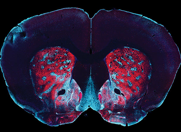
Studies have shown that neurons in the striatum (see image below), a region deep in the brain involved in both motivation and movement, activate as we grapple with these decisions. Graybiel traced this activity even further, to tiny compartments within the striatum called striosomes.

(She discovered striosomes many years ago and has been studying their function for decades.)

A motivational switch

In 2015, Graybiel’s team manipulated striosome signaling within genetically engineered mice and changed the way animals behave in approach-avoidance conflict situations. Taking cues from an assessment used to evaluate approach-avoidance behavior in patients, they presented mice with opportunities to obtain chocolate while experiencing unwelcome exposure in a brightly lit area.

Experimentally activating neurons in striosomes had a dramatic effect, causing mice to venture into brightly lit areas that they would normally avoid. With striosomal circuits switched on, “this animal all of a sudden is like a different creature,” Graybiel says.

Two years later, they found that chronic stress and other factors can also disrupt this signaling and change the choices animals make.

An image of the mouse striatum showing clusters of striosomes (red and yellow). Image: Graybiel lab

Age of ennui

This November, Alexander Friedman, who worked as a research scientist in the Graybiel lab, and Hueske reported in Cell that they found an age-related decline in motivation-modulated learning in mice and rats. Neurons within striosomes became more active than the cells that surround them as animals learned to assign positive and negative values to potential choices. And older mice were less engaged than their younger counterparts in the type of learning required to make these cost-benefit analyses. A similar lack of motivation was observed in a mouse model of Huntington’s disease, a neurodegenerative disorder that is often associated with mood
disturbances in patients.

“This coincides with our previous findings that striosomes are critically important for decisions that involve a conflict.”

“This coincides with our previous findings that striosomes are critically important for decisions that involve a conflict,” says Friedman, who is now an assistant professor at the University of Texas at El Paso.

Graybiel’s team is continuing to investigate these uniquely positioned compartments in the brain, expecting to shed light on the mechanisms that underlie both learning and motivation.

“There’s no learning without motivation, and in fact, motivation can be influenced by learning,” Hueske says. “The more you learn, the more excited you might be to engage in the task. So the two are intertwined.”

The aging brain

Researchers in John Gabrieli’s lab are also seeking to understand the circuits that link motivation to learning, and recently, his team reported that they, too, had found an age-related decline in motivation-modulated learning.

Studies in young adults have shown that memory improves when the brain circuits that process motivation and memory interact. Gabrieli and neurologist Maiya Geddes, who worked in Gabrieli’s lab as a postdoctoral fellow, wondered whether this holds true in older adults, particularly as memory declines.

To find out, the team recruited 40 people to participate in a brain imaging study. About half of the participants were between the ages of 18 and 30, while the others were between the ages of 49 and 84. While inside an fMRI scanner, each participant was asked to commit certain words to memory and told their success would determine how much money they received for participating in the experiment.

Diminished drive

MRI scan
Younger adults show greater activation in the reward-related regions of the brain during incentivized memory tasks compared to older adults. Image: Maiya Geddes

Not surprisingly, when participants were asked 24 hours later to recall the words, the younger group performed better overall than the older group. In young people, incentivized memory tasks triggered activity in parts of the brain involved in both memory and motivation. But in older adults, while these two parts of the brain could be activated independently, they did not seem to be communicating with one another.

“It seemed that the older adults, at least in terms of their brain response, did care about the kind of incentives that we were offering,” says Geddes, who is now an assistant professor at McGill University. “But for whatever reason, that wasn’t allowing them to benefit in terms of improved memory performance.”

Since the study indicates the brain still can anticipate potential rewards, Geddes is now exploring whether other sources of motivation, such as social rewards, might more effectively increase healthful decisions and behaviors in older adults.

Circuit control

Understanding how the brain generates and responds to motivation is not only important for improving learning strategies. Lifestyle choices such as exercise and social engagement can help people preserve cognitive function and improve their quality of life as they age, and Gabrieli says activating the right motivational circuits could help encourage people to implement healthy changes.

By pinpointing these motivational circuits in mice, Graybiel hopes that her research will lead to better treatment strategies for people struggling with motivational challenges, including Parkinson’s disease. Her team is now exploring whether striosomes serve as part of a value-sensitive switch, linking our intentions to dopamine-containing neurons in the midbrain that can modulate our actions.

“Perhaps this motivation is critical for the conflict resolution, and striosomes combine two worlds, dopaminergic motivation and cortical knowledge, resulting in motivation to learn,” Friedman says.

“Now we know that these challenges have a biological basis, and that there are neural circuits that can promote or reduce our feeling of motivational energy,” explains Graybiel. “This realization in itself is a major step toward learning how we can control these circuits both behaviorally and by highly selective therapeutic targeting.”

Stars, brains, and enzymes: a celebration of MIT science

“Our topic tonight, science and discovery, lives at the heart of MIT.” In his welcoming remarks for the first virtual MIT Better World gathering, W. Eric L. Grimson, MIT chancellor for academic advancement, detailed some of the ways MIT excels as a hub of scientific research and innovation. “Institute researchers are plumbing the secrets of the universe; modeling climate at a local, regional, and global scale; striving to understand how brains and bodies give rise to cognition and mind; and racing to find treatments and cures for diseases ranging from the acute, like Covid-19, to the chronic, like cancers and maladies of the aging brain,” said Grimson, who is also the Bernard M. Gordon Professor of Medical Engineering.

Members of the MIT community from around the globe were invited to attend the MIT Better World (Science) event, held online in November, to hear from Institute leaders, faculty, students, and alumni about the pursuit of scientific knowledge. Alumni in more than 80 countries registered to attend, and the evening put a special emphasis on Canada, which is home to a group of alumni and friends who served as virtual hosts, and to which Grimson and all of the opening session speakers captured in the video above have personal ties.

Grimson’s remarks were followed by presentations from the new dean of the MIT School of Science, Nergis Mavalvala; as well as Rebecca Saxe, the John W. Jarve (1978) Professor in Brain and Cognitive Sciences and associate investigator at the McGovern Institute for Brain Research; and microbiology PhD student Linda Zhong-Johnson.

Mavalvala, the Curtis (1963) and Kathleen Marble Professor of Astrophysics, described how she and colleagues have worked to improve the sensitivity of instruments used to detect gravitational waves through LIGO—the landmark research endeavor that has revealed, among other recent discoveries, that colliding neutron stars are the “factories” in which heavy elements like gold and platinum are manufactured. Having begun the role of School of Science dean this fall, Mavalvala now takes joy in enabling discoveries across the MIT community, including those focused on our own corner of the universe. “It’s a vast world out there, and for us to make a better world, we must first understand that world. At MIT, that’s just what we do.”

Saxe, who uses brain imaging to study human social cognition, described prescient experiments on social isolation conducted by her lab between 2017 and 2019. “Sometimes we do science just out of curiosity,” said Saxe as she explained why she, former postdoc Livia Tomova, and fellow researchers pursued a project with uncertain applications — only to find themselves writing what Saxe now calls “the most timely and relevant paper in my life” in March, just as the Covid-19 pandemic triggered widespread isolation measures.

The third speaker, Linda Zhong-Johnson, discussed her PhD research in the labs of Anthony J. Sinskey, professor of biology, and Christopher A. Voigt, the Daniel I.C. Wang Professor of Advanced Biotechnology. Her goal is to reduce the amount of plastic in landfills and oceans by studying enzymes that could digest polyethylene terephthalate, or PET, the plastic used to make most water bottles. “We’re getting closer to the answer,” she said. “I’m grateful to be at MIT, where we have the mandate and resources to keep exploring.”

More virtual MIT Better World events on the topics of health and sustainability are planned for this coming February and March. Meanwhile, watch the full session (above) and a range of breakout sessions on topics such as the politics of molecular medicine and the Mars 2020 mission, and learn more about the MIT Campaign for a Better World at betterworld.mit.edu.

A hunger for social contact

Since the coronavirus pandemic began in the spring, many people have only seen their close friends and loved ones during video calls, if at all. A new study from MIT finds that the longings we feel during this kind of social isolation share a neural basis with the food cravings we feel when hungry.

The researchers found that after one day of total isolation, the sight of people having fun together activates the same brain region that lights up when someone who hasn’t eaten all day sees a picture of a plate of cheesy pasta.

“People who are forced to be isolated crave social interactions similarly to the way a hungry person craves food.”

“Our finding fits the intuitive idea that positive social interactions are a basic human need, and acute loneliness is an aversive state that motivates people to repair what is lacking, similar to hunger,” says Rebecca Saxe, the John W. Jarve Professor of Brain and Cognitive Sciences at MIT, a member of MIT’s McGovern Institute for Brain Research, and the senior author of the study.

The research team collected the data for this study in 2018 and 2019, long before the coronavirus pandemic and resulting lockdowns. Their new findings, described today in Nature Neuroscience, are part of a larger research program focusing on how social stress affects people’s behavior and motivation.

Former MIT postdoc Livia Tomova, who is now a research associate at Cambridge University, is the lead author of the paper. Other authors include Kimberly Wang, a McGovern Institute research associate; Todd Thompson, a McGovern Institute scientist; Atsushi Takahashi, assistant director of the Martinos Imaging Center; Gillian Matthews, a research scientist at the Salk Institute for Biological Studies; and Kay Tye, a professor at the Salk Institute.

Social craving

The new study was partly inspired by a recent paper from Tye, a former member of MIT’s Picower Institute for Learning and Memory. In that 2016 study, she and Matthews, then an MIT postdoc, identified a cluster of neurons in the brains of mice that represent feelings of loneliness and generate a drive for social interaction following isolation. Studies in humans have shown that being deprived of social contact can lead to emotional distress, but the neurological basis of these feelings is not well-known.

“We wanted to see if we could experimentally induce a certain kind of social stress, where we would have control over what the social stress was,” Saxe says. “It’s a stronger intervention of social isolation than anyone had tried before.”

To create that isolation environment, the researchers enlisted healthy volunteers, who were mainly college students, and confined them to a windowless room on MIT’s campus for 10 hours. They were not allowed to use their phones, but the room did have a computer that they could use to contact the researchers if necessary.

“There were a whole bunch of interventions we used to make sure that it would really feel strange and different and isolated,” Saxe says. “They had to let us know when they were going to the bathroom so we could make sure it was empty. We delivered food to the door and then texted them when it was there so they could go get it. They really were not allowed to see people.”

After the 10-hour isolation ended, each participant was scanned in an MRI machine. This posed additional challenges, as the researchers wanted to avoid any social contact during the scanning. Before the isolation period began, each subject was trained on how to get into the machine, so that they could do it by themselves, without any help from the researcher.

“Normally, getting somebody into an MRI machine is actually a really social process. We engage in all kinds of social interactions to make sure people understand what we’re asking them, that they feel safe, that they know we’re there,” Saxe says. “In this case, the subjects had to do it all by themselves, while the researcher, who was gowned and masked, just stood silently by and watched.”

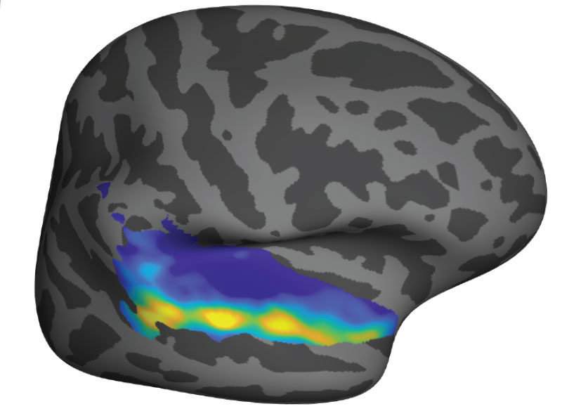
Each of the 40 participants also underwent 10 hours of fasting, on a different day. After the 10-hour period of isolation or fasting, the participants were scanned while looking at images of food, images of people interacting, and neutral images such as flowers. The researchers focused on a part of the brain called the substantia nigra, a tiny structure located in the midbrain, which has previously been linked with hunger cravings and drug cravings. The substantia nigra is also believed to share evolutionary origins with a brain region in mice called the dorsal raphe nucleus, which is the area that Tye’s lab showed was active following social isolation in their 2016 study.

The researchers hypothesized that when socially isolated subjects saw photos of people enjoying social interactions, the “craving signal” in their substantia nigra would be similar to the signal produced when they saw pictures of food after fasting. This was indeed the case. Furthermore, the amount of activation in the substantia nigra was correlated with how strongly the patients rated their feelings of craving either food or social interaction.

Degrees of loneliness

The researchers also found that people’s responses to isolation varied depending on their normal levels of loneliness. People who reported feeling chronically isolated months before the study was done showed weaker cravings for social interaction after the 10-hour isolation period than people who reported a richer social life.

“For people who reported that their lives were really full of satisfying social interactions, this intervention had a bigger effect on their brains and on their self-reports,” Saxe says.

The researchers also looked at activation patterns in other parts of the brain, including the striatum and the cortex, and found that hunger and isolation each activated distinct areas of those regions. That suggests that those areas are more specialized to respond to different types of longings, while the substantia nigra produces a more general signal representing a variety of cravings.

Now that the researchers have established that they can observe the effects of social isolation on brain activity, Saxe says they can now try to answer many additional questions. Those questions include how social isolation affect people’s behavior, whether virtual social contacts such as video calls help to alleviate cravings for social interaction, and how isolation affects different age groups.

The researchers also hope to study whether the brain responses that they saw in this study could be used to predict how the same participants responded to being isolated during the lockdowns imposed during the early stages of the coronavirus pandemic.

The research was funded by a SFARI Explorer Grant from the Simons Foundation, a MINT grant from the McGovern Institute, the National Institutes of Health, including an NIH Pioneer Award, a Max Kade Foundation Fellowship, and an Erwin Schroedinger Fellowship from the Austrian Science Fund.

Face-specific brain area responds to faces even in people born blind

More than 20 years ago, neuroscientist Nancy Kanwisher and others discovered that a small section of the brain located near the base of the skull responds much more strongly to faces than to other objects we see. This area, known as the fusiform face area, is believed to be specialized for identifying faces.

Now, in a surprising new finding, Kanwisher and her colleagues have shown that this same region also becomes active in people who have been blind since birth, when they touch a three-dimensional model of a face with their hands. The finding suggests that this area does not require visual experience to develop a preference for faces.

“That doesn’t mean that visual input doesn’t play a role in sighted subjects — it probably does,” she says. “What we showed here is that visual input is not necessary to develop this particular patch, in the same location, with the same selectivity for faces. That was pretty astonishing.”

Kanwisher, the Walter A. Rosenblith Professor of Cognitive Neuroscience and a member of MIT’s McGovern Institute for Brain Research, is the senior author of the study. N. Apurva Ratan Murty, an MIT postdoc, is the lead author of the study, which appears this week in the Proceedings of the National Academy of Sciences. Other authors of the paper include Santani Teng, a former MIT postdoc; Aude Oliva, a senior research scientist, co-director of the MIT Quest for Intelligence, and MIT director of the MIT-IBM Watson AI Lab; and David Beeler and Anna Mynick, both former lab technicians.

Selective for faces

Studying people who were born blind allowed the researchers to tackle longstanding questions regarding how specialization arises in the brain. In this case, they were specifically investigating face perception, but the same unanswered questions apply to many other aspects of human cognition, Kanwisher says.

“This is part of a broader question that scientists and philosophers have been asking themselves for hundreds of years, about where the structure of the mind and brain comes from,” she says. “To what extent are we products of experience, and to what extent do we have built-in structure? This is a version of that question asking about the particular role of visual experience in constructing the face area.”

The new work builds on a 2017 study from researchers in Belgium. In that study, congenitally blind subjects were scanned with functional magnetic resonance imaging (fMRI) as they listened to a variety of sounds, some related to faces (such as laughing or chewing), and others not. That study found higher responses in the vicinity of the FFA to face-related sounds than to sounds such as a ball bouncing or hands clapping.

In the new study, the MIT team wanted to use tactile experience to measure more directly how the brains of blind people respond to faces. They created a ring of 3D-printed objects that included faces, hands, chairs, and mazes, and rotated them so that the subject could handle each one while in the fMRI scanner.

They began with normally sighted subjects and found that when they handled the 3D objects, a small area that corresponded to the location of the FFA was preferentially active when the subjects touched the faces, compared to when they touched other objects. This activity, which was weaker than the signal produced when sighted subjects looked at faces, was not surprising to see, Kanwisher says.

“We know that people engage in visual imagery, and we know from prior studies that visual imagery can activate the FFA. So the fact that you see the response with touch in a sighted person is not shocking because they’re visually imagining what they’re feeling,” she says.

The researchers then performed the same experiments, using tactile input only, with 15 subjects who reported being blind since birth. To their surprise, they found that the brain showed face-specific activity in the same area as the sighted subjects, at levels similar to when sighted people handled the 3D-printed faces.

“When we saw it in the first few subjects, it was really shocking, because no one had seen individual face-specific activations in the fusiform gyrus in blind subjects previously,” Murty says.

Patterns of connection

The researchers also explored several hypotheses that have been put forward to explain why face-selectivity always seems to develop in the same region of the brain. One prominent hypothesis suggests that the FFA develops face-selectivity because it receives visual input from the fovea (the center of the retina), and we tend to focus on faces at the center of our visual field. However, since this region developed in blind people with no foveal input, the new findings do not support this idea.

Another hypothesis is that the FFA has a natural preference for curved shapes. To test that idea, the researchers performed another set of experiments in which they asked the blind subjects to handle a variety of 3D-printed shapes, including cubes, spheres, and eggs. They found that the FFA did not show any preference for the curved objects over the cube-shaped objects.

The researchers did find evidence for a third hypothesis, which is that face selectivity arises in the FFA because of its connections to other parts of the brain. They were able to measure the FFA’s “connectivity fingerprint” — a measure of the correlation between activity in the FFA and activity in other parts of the brain — in both blind and sighted subjects.

They then used the data from each group to train a computer model to predict the exact location of the brain’s selective response to faces based on the FFA connectivity fingerprint. They found that when the model was trained on data from sighted patients, it could accurately predict the results in blind subjects, and vice versa. They also found evidence that connections to the frontal and parietal lobes of the brain, which are involved in high-level processing of sensory information, may be the most important in determining the role of the FFA.

“It’s suggestive of this very interesting story that the brain wires itself up in development not just by taking perceptual information and doing statistics on the input and allocating patches of brain, according to some kind of broadly agnostic statistical procedure,” Kanwisher says. “Rather, there are endogenous constraints in the brain present at birth, in this case, in the form of connections to higher-level brain regions, and these connections are perhaps playing a causal role in its development.”

The research was funded by the National Institutes of Health Shared Instrumentation Grant to the Athinoula Martinos Center at MIT, a National Eye Institute Training Grant, the Smith-Kettlewell Eye Research Institute’s Rehabilitation Engineering Research Center, an Office of Naval Research Vannevar Bush Faculty Fellowship, an NIH Pioneer Award, and a National Science Foundation Science and Technology Center Grant.

Full paper at PNAS

Nine MIT School of Science professors receive tenure for 2020

Beginning July 1, nine faculty members in the MIT School of Science have been granted tenure by MIT. They are appointed in the departments of Brain and Cognitive Sciences, Chemistry, Mathematics, and Physics.

Physicist Ibrahim Cisse investigates living cells to reveal and study collective behaviors and biomolecular phase transitions at the resolution of single molecules. The results of his work help determine how disruptions in genes can cause diseases like cancer. Cisse joined the Department of Physics in 2014 and now holds a joint appointment with the Department of Biology. His education includes a bachelor’s degree in physics from North Carolina Central University, concluded in 2004, and a doctoral degree in physics from the University of Illinois at Urbana-Champaign, achieved in 2009. He followed his PhD with a postdoc at the École Normale Supérieure of Paris and a research specialist appointment at the Howard Hughes Medical Institute’s Janelia Research Campus.

Jörn Dunkel is a physical applied mathematician. His research focuses on the mathematical description of complex nonlinear phenomena in a variety of fields, especially biophysics. The models he develops help predict dynamical behaviors and structure formation processes in developmental biology, fluid dynamics, and even knot strengths for sailing, rock climbing and construction. He joined the Department of Mathematics in 2013 after completing postdoctoral appointments at Oxford University and Cambridge University. He received diplomas in physics and mathematics from Humboldt University of Berlin in 2004 and 2005, respectively. The University of Augsburg awarded Dunkel a PhD in statistical physics in 2008.

A cognitive neuroscientist, Mehrdad Jazayeri studies the neurobiological underpinnings of mental functions such as planning, inference, and learning by analyzing brain signals in the lab and using theoretical and computational models, including artificial neural networks. He joined the Department of Brain and Cognitive Sciences in 2013. He achieved a BS in electrical engineering from the Sharif University of Technology in 1994, an MS in physiology at the University of Toronto in 2001, and a PhD in neuroscience from New York University in 2007. Prior to joining MIT, he was a postdoc at the University of Washington. Jazayeri is also an investigator at the McGovern Institute for Brain Research.

Yen-Jie Lee is an experimental particle physicist in the field of proton-proton and heavy-ion physics. Utilizing the Large Hadron Colliders, Lee explores matter in extreme conditions, providing new insight into strong interactions and what might have existed and occurred at the beginning of the universe and in distant star cores. His work on jets and heavy flavor particle production in nuclei collisions improves understanding of the quark-gluon plasma, predicted by quantum chromodynamics (QCD) calculations, and the structure of heavy nuclei. He also pioneered studies of high-density QCD with electron-position annihilation data. Lee joined the Department of Physics in 2013 after a fellowship at CERN and postdoc research at the Laboratory for Nuclear Science at MIT. His bachelor’s and master’s degrees were awarded by the National Taiwan University in 2002 and 2004, respectively, and his doctoral degree by MIT in 2011. Lee is a member of the Laboratory for Nuclear Science.

Josh McDermott investigates the sense of hearing. His research addresses both human and machine audition using tools from experimental psychology, engineering, and neuroscience. McDermott hopes to better understand the neural computation underlying human hearing, to improve devices to assist hearing impaired, and to enhance machine interpretation of sounds. Prior to joining MIT’s Department of Brain and Cognitive Sciences, he was awarded a BA in 1998 in brain and cognitive sciences by Harvard University, a master’s degree in computational neuroscience in 2000 by University College London, and a PhD in brain and cognitive sciences in 2006 by MIT. Between his doctoral time at MIT and returning as a faculty member, he was a postdoc at the University of Minnesota and New York University, and a visiting scientist at Oxford University. McDermott is also an associate investigator at the McGovern Institute for Brain Research and an investigator in the Center for Brains, Minds and Machines.

Solving environmental challenges by studying and manipulating chemical reactions is the focus of Yogesh Surendranath’s research. Using chemistry, he works at the molecular level to understand how to efficiently interconvert chemical and electrical energy. His fundamental studies aim to improve energy storage technologies, such as batteries, fuel cells, and electrolyzers, that can be used to meet future energy demand with reduced carbon emissions. Surendranath joined the Department of Chemistry in 2013 after a postdoc at the University of California at Berkeley. His PhD was completed in 2011 at MIT, and BS in 2006 at the University of Virginia. Suendranath is also a collaborator in the MIT Energy Initiative.

A theoretical astrophysicist, Mark Vogelsberger is interested in large-scale structures of the universe, such as galaxy formation. He combines observational data, theoretical models, and simulations that require high-performance supercomputers to improve and develop detailed models that simulate galaxy diversity, clustering, and their properties, including a plethora of physical effects like magnetic fields, cosmic dust, and thermal conduction. Vogelsberger also uses simulations to generate scenarios involving alternative forms of dark matter. He joined the Department of Physics in 2014 after a postdoc at the Harvard-Smithsonian Center for Astrophysics. Vogelsberger is a 2006 graduate of the University of Mainz undergraduate program in physics, and a 2010 doctoral graduate of the University of Munich and the Max Plank Institute for Astrophysics. He is also a principal investigator in the MIT Kavli Institute for Astrophysics and Space Research.

Adam Willard is a theoretical chemist with research interests that fall across molecular biology, renewable energy, and material science. He uses theory, modeling, and molecular simulation to study the disorder that is inherent to systems over nanometer-length scales. His recent work has highlighted the fundamental and unexpected role that such disorder plays in phenomena such as microscopic energy transport in semiconducting plastics, ion transport in batteries, and protein hydration. Joining the Department of Chemistry in 2013, Willard was formerly a postdoc at Lawrence Berkeley National Laboratory and then the University of Texas at Austin. He holds a PhD in chemistry from the University of California at Berkeley, achieved in 2009, and a BS in chemistry and mathematics from the University of Puget Sound, granted in 2003.

Lindley Winslow seeks to understand the fundamental particles shaped the evolution of our universe. As an experimental particle and nuclear physicist, she develops novel detection technology to search for axion dark matter and a proposed nuclear decay that makes more matter than antimatter. She started her faculty position in the Department of Physics in 2015 following a postdoc at MIT and a subsequent faculty position at the University of California at Los Angeles. Winslow achieved her BA in physics and astronomy in 2001 and PhD in physics in 2008, both at the University of California at Berkeley. She is also a member of the Laboratory for Nuclear Science.

Universal musical harmony

Many forms of Western music make use of harmony, or the sound created by certain pairs of notes. A longstanding question is why some combinations of notes are perceived as pleasant while others sound jarring to the ear. Are the combinations we favor a universal phenomenon? Or are they specific to Western culture?

Through intrepid research trips to the remote Bolivian rainforest, the McDermott lab at the McGovern Institute has found that aspects of the perception of note combinations may be universal, even though the aesthetic evaluation of note combination as pleasant or unpleasant is culture-specific.

“Our work has suggested some universal features of perception that may shape musical behavior around the world,” says McGovern Associate Investigator Josh McDermott, senior author of the Nature Communications study. “But it also indicates the rich interplay with cultural influences that give rise to the experience of music.”

Remote learning

Questions about the universality of musical perception are difficult to answer, in part because of the challenge in finding people with little exposure to Western music. McDermott, who is also an associate professor in MIT’s Department of Brain and Cognitive Sciences and an investigator in the Center for Brains Minds and Machines, has found a way to address this problem. His lab has performed a series of studies with the participation of an indigenous population, the Tsimane’, who live in relative isolation from Western culture and have had little exposure to Western music. Accessing the Tsimane’ villages is challenging, as they are scattered throughout the rainforest and only reachable during the dry part of the year.

Left to right Josh McDermott (in vehicle), Alex Durango, Sophie Dolan and Malinda McPherson experiencing a travel delay en route to a Tsimane’ village after a heavy rainfall. Photo: Malinda McPherson

“When we enter a village there is always a crowd of curious children to greet us,” says Malinda McPherson, a graduate student in the lab and lead author of the study. “Tsimane’ are friendly and welcoming, and we have visited some villages several times, so now many people recognize us.”

In a study published in 2019, McDermott’s team found evidence that the brain’s ability to detect musical octaves is not universal, but is gained through cultural experience. And in 2016 they published findings suggesting that the preference for consonance over dissonance is culture-specific. In their new study, the team decided to explore whether aspects of the perception of consonance and dissonance might nonetheless be universally present across cultures.

Music lessons

In Western music, harmony is the sound of two or more notes heard simultaneously. Think of the Leonard Cohen song, Hallelujah, where he sings about harmony (“the fourth, the fifth, the minor fall and the major lift”). A combination of two notes is called an interval, and intervals that are perceived to be the most pleasant (or consonant, like the fourth and the fifth, for example) to the Western ear are generally represented by smaller integer ratios.

Intervals that are related by low integer ratios have fascinated scientists for centuries.

“Such intervals are central to Western music, but are also believed to be a common feature of many musical systems around the world,” McPherson explains. “So intervals are a natural target for cross-cultural research, which can help identify aspects of perception that are and aren’t independent of cultural experience.”

Scientists have been drawn to low integer ratios in music in part because they relate to the frequencies in voices and many instruments, known as ‘overtones’. Overtones from sounds like voices form a particular pattern known as the harmonic series. As it happens, the combination of two concurrent notes related by a low integer ratio partially reproduces this pattern. Because the brain presumably evolved to represent natural sounds, such as voices, it has seemed plausible that intervals with low integer ratios might have special perceptual status across cultures.

Since the Tsimane’ do not generally sing or play music together, meaning they have not been trained to hear or sing in harmony, McPherson and her colleagues were presented with a unique opportunity to explore whether there is anything universal about the perception of musical intervals.

Taking notes

In order to probe the perception of musical intervals, McDermott and colleagues took advantage of the fact that ears accustomed to Western musical harmony often have difficulty picking apart two “consonant” notes when they are played at the same time. This auditory confusion is known as “fusion” in the field. By contrast, two “dissonant” notes are easier to hear as separate.

The tendency of “consonant” notes to be heard by Westerners as fused could reflect their common occurrence in Western music. But it could also be driven by the resemblance of low-integer-ratio note combinations to the harmonic series. This similarity of consonant intervals to the acoustic structure of typical natural sounds raises the possibility that the human brain is biologically tuned to “fuse” consonant notes.

Graduate student and lead author, Malinda McPherson, works with a participant and translator in the field. Photo: Malinda McPherson

To explore this question, the team ran identical sets of experiments on two participant groups: US non-musicians residing in the Boston metropolitan area and Tsimane’ residing in villages in the Amazon rain forest. Listeners heard two concurrent notes separated by a particular musical interval (consonant or dissonant), and were asked to judge whether they heard one or two sounds. The experiment was performed with both synthetic and natural sounds.

They found that like the Boston cohort, the Tsimane’ were more likely to mistake two notes as a single sound if they were consonant than if they were dissonant.

“I was surprised by how similar some of the results in Tsimane’ participants were to those in US participants,” says McPherson, “particularly given the striking differences that we consistently see in preferences for musical intervals.”

When it came to whether consonant intervals were more pleasant than dissonant intervals, the results told a very different story. While the US study participants found consonant intervals more pleasant than dissonant intervals, the Tsimane’ showed no preference, implying that our sense of what is pleasant is shaped by culture.

“The fusion results provide an example of a perceptual effect that could influence musical systems, for instance by creating a natural perceptual contrast to exploit,” explains McDermott. “Hopefully our work helps to show how one can conduct rigorous perceptual experiments in the field and learn things that would be hidden if we didn’t consider populations in other parts of the world.”

Saxe Lab examines social impact of COVID-19

After being forced to relocate from their MIT dorms during the COVID19 crisis, two members of the Saxe lab are now applying their psychology skills to study the impact of mandatory relocation and social isolation on mental health.

“When ‘social distancing’ measures hit MIT, we tried to process how the implementation of these policies would impact the landscape of our social lives,” explains graduate student Heather Kosakowski, who conceived of the study late one evening with undergraduate Michelle Hung.  This landscape is broad, examining the effects of being uprooted and physically relocated from a place, but also changes in social connections, including friendships and even dating life.

MIT undergrad Michelle Hung in the Saxe lab. Photo: Michelle Hung

“I started speculating about how my life and the lives of other MIT students would change,” says Hung. “I was overwhelmed, sad, and scared. But then we realized that we were actually equipped to find the answers to our questions by conducting a study.”

Together, Kosakowski and Hung developed a survey to measure how the social behavior of MIT students, postdocs, and staff is changing over the course of the pandemic. Survey questions were designed to measure loneliness and other aspects of mental health. The survey was sent to members of the MIT community and shared on social media in mid-March, when the pandemic hit the US, and MIT made the unprecedented decision to send students home, shift to online instruction, and dramatically ramp down operations on campus.

More than 500 people responded to the initial survey, ranging in age from 18 to 60, living in cities and countries around the world. Many but not all of those who responded were affiliated with MIT. Kosakowski and Hung are sending follow-up surveys to participants every two weeks and the team plans to collect data for the duration of the pandemic.

“Throwing myself into creating the survey was a way to cope with feeling sad about leaving a community I love,” explains Hung, who flew home to California in March and admits that she struggles with feelings of loneliness now that she’s off campus.

Although it is too soon to form any conclusions about their research, Hung predicts that feelings of loneliness may actually diminish over the course of the pandemic.

“Humans have an impressive ability to adapt to change,” she says. “And I think in this virtual world, people will find novel ways to stay connected that we couldn’t have predicted.”

Whether we find ourselves feeling more or less lonely as this COVID-19 crisis comes to an end, both Kosakowski and Hung agree that it will fundamentally change life as we know it.

The Saxe lab is looking for more survey participants. To learn more about this study or to participate in the survey, click here.