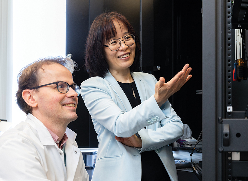
Michale Fee, the Glen V. and Phyllis F. Dorflinger Professor of Neuroscience and head of the Department of Brain and Cognitive Sciences, and Fan Wang, a professor of brain and cognitive sciences, have been elected to join the National Academy of Sciences (NAS). Fee and Wang, who are also investigators at the McGovern Institute for Brain Research, were elected by current NAS members in recognition of their “distinguished and continuing achievements in original research.”
The NAS is a private, nonprofit institution that was established under a congressional charter signed by President Abraham Lincoln in 1863. It recognizes achievement in science by election to membership, and — with the National Academy of Engineering and the National Academy of Medicine — provides science, engineering, and health policy advice to the federal government and other organizations. This year, the NAS elected 120 members and 25 international members, including six MIT faculty, bringing the total number of active members to 2,705.
“Election to the National Academy of Sciences by one’s peers is a great honor for a scientist in the United States,” says McGovern Institute Director Robert Desimone. “Michale and Fan represent the very best of our research community and we are tremendously proud of their accomplishments and this well-deserved recognition.”
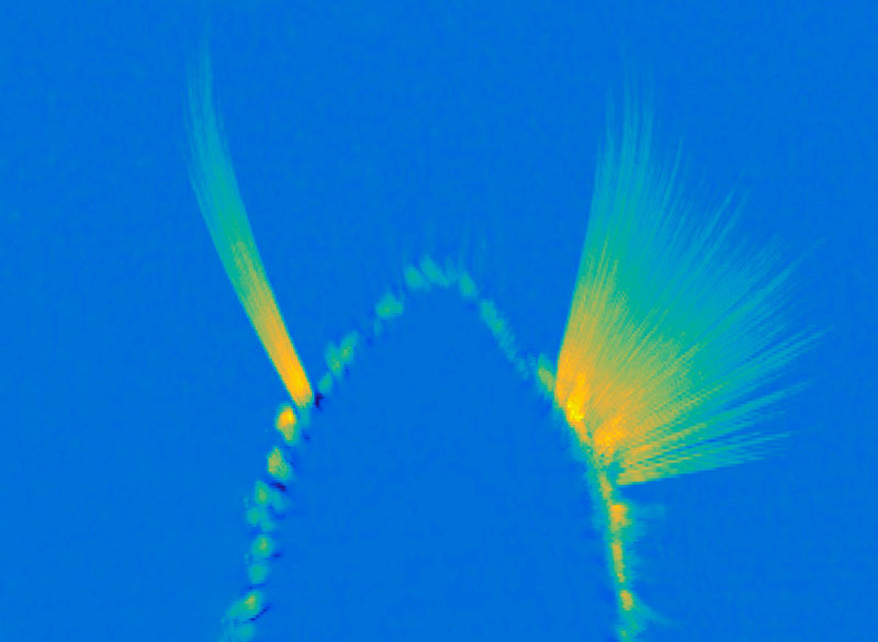
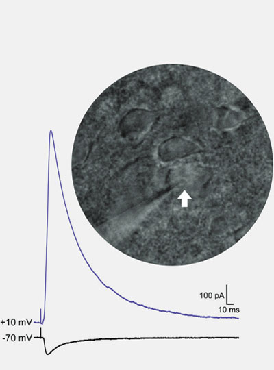
Michale Fee’s research explores how the brain learns and generates complex sequential behaviors. Using the zebra finch as a model system, Fee investigates the neural mechanisms underlying birdsong—a behavior that young birds learn from their fathers through trial and error, much as human infants learn to speak through babbling. His work has revealed that a brain region called the higher vocal center (HVC) functions like an orchestra conductor, precisely controlling the tempo and timing of song production. Other work from his lab has shown how this same circuit helps to store a memory of the father’s song, how baby birds babble in order to practice their song, and how this vocal practice is translated to song learning by listening to themselves sing.
These findings extend far beyond birdsong—the neural circuits controlling birdsong learning are closely related to human brain circuits disrupted in Parkinson’s and Huntington’s disease. Insights from Fee’s research could reveal new clues to the causes and potential treatments of these complex brain disorders.
Fee’s appointment in 2021 as head of the Department of Brain and Cognitive Sciences continues the department’s tradition of being led by scientists whose exemplary work makes MIT a world leader in brain science.
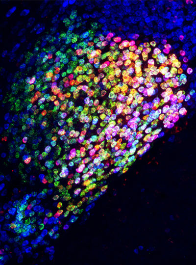
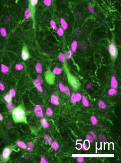
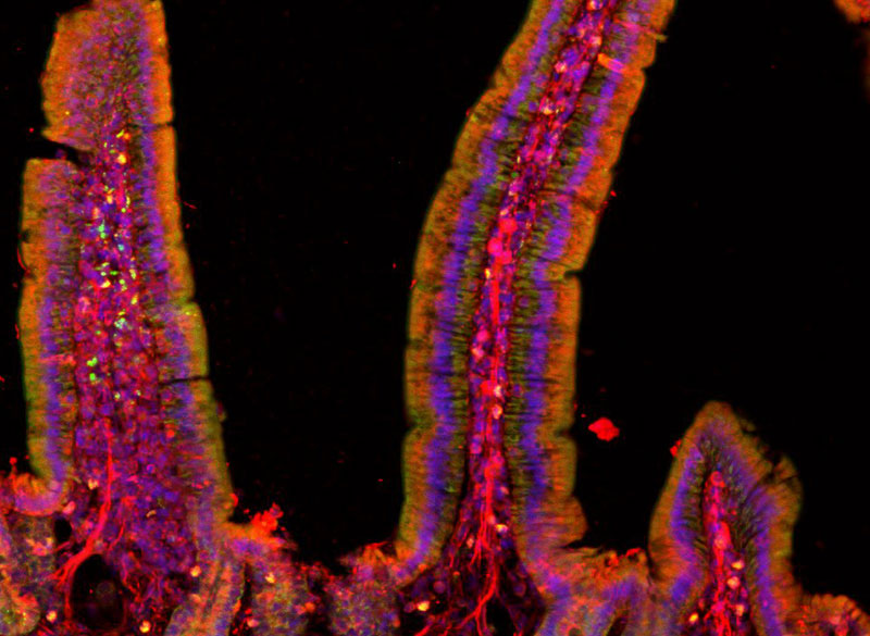
Fan Wang investigates the neural circuits that govern the dynamic interactions between brain and body, exploring how the brain generates sensory perceptions and controls movement. Wang, who is also the co-director of the K. Lisa Yang and Hock E. Tan Center for Molecular Therapeutics, uses cutting-edge techniques including optogenetics, in vivo electrophysiology, and in vivo imaging, to make discoveries with profound clinical implications.
By developing innovative tools to study how brain circuits work, Wang discovered distinct populations of neurons activated by anesthesia that can suppress pain without blocking sensation, and can calm anxiety by regulating automatic body functions like heart rate. She also identified the brain circuits controlling rhythmic movements essential for exploration and communication. Together, these findings reveal how emotion, physiology, movement, and consciousness are deeply interconnected.
Wang combines rigorous basic neuroscience with a commitment to translating her discoveries into therapies that relieve human suffering. Her election to the NAS recognizes her contributions to understanding the brain-body connection and therapeutic potential of her groundbreaking research.
The formal induction ceremony for new NAS members, during which they sign the ledger whose first signatory is Abraham Lincoln, will be held at the Academy’s annual meeting in Washington D.C. next spring.