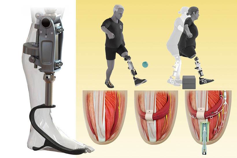
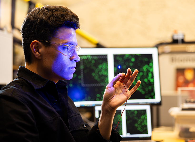
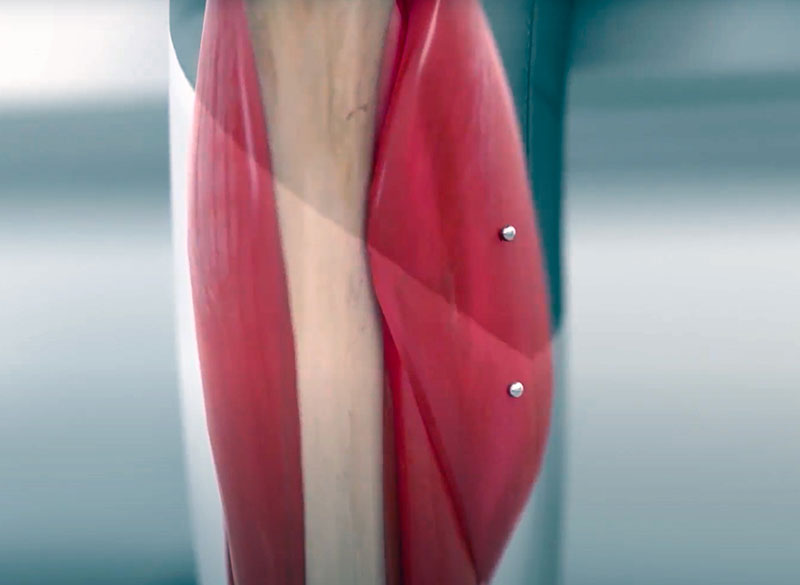
New MIT research offers a glimpse into this future. In a study published today in Nature Communications, the researchers introduce a novel myoneural actuator (MNA) that reprograms living muscles into fatigue-resistant, computer-controlled motors that can be implanted inside the body to restore movement in organs.
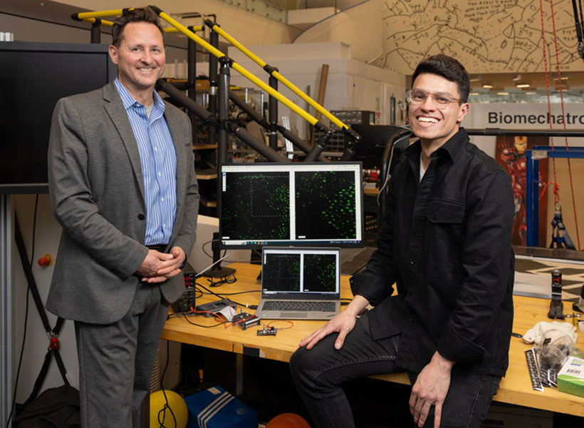
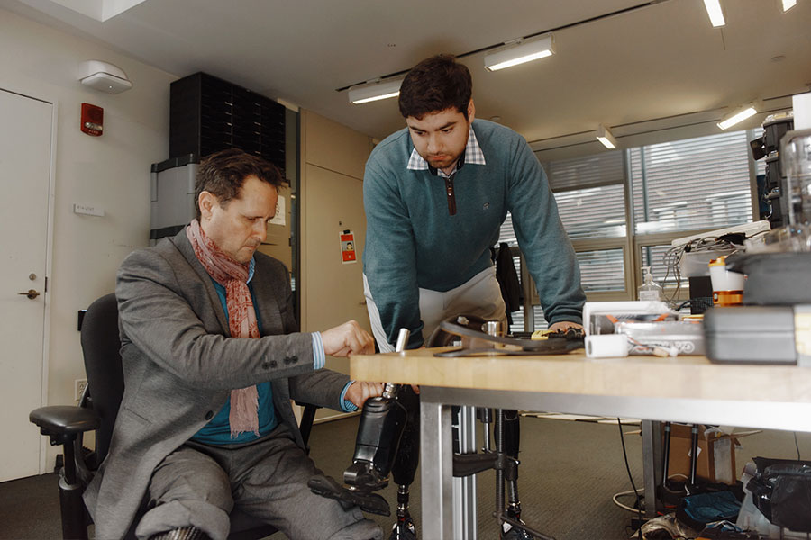
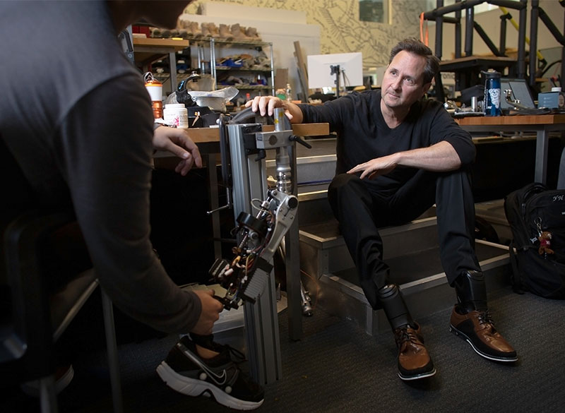
“We’ve built an interface that leverages natural pathways used by the nervous system so that we can seamlessly control organs in the body, while also enabling the transmission of sensory feedback to the brain,” says Hugh Herr, senior author of the study, a professor of Media Arts and Sciences at the MIT Media Lab, co-director of the K. Lisa Yang Center for Bionics, and an associate member of the McGovern Institute for Brain Research at MIT. The study was co-led by Herr’s postdoctoral associate Guillermo Herrera-Arcos and former postdoc Hyungeun Song.
By repurposing existing muscle in the body, the researchers have developed the first “living” implant that uses rewired sensory nerves to revive paralyzed organs — which may present a new genre of medicine where a person’s own tissue becomes the hardware.
Rewiring the brain-body interface
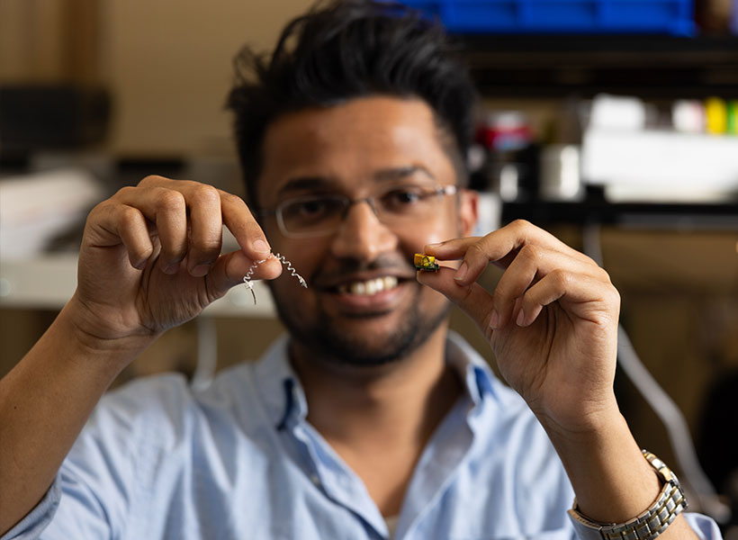
Many scientists have toiled to restore function in paralyzed organs, but it’s extremely challenging to design a technology that both communicates with the nervous system and doesn’t fatigue over time. Some have tried to insert miniaturized actuators — small machines that can power bionic limbs — into the body. However, Herrera-Arcos says “it’s hard to make actuators at the centimeter level and they aren’t very efficient.” Others have focused on creating muscle tissue in the lab, but building muscles cell by cell is time-intensive and far from ready for human use.
Herr’s team tried something different.
“We engineered existing muscles to become an actuator, or motor, that reinstates motion in organs,” says Song.
To do this, the researchers had to navigate the delicate dynamics within the nervous system. The actuator would have to interface with the nervous system to work properly, but it must also somehow evade the brain’s control. “You don’t want the brain to consciously control the muscle actuator because you want the actuator to automatically control an organ, like the heart,” explains Herrera-Arcos. Establishing a computer-controlled muscle to move organs could ensure automatic function and also bypass damaged brain pathways.
Incorporating motor neurons into the actuator may help generate movement, but these neurons are directly controlled by the brain. “Sensory neurons, however, are wired to receive, not to command,” explains Song. “We thought we could leverage this dynamic and reroute motor signals through sensory fibers, making a computer — rather than the brain — the muscle’s new command center.”
To achieve this, sensory nerves would need to fuse fluidly with muscle, and scientists had not yet determined if this was possible. Remarkably, when the team replaced motor nerves in rodent muscle with sensory ones, “the sensory nerves reinnervated the muscles and formed functional synapses. It’s a tremendous discovery,” says Herrera-Arcos.
Sensory neurons not only enabled the use of a digital controller but also helped curb muscle fatigue — increasing fatigue resistance in rodent muscle by 260 percent compared to native muscles. That’s because muscle fatigue depends largely on the diameter of the axons, or cable-like projections that innervate muscles. Motor neuron axons vary greatly in size, and when a motor nerve is electrically stimulated, the largest axons fire first — exhausting the muscle quickly. However, sensory axons are all nearly the same size, so the signal is broadcast more evenly across muscle fibers, avoiding fatigue, explains Herrera-Arcos.
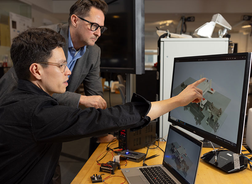
Designing a biohybrid system
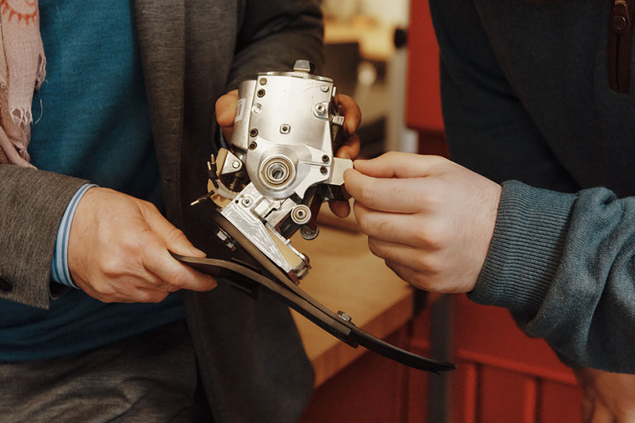
They combined all of these elements into a fatigue-resistant biohybrid motor called a myoneural actuator (MNA). By wrapping their actuator around a paralyzed intestine in a rodent, the researchers reinstated the organ’s squeezing motion. They also successfully controlled rodent calf muscles in an experiment designed to mimic residual muscle in human lower-limb amputations. Importantly, the MNA system transmitted sensory signals to the brain. “This suggests that our technology could seamlessly link organs to the brain. For example, we might be able to make a paralyzed stomach relay hunger,” explains Song.
Bringing their MNA to clinic will require further testing in larger animal models, and eventually, humans. But if it passes the regulatory gauntlet, their system could pave a smoother and safer path toward reviving static organs. Implanting MNAs would require a surgery that is already commonplace in clinic, the researchers say, and their system might be simpler and safer to implement than mechanical devices or organ transplants that introduce foreign material into the body.
The team is hopeful that their new technology could improve the lives of millions living with organ dysfunctions. “Today’s solutions are mostly synthetic: pacemakers and other mechanical assist devices. A living muscle actuator implanted alongside a weakened organ would be part of the body itself. That is a category of medicine different from anything seen in clinic,” explains Herrera-Arcos.
Song says that skin is of special interest. “Hypothetically, we could wrap MNAs around skin grafts to relay tactile feedback, such as strain or tension, which is currently missing for users of prostheses.” Their technology could even augment virtual reality systems, too. “The idea is that, if we couple the MNA system to skin and muscles, a person could feel what their virtual avatar is touching even though their real body isn’t moving,” says Song.
“Our research is on the brink of giving new life to various parts and extensions of the body,” adds Herrera-Arcos. “It’s exciting to think that our system could enhance human potential in ways that once only belonged to the realm of science fiction.”
This research was funded in part by the Yang Tan Collective at MIT, K. Lisa Yang Center for Bionics at MIT, Nakos Family Bionics Research Fund at MIT, and the Carl and Ruth Shapiro Foundation.