Mapping healthy cells’ connections in the brain

Portrait of scientist in a suit and tie.
McGovern Institute Principal Research Scientist Ian Wickersham. Photo: Caitlin Cunningham

A new tool developed by researchers at MIT’s McGovern Institute gives neuroscientists the power to find connected neurons within the brain’s tangled network of cells, and then follow or manipulate those neurons over a prolonged period. Its development, led by Principal Research Scientist Ian Wickersham, transforms a powerful tool for exploring the anatomy of the brain into a sophisticated system for studying brain function.

Wickersham and colleagues have designed their system to enable long-term analysis and experiments on groups of neurons that reach through the brain to signal to select groups of cells. It is described in the January 11, 2024, issue of the journal Nature Neuroscience. “This second-generation system will allow imaging, recording, and control of identified networks of synaptically-connected neurons in the context of behavioral studies and other experimental designs lasting weeks, months, or years,” Wickersham says.

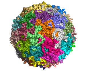
The system builds on an approach to anatomical tracing that Wickersham developed in 2007, as a graduate student in Edward Callaway’s lab at the Salk Institute for Biological Studies. Its key is a modified version of a rabies virus, whose natural—and deadly—life cycle involves traveling through the brain’s neural network.

Viral tracing

The rabies virus is useful for tracing neuronal connections because once it has infected the nervous system, it spreads through the neural network by co-opting the very junctions that neurons use to communicate with one another. Hopping across those junctions, or synapses, the virus can pass from cell to cell. Traveling in the opposite direction of neuronal signals, it reaches the brain, where it continues to spread.

Labeled illustration of rabies virus
Simplified illustration of rabies virus. Image: istockphoto

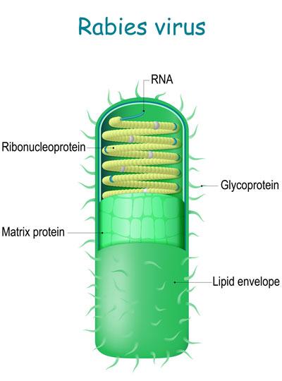
To use the rabies virus to identify specific connections within the brain, Wickersham modified it to limit its spread. His original tracing system uses a rabies virus that lacks an essential gene. When researchers deliver the modified virus to the neurons whose connections they want to map, they also instruct those neurons to make the protein encoded by the virus’s missing gene. That allows the virus to replicate and travel across the synapses that link an infected cell to others in the network. Once it is inside a new cell, the virus is deprived of the critical protein and can go no farther.

Under a microscope, a fluorescent protein delivered by the modified virus lights up, exposing infected cells: those to which the virus was originally delivered as well as any neurons that send it direct inputs. Because the virus crosses only one synapse after leaving the cell it originally infected, the technique is known as monosynaptic tracing.

Labs around the world now use this method to identify which brain cells send signals to a particular set of neurons. But while the virus used in the original system can’t spread through the brain like a natural rabies virus, it still sickens the cells it does infect. Infected cells usually die in about two weeks, and that has limited scientists’ ability to conduct further studies of the cells whose connections they trace. “If you want to then go on to manipulate those connected populations of cells, you have a very short time window,” Wickersham says.

Reducing toxicity

To keep cells healthy after monosynaptic tracing, Wickersham, postdoctoral researcher Lei Jin, and colleagues devised a new approach. They began by deleting a second gene from the modified virus they use to label cells. That gene encodes an enzyme the rabies virus needs to produce the proteins encoded in its own genome. As with the original system, neurons are instructed to create the virus’s missing proteins, equipping the virus to replicate inside those cells. In this case, this is done in mice that have been genetically modified to produce the second deleted viral gene in specific sets of neurons.

Brightly colored neurons under a microscope.
The initially-infected “starter cells” at the injection site in the substantia nigra, pars compacta. Blue: tyrosine hydroxylase immunostaining, showing dopaminergic cells; green: enhanced green fluorescent protein showing neurons able to be initially infected with the rabies virus; red: the red fluorescent protein tdTomato, reporting the presence of the second-generation rabies virus. Image: Ian Wickersham, Lei Jin

To limit toxicity, Wickersham and his team built in a control that allows researchers to switch off cells’ production of viral proteins once the virus has had time to replicate and begin its spread to connected neurons. With those proteins no longer available to support the viral life cycle, the tracing tool is rendered virtually harmless. After following mice for up to 10 weeks, the researchers detected minimal toxicity in neurons where monosynaptic tracing was initiated. And, Wickersham says, “as far as we can tell, the trans-synaptically labeled cells are completely unscathed.”

Neurons illuminated in red under a microscope
Transsynaptically labeled cells in the striatum, which provides input to the dopaminergic cells of the substantia nigra. These cells show no morphological abnormalities or any other indication of toxicity five weeks after the rabies virus injection. Image: Ian Wickersham, Lei Jin

That means neuroscientists can now pair monosynaptic tracing with many of neuroscience’s most powerful tools for functional studies. To facilitate those experiments, Wickersham’s team encoded enzymes called recombinases into their connection-tracing rabies virus, which enables the introduction of genetically encoded research tools to targeted cells. After tracing cells’ connections, researchers will be able to manipulate those neurons, follow their activity, and explore their contributions to animal behavior. Such experiments will deepen scientists’ understanding of the inputs select groups of neurons receive from elsewhere in the brain, as well as the cells that are sending those signals.

Jin, who is now a principal investigator at Lingang Laboratory in Shanghai, says colleagues are already eager to begin working with the new non-toxic tracing system. Meanwhile, Wickersham’s group has already started experimenting with a third-generation system, which they hope will improve efficiency and be even more powerful.

Four McGovern Investigators receive NIH BRAIN Initiative grants

In the human brain, 86 billion neurons form more than 100 trillion connections with other neurons at junctions called synapses. Scientists at the McGovern Institute are working with their collaborators to develop technologies to map these connections across the brain, from mice to humans.

Today, the National Institutes of Health (NIH) announced a new program to support research projects that have the potential to reveal an unprecedented and dynamic picture of the connected networks in the brain. Four of these NIH-funded research projects will take place in McGovern labs.

BRAIN Initiative

In 2013, the Obama administration announced the Brain Research Through Advancing Innovative Neurotechnologies® (BRAIN) Initiative, a public-private research effort to support the development and application of new technologies to understand brain function.

Today, the NIH announced its third project supported by the BRAIN Initiative, called BRAIN Initiative Connectivity Across Scales (BRAIN CONNECTS). The new project complements two previous large-scale projects, which together aim to transform neuroscience research by generating wiring diagrams that can span entire brains across multiple species. These detailed wiring diagrams can help uncover the logic of the brain’s neural code, leading to a better understanding of how this circuitry makes us who we are and how it could be rewired to treat brain diseases.

BRAIN CONNECTS at McGovern

The initial round of BRAIN CONNECTS awards will support researchers at more than 40 university and research institutions across the globe with 11 grants totaling $150 million over five years. Four of these grants have been awarded to McGovern researchers Guoping Feng, Ila Fiete, Satra Ghosh, and Ian Wickersham, whose projects are outlined below:

BRAIN CONNECTS: Comprehensive regional projection map of marmoset with single axon and cell type resolution
Team: Guoping Feng (McGovern Institute, MIT), Partha Mitra (Cold Spring Harbor Laboratory), Xiao Wang (Broad Institute), Ian Wickersham (McGovern Institute, MIT)

Summary: This project will establish an integrated experimental-computational platform to create the first comprehensive brain-wide mesoscale connectivity map in a non-human primate (NHP), the common marmoset (Callithrix jacchus). It will do so by tracing axonal projections of RNA barcode-identified neurons brain-wide in the marmoset, utilizing a sequencing-based imaging method that also permits simultaneous transcriptomic cell typing of the identified neurons. This work will help bridge the gap between brain-wide mesoscale connectivity data available for the mouse from a decade of mapping efforts using modern techniques and the absence of comparable data in humans and NHPs.

BRAIN CONNECTS: A center for high-throughput integrative mouse connectomics
Team: Jeff Lichtman (Harvard University), Ila Fiete (McGovern Institute, MIT), Sebastian Seung (Princeton University), David Tank (Princeton University), Hongkui Zeng (Allen Institute), Viren Jain (Google), Greg Jeffries (Oxford University)

Summary: This project aims to produce a large-scale synapse-level brain map (connectome) that includes all the main areas of the mouse hippocampus. This region is of clinical interest because it is an essential part of the circuit underlying spatial navigation and memory and the earliest impairments and degeneration related to Alzheimer’s disease.

BRAIN CONNECTS: The center for Large-scale Imaging of Neural Circuits (LINC)
Team: Anastasia Yendiki (MGH), Satra Ghosh (McGovern, MIT), Suzanne Haber (University of Rochester), Elizabeth Hillman (Columbia University)

Summary: This project will generate connectional diagrams of the monkey and human brain at unprecedented resolutions. These diagrams will be linked both to the neuroanatomic literature and to in vivo neuroimaging techniques, bridging between the rigor of the former and the clinical relevance of the latter. The data to be generated by this project will advance our understanding of brain circuits that are implicated in motor and psychiatric disorders, and that are targeted by deep-brain stimulation to treat these disorders.

BRAIN CONNECTS: Mapping brain-wide connectivity of neuronal types using barcoded connectomics
Team: Xiaoyin Chen (Allen Institute), Ian Wickersham (McGovern Institute, MIT), and Justus Kebschull of JHU

Summary: This project aims to optimize and develop barcode sequencing-based neuroanatomical techniques to achieve brain-wide, high-throughput, highly multiplexed mapping of axonal projections and synaptic connectivity of neuronal types at cellular resolution in primate brains. The team will work together to apply these techniques to generate an unprecedented multi-resolution map of brain-wide projections and synaptic inputs of neurons in the macaque visual cortex at cellular resolution.


Circuit that focuses attention brings in wide array of inputs

In a new brain-wide circuit tracing study, scientists at MIT’s Picower Institute for Learning and Memory focused selective attention on a circuit that governs, fittingly enough, selective attention. The comprehensive maps they produced illustrate how broadly the mammalian brain incorporates and integrates information to focus its sensory resources on its goals.

Working in mice, the team traced thousands of inputs into the circuit, a communication loop between the anterior cingulate cortex (ACC) and the lateral posterior (LP) thalamus. In primates the LP is called the pulvinar. Studies in humans and nonhuman primates have indicated that the byplay of these two regions is critical for brain functions like being able to focus on an object of interest in a crowded scene, says study co-lead author Yi Ning Leow, a graduate student in the lab of senior author Mriganka Sur, the Newton Professor in MIT’s Department of Brain and Cognitive Sciences. Research has implicated dysfunction in the circuit in attention-affecting disorders such as autism and attention deficit/hyperactivity disorder.

The new study in the Journal of Comparative Neurology extends what’s known about the circuit by detailing it in mice, Leow says, importantly showing that the mouse circuit is closely analogous to the primate version even if the LP is proportionately smaller and less evolved than the pulvinar.

“In these rodent models we were able to find very similar circuits,” Leow says. “So we can possibly study these higher-order functions in mice as well. We have a lot more genetic tools in mice so we are better able to look at this circuit.”

The study, also co-led by former MIT undergraduate Blake Zhou, therefore provides a detailed roadmap in the experimentally accessible mouse model for understanding how the ACC and LP cooperate to produce selective attention. For instance, now that Leow and Zhou have located all the inputs that are wired into the circuit, Leow is tapping into those feeds to eavesdrop on the information they are carrying. Meanwhile, she is correlating that information flow with behavior.

“This study lays the groundwork for understanding one of the most important, yet most elusive, components of brain function, namely our ability to selectively attend to one thing out of several, as well as switch attention,” Sur says.

Using virally mediated circuit-tracing techniques pioneered by co-author Ian Wickersham, principal research scientist in brain and cognitive sciences and the McGovern Institute for Brain Research at MIT, the team found distinct sources of input for the ACC and the LP. Generally speaking, the detailed study finds that the majority of inputs to the ACC were from frontal cortex areas that typically govern goal-directed planning, and from higher visual areas. The bulk of inputs to the LP, meanwhile, were from deeper regions capable of providing context such as the mouse’s needs, location and spatial cues, information about movement, and general information from a mix of senses.

So even though focusing attention might seem like a matter of controlling the senses, Leow says, the circuit pulls in a lot of other information as well.

“We’re seeing that it’s not just sensory — there are so many inputs that are coming from non-sensory areas as well, both sub-cortically and cortically,” she says. “It seems to be integrating a lot of different aspects that might relate to the behavioral state of the animal at a given time. It provides a way to provide a lot of internal and special context for that sensory information.”

Given the distinct sets of inputs to each region, the ACC may be tasked with focusing attention on a desired object, while the LP is modulating how the ACC goes about making those computations, accounting for what’s going on both inside and outside the animal. Decoding just what that incoming contextual information is, and what the LP tells the ACC, are the key next steps, Leow says. Another clear set of questions the study raises are what are the circuit’s outputs. In other words, after it integrates all this information, what does it do with it?

The paper’s other authors are Heather Sullivan and Alexandria Barlowe.

A National Science Scholarship, the National Institutes of Health, and the JPB Foundation provided support for the study.

Powered by viruses

View the interactive version of this story in our Winter 2021 issue of Brain Scan.

Viruses are notoriously adept invaders. The efficiency with which these unseen threats infiltrate tissues, evade immune systems, and occupy the cells of their hosts can be alarming — but it’s exactly why most McGovern neuroscientists keep a stash of viruses in the freezer.

In the hands of neuroscientists, viruses become vital tools for delivering cargo to cells.

With a bit of genetic manipulation, they can instruct neurons to produce proteins that illuminate complex circuitry, report on activity, or place certain cells under scientists’ control. They can even deliver therapies designed to correct genetic defects in patients.

“We rely on the virus to deliver whatever we want,” says McGovern Investigator Guoping Feng. “This is one of the most important technologies in neuroscience.”

Tracing connections

In Ian Wickersham’s lab, researchers are adapting a virus that, in its natural form, is devastating to the mammalian nervous system. Once it gains access to a neuron, the rabies virus spreads to connected cells, killing them within weeks. “That makes it a very dangerous pathogen, but also a very powerful tool for neuroscience,” says Wickersham, a Principal Research Scientist at the Institute.

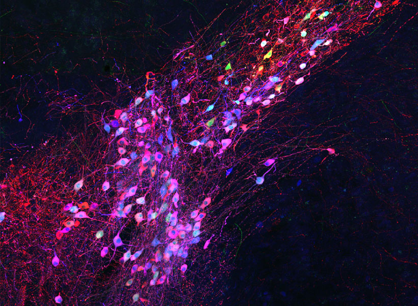
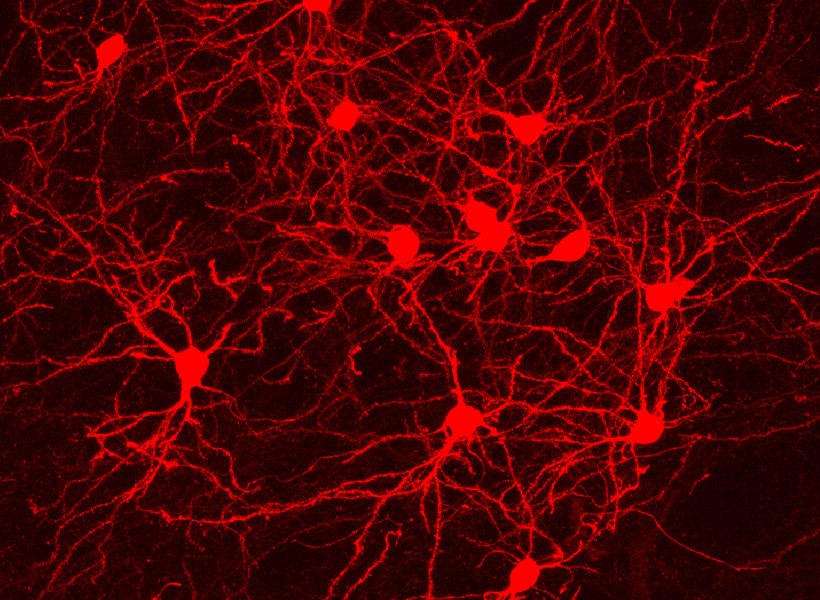
Taking advantage of its pernicious spread, neuroscientists use a modified version of the rabies virus to introduce a fluorescent protein to infected cells and visualize their connections (above). As a graduate student in Edward Callaway’s lab at the Salk Institute for Biological Studies, Wickersham figured out how to limit the virus’s passage through the nervous system, allowing it to access cells that are directly connected to the neuron it initially infects, but go no further. Rabies virus travels across synapses in the opposite direction of neuronal signals, so researchers can deliver it to a single cell or set of cells, then see exactly where those cells’ inputs are coming from.

Labs around the world use Wickersham’s modified rabies virus to trace neuronal anatomy in the brains of mice. While his team tinkers to make the virus even more powerful, his collaborators have deployed it to map a variety of essential connections, offering clues into how the brain controls movement, detects odors, and retrieves memories.

With the newest tracing tool from the Wickersham lab, moving from anatomical studies to experiments that reveal circuit function is seamless, because the lab has further modified their virus so that it cannot kill cells. Researchers can label connected cells, then proceed to monitor their signals or manipulate their activity in the same animals.

Researchers usually conduct these experiments in genetically modified mice to control the subset of cells that activate the tracing system. It’s the same approach used to restrict most virally-delivered tools to specific neurons, which is crucial, Feng says. When introducing a fluorescent protein for imaging, for example, “we don’t want the gene we deliver to be activated everywhere, otherwise the whole brain will be lighting up,” he says.

Selective targets

In Feng’s lab, research scientist Martin Wienisch is working to make it easier to control this aspect of delivery. Rather than relying on the genetic makeup of an entire animal to determine where a virally-transported gene is switched on, instructions can be programmed directly into the virus, borrowing regulatory sequences that cells already know how to interpret.

Wienisch is scouring the genomes of individual neurons to identify short segments of regulatory DNA called enhancers. He’s focused on those that selectively activate gene expression in just one of hundreds of different neuron types, particularly in animal models that are not very amenable to genetic engineering. “In the real brain, many elements interact to drive cell specific expression. But amazingly sometimes a single enhancer is all we need to get the same effect,” he says.

Researchers are already using enhancers to confine viral tools to select groups of cells, but Wienisch, who is collaborating with Fenna Krienen in Steve McCarroll’s lab at Harvard University, aims to create a comprehensive library. The enhancers they identify will be paired with a variety of genetically-encoded tools and packaged into adeno-associated viruses (AAV), the most widely used vectors in neuroscience. The Feng lab plans to use these tools to better understand the striatum, a part of the primate brain involved in motivation and behavioral choices. “Ideally, we would have a set of AAVs with enhancers that would give us selective access to all the different cell types in the striatum,” Wienisch says.

Enhancers will also be useful for delivering potential gene therapies to patients, Wienisch says. For many years, the Feng lab has been studying how a missing copy of a gene called Shank3 impairs neurons’ ability to communicate, leading to autism and intellectual disability. Now, they are investigating whether they can overcome these deficits by delivering a functional copy of Shank3 to the brain cells that need it. Widespread activation of the therapeutic gene might do more harm than good, but incorporating the right enhancer could ensure it is delivered to the appropriate cells at the right dose, Wienisch says.

Like most gene therapies in development, the therapeutic Shank3, which is currently being tested in animal models, is packaged into an AAV. AAVs safely and efficiently infect human cells, and by selecting the right type, therapies can be directed to specific cells. But AAVs are small viruses, capable of carrying only small genes. Xian Gao, a postdoctoral researcher in the Feng lab, has pared Shank3 down to its most essential components, creating a “minigene” that can be packaged inside the virus, but some things are difficult to fit inside an AAV. Therapies that aim to correct mutations using the CRISPR gene editing system, for example, often exceed the carrying capacity of an AAV.

Expanding options

“There’s been a lot of really phenomenal advances in our gene editing toolkit,” says Victoria Madigan, a postdoctoral researcher in McGovern Investigator Feng Zhang’s lab, where researchers are developing enzymes to more precisely modify DNA. “One of the main limitations of employing these enzymes clinically has been their delivery.”

To open up new options for gene therapy, Zhang and Madigan are working with a group of viruses called densoviruses. Densoviruses and AAVs belong to the same family, but about 50 percent more DNA can be packed inside the outer shell of some densoviruses.

A molecular model of Galleria mellonella densovirus. Image: Victoria Madigan / Zhang Lab

They are an esoteric group of viruses, Madigan says, infecting only insects and crustaceans and perhaps best known for certain members’ ability to devastate shrimp farms. While densoviruses haven’t received a lot of attention from scientists, their similarities to AAV have given the team clues about how to alter their outer capsids to enable them to enter human cells, and even direct them to particular cell types. The fact that they don’t naturally infect people also makes densoviruses promising candidates for clinical use, Madigan says, because patients’ immune systems are unlikely to be primed to reject them. AAV infections, in contrast, are so common that patients are often excluded from clinical trials for AAV-based therapies due to the presence of neutralizing antibodies against the vector.

Ultimately, densoviruses could enable major advances in gene therapy, making it possible to safely deliver sophisticated gene editing systems to patients’ cells, Madigan says — and that’s good reason for scientists to continue exploring the vast diversity in the viral world. “There’s something to be said for looking into viruses that are understudied as new tools,” she says. “There’s a lot of interesting stuff out there — a lot of diversity and thousands of years of evolution.”

Ian Wickersham

Making Connections

Ian Wickersham develops genetic tools that provide more powerful and precise ways to study the organization of the brain. His lab invents techniques for targeting neurons based on their synaptic connectivity and gene expression patterns in order to cause them to express genes that allow the neurons to be studied and controlled by neuroscientists and clinicians. The goal of Wickersham’s work is to provide neuroscience with more effective ways of studying the brain, and potentially to provide clinical neurology with more effective ways of treating disorders of the brain.

Viral tool traces long-term neuron activity

For the past decade, neuroscientists have been using a modified version of the rabies virus to label neurons and trace the connections between them. Although this technique has proven very useful, it has one major drawback: The virus is toxic to cells and can’t be used for studies longer than about two weeks.

Researchers at MIT and the Allen Institute for Brain Science have now developed a new version of this virus that stops replicating once it infects a cell, allowing it to deliver its genetic cargo without harming the cell. Using this technique, scientists should be able to study the infected neurons for several months, enabling longer-term studies of neuron functions and connections.

“With the first-generation vectors, the virus is replicating like crazy in the infected neurons, and that’s not good for them,” says Ian Wickersham, a principal research scientist at MIT’s McGovern Institute for Brain Research and the senior author of the new study. “With the second generation, infected cells look normal and act normal for at least four months — which was as long as we tracked them — and probably for the lifetime of the animal.”

Soumya Chatterjee of the Allen Institute is the lead author of the paper, which appears in the March 5 issue of Nature Neuroscience.

Viral tracing

Rabies viruses are well-suited for tracing neural connections because they have evolved to spread from neuron to neuron through junctions known as synapses. The viruses can also spread from the terminals of axons back to the cell body of the same neuron. Neuroscientists can engineer the viruses to carry genes for fluorescent proteins, which are useful for imaging, or for light-sensitive proteins that can be used to manipulate neuron activity.

In 2007, Wickersham demonstrated that a modified version of the rabies virus could be used to trace synapses between only directly connected neurons. Before that, researchers had been using the rabies virus for similar studies, but they were unable to keep it from spreading throughout the entire brain.

By deleting one of the virus’ five genes, which codes for a glycoprotein normally found on the surface of infected cells, Wickersham was able to create a version that can only spread to neurons in direct contact with the initially infected cell. This 2007 modification enabled scientists to perform “monosynaptic tracing,” a technique that allows them to identify connections between the infected neuron and any neuron that provides input to it.

This first generation of the modified rabies virus is also used for a related technique known as retrograde targeting, in which the virus can be injected into a cluster of axon terminals and then travel back to the cell bodies of those axons. This can help researchers discover the location of neurons that send impulses to the site of the virus injection.

Researchers at MIT have used retrograde targeting to identify populations of neurons of the basolateral amygdala that project to either the nucleus accumbens or the central medial amygdala. In that type of study, researchers can deliver optogenetic proteins that allow them to manipulate the activity of each population of cells. By selectively stimulating or shutting off these two separate cell populations, researchers can determine their functions.

Reduced toxicity

To create the second-generation version of this viral tool, Wickersham and his colleagues deleted the gene for the polymerase enzyme, which is necessary for transcribing viral genes. Without this gene, the virus becomes less harmful and infected cells can survive much longer. In the new study, the researchers found that neurons were still functioning normally for up to four months after infection.

“The second-generation virus enters a cell with its own few copies of the polymerase protein and is able to start transcribing its genes, including the transgene that we put into it. But then because it’s not able to make more copies of the polymerase, it doesn’t have this exponential takeover of the cell, and in practice it seems to be totally nontoxic,” Wickersham says.

The lack of polymerase also greatly reduces the expression of whichever gene the researchers engineer into the virus, so they need to employ a little extra genetic trickery to achieve their desired outcome. Instead of having the virus deliver a gene for a fluorescent or optogenetic protein, they engineer it to deliver a gene for an enzyme called Cre recombinase, which can delete target DNA sequences in the host cell’s genome.

This virus can then be used to study neurons in mice whose genomes have been engineered to include a gene that is turned on when the recombinase cuts out a small segment of DNA. Only a small amount of recombinase enzyme is needed to turn on the target gene, which could code for a fluorescent protein or another type of labeling molecule. The second-generation viruses can also work in regular mice if the researchers simultaneously inject another virus carrying a recombinase-activated gene for a fluorescent protein.

The new paper shows that the second-generation virus works well for retrograde labeling, not tracing synapses between cells, but the researchers have also now begun using it for monosynaptic tracing.

The research was funded by the National Institute of Mental Health, the National Institute on Aging, and the National Eye Institute.

Fifteen MIT scientists receive NIH BRAIN Initiative grants

Today, the National Institutes of Health (NIH) announced their first round of BRAIN Initiative award recipients. Six teams and 15 researchers from the Massachusetts Institute of Technology were recipients.

Mriganka Sur, principal investigator at the Picower Institute for Learning and Memory and the Paul E. Newton Professor of Neuroscience in MIT’s Department of Brain and Cognitive Sciences (BCS) leads a team studying cortical circuits and information flow during memory-guided perceptual decisions. Co-principal investigators include Emery Brown, BCS professor of computational neuroscience and the Edward Hood Taplin Professor of Medical Engineering; Kwanghun Chung, Picower Institute principal investigator and assistant professor in the Department of Chemical Engineering and the Institute for Medical Engineering and Science (IMES); and Ian Wickersham, research scientist at the McGovern Institute for Brain Research and head of MIT’s Genetic Neuroengineering Group.

Elly Nedivi, Picower Institute principal investigator and professor in BCS and the Department of Biology, leads a team studying new methods for high-speed monitoring of sensory-driven synaptic activity across all inputs to single living neurons in the context of the intact cerebral cortex. Her co-principal investigator is Peter So, professor of mechanical and biological engineering, and director of the MIT Laser Biomedical Research Center.

Ian Wickersham will lead a team looking at novel technologies for nontoxic transsynaptic tracing. His co-principal investigators include Robert Desimone, director of the McGovern Institute and the Doris and Don Berkey Professor of Neuroscience in BCS; Li-Huei Tsai, director of the Picower Institute and the Picower Professor of Neuroscience in BCS; and Kay Tye, Picower Institute principal investigator and assistant professor of neuroscience in BCS.

Robert Desimone will lead a team studying vascular interfaces for brain imaging and stimulation. Co-principal investigators include Ed Boyden, associate professor at the MIT Media Lab, McGovern Institute, and departments of BCS and Biological Engineering; head of MIT’s Synthetic Neurobiology Group, and co-director of MIT’s Center for Neurobiological Engineering; and Elazer Edelman, the Thomas D. and Virginia W. Cabot Professor of Health Sciences and Technology in IMES and director of the Harvard-MIT Biomedical Engineering Center. Collaborators on this project include: Rodolfo Llinas (New York University), George Church (Harvard University), Jan Rabaey (University of California at Berkeley), Pablo Blinder (Tel Aviv University), Eric Leuthardt (Washington University/St. Louis), Michel Maharbiz (Berkeley), Jose Carmena (Berkeley), Elad Alon (Berkeley), Colin Derdeyn (Washington University in St. Louis), Lowell Wood (Bill and Melinda Gates Foundation), Xue Han (Boston University), and Adam Marblestone (MIT).

Ed Boyden will be co-principal investigator with Mark Bathe, associate professor of biological engineering, and Peng Yin of Harvard on a project to study ultra-multiplexed nanoscale in situ proteomics for understanding synapse types.

Alan Jasanoff, associate professor of biological engineering and director of the MIT Center for Neurobiological Engineering, will lead a team looking at calcium sensors for molecular fMRI. Stephen Lippard, the Arthur Amos Noyes Professor of Chemistry, is co-principal investigator.

In addition, Sur and Wickersham also received BRAIN Early Concept Grants for Exploratory Research (EAGER) from the National Science Foundation (NSF). Sur will focus on massive-scale multi-area single neuron recordings to reveal circuits underlying short-term memory. Wickersham, in collaboration with Li-Huei Tsai, Kay Tye, and Robert Desimone, will develop cell-type specific optogenetics in wild-type animals. Additional information about NSF support of the BRAIN initiative can be found at NSF.gov/brain.

The BRAIN Initiative, spearheaded by President Obama in April 2013, challenges the nation’s leading scientists to advance our sophisticated understanding of the human mind and discover new ways to treat, prevent, and cure neurological disorders like Alzheimer’s, schizophrenia, autism, and traumatic brain injury. The scientific community is charged with accelerating the invention of cutting-edge technologies that can produce dynamic images of complex neural circuits and illuminate the interaction of lightning-fast brain cells. The new capabilities are expected to provide greater insights into how brain functionality is linked to behavior, learning, memory, and the underlying mechanisms of debilitating disease. BRAIN was launched with approximately $100 million in initial investments from the NIH, the National Science Foundation, and the Defense Advanced Research Projects Agency (DARPA).

BRAIN Initiative scientists are engaged in a challenging and transformative endeavor to explore how our minds instantaneously processes, store, and retrieve vast quantities of information. Their discoveries will unlock many of the remaining mysteries inherent in the brain’s billions of neurons and trillions of connections, leading to a deeper understanding of the underlying causes of many neurological and psychiatric conditions. Their findings will enable scientists and doctors to develop the groundbreaking arsenal of tools and technologies required to more effectively treat those suffering from these devastating disorders.