A social side to face recognition by infants
Rebecca Saxe and team discuss how face-selective areas of the brain might emerge.

When interacting with an infant you have likely noticed that the human face holds a special draw from a very young age. But how does this relate to face recognition by adults, which is known to map to specific cortical regions? Rebecca Saxe, Associate Investigator at MIT’s McGovern Institute and John W. Jarve (1978) Professor in Brain and Cognitive Sciences, and her team have now considered two emerging theories regarding early face recognition, and come up with a third proposition, arguing that when a baby looks at a face, the response is also social, and that the resulting contingent interactions are key to subsequent development of organized face recognition areas in the brain.
By a certain age you are highly skilled at recognizing and responding to faces, and this correlates with activation of a number of face-selective regions of the cortex. This is incredibly important to reading the identities and intentions of other people, and selective categorical representation of faces in cortical areas is a feature shared by our primate cousins. While brain imaging tells us where face-responsive regions are in the adult cortex, how and when they emerge remains unclear.
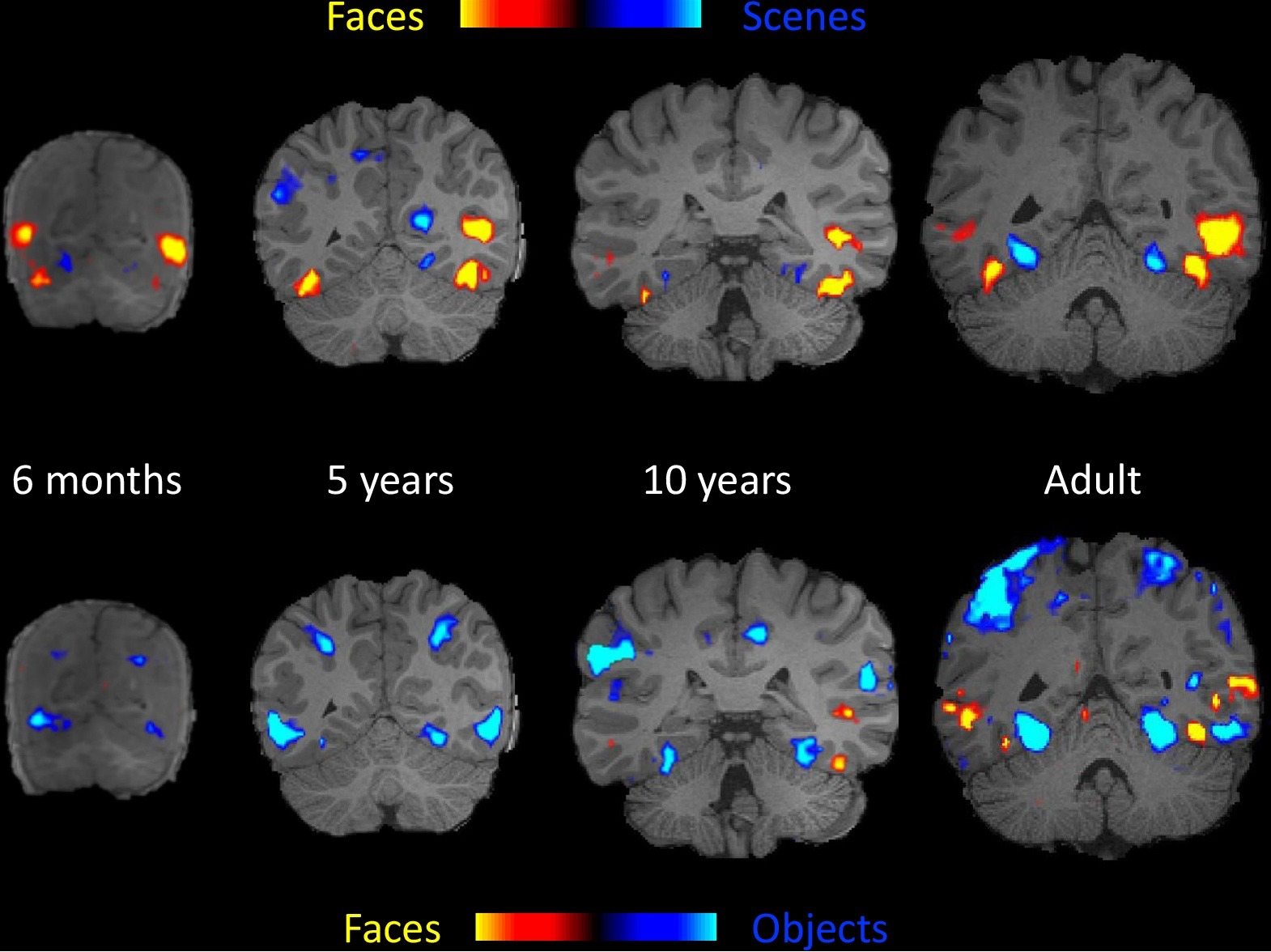
In 2017, functional magnetic resonance imaging (fMRI) studies of human and macaque infants provided the first glimpse of how the youngest brains respond to faces. The scans showed that in 4-6 month human infants and equivalently aged macaques, regions known to be face-responsive in the adult brain are activated when shown movies of faces, but not in a selective fashion. Essentially fMRI argues that these specific, cortical regions are activated by faces, but a chair will do just as well. Upon further experience of faces over time, the specific cortical regions in macaques became face-selective, no longer responding to other objects.
There are two prevailing ideas in the field of how face preference, and eventually selectivity, arise through experience. These ideas are now considered in turn by Saxe and her team in an opinion piece in the September issue of Trends in Cognitive Sciences, and then a third, new theory proposed. The first idea centers on the way we dote over babies, centering our own faces right in their field of vision. The idea is that such frequent exposures to low level face features (curvilinear shape etc.) will eventually lead to co-activation of neurons that are responsive to all of the different aspects of facial features. If these neurons stimulated by different features are co-activated, and there’s a brain region where these neurons are also found together, this area with be stimulated eventually reinforcing emergence of a face category-specific area.
A second idea is that babies already have an innate “face template,” just as a duckling or chick already knows to follow its mother after hatching. So far there is little evidence for the second proposition, and the first fails to explain why babies seek out a face, rather than passively look upon and eventually “learn” the overlapping features that represent “face.”
Saxe, along with postdoc Lindsey Powell and graduate student Heather Kosakowski, instead now argue that the role a face plays in positive social interactions comes to drive organization of face-selective cortical regions. Taking the next step, the researchers propose that a prime suspect for linking social interactions to the development of face-selective areas is the medial prefrontal cortex (mPFC), a region linked to social cognition and behavior.
“I was asked to give a talk at a conference, and I wanted to talk about both the development of cortical face areas and the social role of the medial prefrontal cortex in young infants,” says Saxe. “I was puzzling over whether these two ideas were related, when I suddenly saw that they could be very fundamentally related.”
The authors argue that this relationship is supported by existing data that has shown that babies prefer dynamic faces and are more interested in faces that engage in a back and forth interaction. Regions of the mPFC are also known to activated during social interactions and known to be activated during exposure to dynamic faces in infants.
Powell is now using functional near infrared spectroscopy (fNIRS), a brain imaging technique that measures changes in blood flow to the brain, to test this hypothesis in infants. “This will allow us to see whether mPFC responses to social cues are linked to the development of face-responsive areas.”
In Daniel Deronda, the novel by George Eliot, the protagonist says “I think my life began with waking up and loving my mother’s face: it was so near to me, and her arms were round me, and she sang to me.” Perhaps this type of positively valenced social interaction, reinforced by the mPFC, is exactly what leads to the particular importance of faces and their selective categorical representation in the human brain. Further testing of the hypothesis proposed by Powell, Kosakowski, and Saxe will tell.


