How do neurons communicate (so quickly)?
We take a closer look at the anatomy of the neuron and the role myelin plays in the rapid transmission of messages between brain cells.

Neurons are the most fundamental unit of the nervous system, and yet, researchers are just beginning to understand how they perform the complex computations that underlie our behavior. We asked Boaz Barak, previously a postdoc in Guoping Feng’s lab at the McGovern Institute and now Senior Lecturer at the School of Psychological Sciences and Sagol School of Neuroscience at Tel Aviv University, to unpack the basics of neuron communication for us.
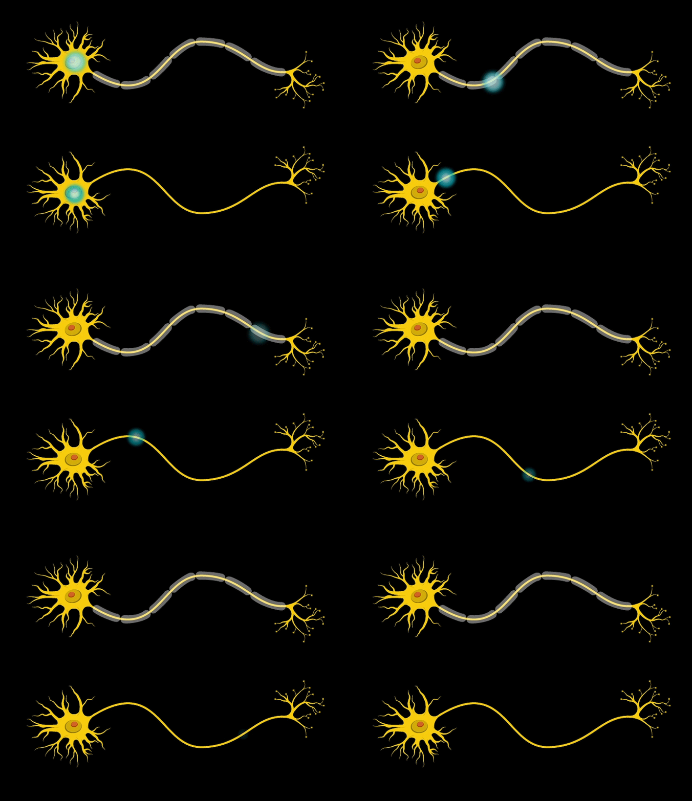
“Neurons communicate with each other through electrical and chemical signals,” explains Barak. “The electrical signal, or action potential, runs from the cell body area to the axon terminals, through a thin fiber called axon. Some of these axons can be very long and most of them are very short. The electrical signal that runs along the axon is based on ion movement. The speed of the signal transmission is influenced by an insulating layer called myelin,” he explains.
Myelin is a fatty layer formed, in the vertebrate central nervous system, by concentric wrapping of oligodendrocyte cell processes around axons. The term “myelin” was coined in 1854 by Virchow (whose penchant for Greek and for naming new structures also led to the terms amyloid, leukemia, and chromatin). In more modern images, the myelin sheath is beautifully visible as concentric spirals surrounding the “tube” of the axon itself. Neurons in the peripheral nervous system are also myelinated, but the cells responsible for myelination are Schwann cells, rather than oligodendrocytes.
“Neurons communicate with each other through electrical and chemical signals,” explains Boaz Barak.
“Myelin’s main purpose is to insulate the neuron’s axon,” Barak says. “It speeds up conductivity and the transmission of electrical impulses. Myelin promotes fast transmission of electrical signals mainly by affecting two factors: 1) increasing electrical resistance, or reducing leakage of the electrical signal and ions along the axon, “trapping” them inside the axon and 2) decreasing membrane capacitance by increasing the distance between conducting materials inside the axon (intracellular fluids) and outside of it (extracellular fluids).”
Adjacent sections of axon in a given neuron are each surrounded by a distinct myelin sheath. Unmyelinated gaps between adjacent ensheathed regions of the axon are called Nodes of Ranvier, and are critical to fast transmission of action potentials, in what is termed “saltatory conduction.” A useful analogy is that if the axon itself is like an electrical wire, myelin is like insulation that surrounds it, speeding up impulse propagation, and overcoming the decrease in action potential size that would occur during transmission along a naked axon due to electrical signal leakage, how the myelin sheath promotes fast transmission that allows neurons to transmit information long distances in a timely fashion in the vertebrate nervous system.
Myelin seems to be critical to healthy functioning of the nervous system; in fact, disruptions in the myelin sheath have been linked to a variety of disorders.

“Abnormal myelination can arise from abnormal development caused by genetic alterations,” Barak explains further. “Demyelination can even occur, due to an autoimmune response, trauma, and other causes. In neurological conditions in which myelin properties are abnormal, as in the case of lesions or plaques, signal transmission can be affected. For example, defects in myelin can lead to lack of neuronal communication, as there may be a delay or reduction in transmission of electrical and chemical signals. Also, in cases of abnormal myelination, it is possible that the synchronicity of brain region activity might be affected, for example, leading to improper actions and behaviors.”
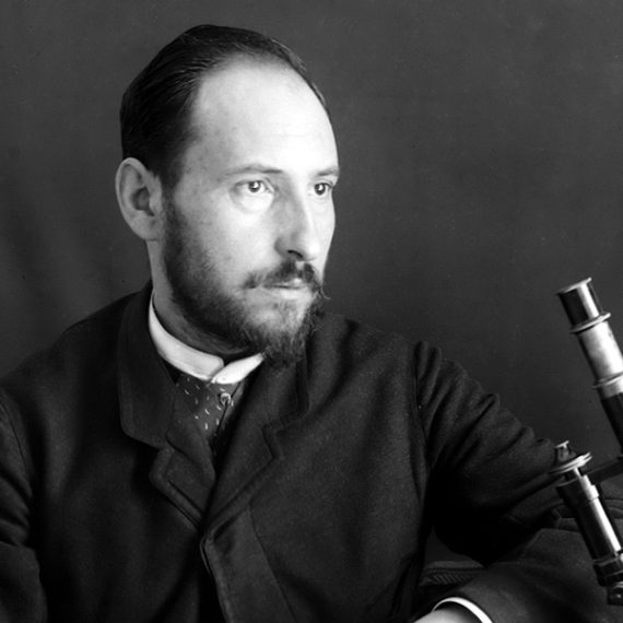
Researchers are still working to fully understand the role of myelin in disorders. Myelin has a long history of being evasive though, with its origins in the central nervous system being unclear for many years. For a period of time, the origin of myelin was thought to be the axon itself, and it was only after initial discovery (by Robertson, 1899), re-discovery (Del Rio-Hortega, 1919), and skepticism followed by eventual confirmation, that the role of oligodendrocytes in forming myelin became clear. With modern imaging and genetic tools, we should be able to increasingly understand its role in the healthy, as well as a compromised, nervous system.
Do you have a question for The Brain? Ask it here.


