How the brain distinguishes oozing fluids from solid objects

Imagine a ball bouncing down a flight of stairs. Now think about a cascade of water flowing down those same stairs. The ball and the water behave very differently, and it turns out that your brain has different regions for processing visual information about each type of physical matter.

In a new study, MIT neuroscientists have identified parts of the brain’s visual cortex that respond preferentially when you look at “things” — that is, rigid or deformable objects like a bouncing ball. Other brain regions are more activated when looking at “stuff” — liquids or granular substances such as sand.

This distinction, which has never been seen in the brain before, may help the brain plan how to interact with different kinds of physical materials, the researchers say.

“When you’re looking at some fluid or gooey stuff, you engage with it in different way than you do with a rigid object. With a rigid object, you might pick it up or grasp it, whereas with fluid or gooey stuff, you probably are going to have to use a tool to deal with it,” says Nancy Kanwisher, the Walter A. Rosenblith Professor of Cognitive Neuroscience; a member of the McGovern Institute for Brain Research and MIT’s Center for Brains, Minds, and Machines; and the senior author of the study.

MIT postdoc Vivian Paulun, who is joining the faculty of the University of Wisconsin at Madison this fall, is the lead author of the paper, which appears today in the journal Current Biology. RT Pramod, an MIT postdoc, and Josh Tenenbaum, an MIT professor of brain and cognitive sciences, are also authors of the study.

Stuff vs. things

Decades of brain imaging studies, including early work by Kanwisher, have revealed regions in the brain’s ventral visual pathway that are involved in recognizing the shapes of 3D objects, including an area called the lateral occipital complex (LOC). A region in the brain’s dorsal visual pathway, known as the frontoparietal physics network (FPN), analyzes the physical properties of materials, such as mass or stability.

Although scientists have learned a great deal about how these pathways respond to different features of objects, the vast majority of these studies have been done with solid objects, or “things.”

“Nobody has asked how we perceive what we call ‘stuff’ — that is, liquids or sand, honey, water, all sorts of gooey things. And so we decided to study that,” Paulun says.

These gooey materials behave very differently from solids. They flow rather than bounce, and interacting with them usually requires containers and tools such as spoons. The researchers wondered if these physical features might require the brain to devote specialized regions to interpreting them.

To explore how the brain processes these materials, Paulun used a software program designed for visual effects artists to create more than 100 video clips showing different types of things or stuff interacting with the physical environment. In these videos, the materials could be seen sloshing or tumbling inside a transparent box, being dropped onto another object, or bouncing or flowing down a set of stairs.

The researchers used functional magnetic resonance imaging (fMRI) to scan the visual cortex of people as they watched the videos. They found that both the LOC and the FPN respond to “things” and “stuff,” but that each pathway has distinctive subregions that respond more strongly to one or the other.

“Both the ventral and the dorsal visual pathway seem to have this subdivision, with one part responding more strongly to ‘things,’ and the other responding more strongly to ‘stuff,’” Paulun says. “We haven’t seen this before because nobody has asked that before.”

Roland Fleming, a professor of experimental psychology at Justus Liebig University of Geissen, described the findings as a “major breakthrough in the scientific understanding of how our brains represent the physical properties of our surrounding world.”

“We’ve known the distinction exists for a long time psychologically, but this is the first time that it’s been really mapped onto separate cortical structures in the brain. Now we can investigate the different computations that the distinct brain regions use to process and represent objects and materials,” says Fleming, who was not involved in the study.

Physical interactions

The findings suggest that the brain may have different ways of representing these two categories of material, similar to the artificial physics engines that are used to create video game graphics. These engines usually represent a 3D object as a mesh, while fluids are represented as sets of particles that can be rearranged.

“The interesting hypothesis that we can draw from this is that maybe the brain, similar to artificial game engines, has separate computations for representing and simulating ‘stuff’ and ‘things.’ And that would be something to test in the future,” Paulun says.

Portrait of smiling woman wearing a grey sweater.
McGovern Institute postdoc Vivian Paulun, who is joining the faculty of the University of Wisconsin at Madison in the fall of 2025, is the lead author of the “things vs. stuff” paper, which appears today in the journal Current Biology. Photo: Steph Stevens

The researchers also hypothesize that these regions may have developed to help the brain understand important distinctions that allow it to plan how to interact with the physical world. To further explore this possibility, the researchers plan to study whether the areas involved in processing rigid objects are also active when a brain circuit involved in planning to grasp objects is active.

They also hope to look at whether any of the areas within the FPN correlate with the processing of more specific features of materials, such as the viscosity of liquids or the bounciness of objects. And in the LOC, they plan to study how the brain represents changes in the shape of fluids and deformable substances.

The research was funded by the German Research Foundation, the U.S. National Institutes of Health, and a U.S. National Science Foundation grant to the Center for Brains, Minds, and Machines.


Adolescents’ willingness to explore is shaped by socioeconomic status

Exploration is essential to learning—and a new study from scientists at MIT’s McGovern Institute suggests that students may be less willing to explore if they come from a low socioeconomic environment. The study, which focused on adolescents and was published July 9, 2025, in the journal Nature Communications, shows how differences in learning strategies might contribute to socioeconomic-related disparities in academic achievement.

Students with low socioeconomic status (SES)—a measure that takes into account parents’ income levels and educational attainment—tend to lag behind their higher-SES peers academically. Limited resources at home can restrict access to educational tools and experiences, likely contributing to these disparities. But the new study, led by McGovern Institute Investigator John Gabrieli, shows that students from low-SES backgrounds may approach learning differently, too.

“We often think about external factors when we think about socioeconomic differences in learning, but kids’ mindsets and internal factors can also play a role,” says Alexandra Decker, a postdoctoral fellow in Gabrieli’s lab who ran the study. Understanding such differences can help educators develop strategies to reduce disparities and help all students succeed.

The value of exploration

Exploration is a vital part of development, particularly during adolescence. By trying new things and testing limits, children begin to find their way in the world, discovering the subjects and experiences that motivate them. That’s important for obtaining new knowledge, both in and out of school. “There’s a lot of research suggesting that exploration is a really important mechanism that children use for learning,” Decker says. “Exploring their environment really broadly and making mistakes helps them get the feedback that they need for learning,” she says.

Because the outcomes of exploration are unknown, this way of interacting with the world involves risk. “If you try something new, the outcome is uncertain, and it could lead to a bad outcome before things get better. You might lose out, at least in the short term. ” Decker says.

At school, students can explore in a variety of ways, such as by asking questions in class or taking on courses in unfamiliar subjects. Both are opportunities to learn something new, though they may seem less safe than sitting quietly and sticking to more comfortable coursework. Decker points out that this kind of exploration might feel particularly risky when students feel they lack the resources to compensate if things don’t go well.

“If you’re in an environment that’s really enriching, you have resources to compensate for challenges that might be accrued through exploring. If you take a new course and you struggle, you can use your resources to get a tutor and overcome these challenges. Your environment can support exploration and its costs,” she says. “But if you’re in an environment where you don’t have resources to compensate for bad outcomes, you might not take that course that could lead to unknown outcomes.”

Risk-benefit analysis

To investigate the relationship between SES and exploration, Gabrieli’s team had students play a computer game in which they earned points for pumping up balloons as much as possible without popping them. The most successful strategy was to explore the limits early on by pumping the first balloons until they popped, thereby learning when to stop with future balloons. A less exploratory approach could keep all the balloons intact, but earn fewer points over the course of the game.

The students who participated in the study were between the ages of 12 and 14 and came from families with a wide range of SES. Those from lower-SES backgrounds were less likely to explore in the balloon pumping task, resulting in lower outcomes in the game. What’s more, the researchers found a relationship between students’ exploration in the game and their real-world academic performance. Those who explored the least in the balloon-popping game had lower grades than students who explored more. For students at lower-SES levels, reduced exploration also correlated to lower scores on standardized tests of academic skills.

The researchers took a closer look at the data to investigate why some students explored more than others in their game. Their analysis indicated that students who were reluctant to explore were more strongly motivated by avoiding losses than students who had pushed the limits as they pumped their balloons.

The finding suggests that potential losses might be particularly distressing to lower-SES students, says Gabrieli, who is also the Grover Hermann Professor of Health Sciences and Technology and a professor of brain and cognitive sciences at MIT. Decker adds students from less affluent backgrounds may have found losses to be more consequential than they are for students whose families have more resources, so it makes sense that those students might take greater pains to avoid them.

This is not the first time Gabrieli’s group has found that evidence of differences in the ways students from different socioeconomic backgrounds make decisions. In a brain imaging study published last year, they found that the brains of adolescents from low-SES backgrounds respond less to rewards than the brains of their higher-SES peers. “How you think about the world—in terms of what’s rewarding, risks worth taking or not taking—seems strongly influenced by the environment that you’re growing up in,” he says.

Decker notes that regardless of SES, students in the study were generally more willing to explore when they had experienced more recent successes in the task. This finding, along with what the team learned about how loss aversion curtails exploration, suggest strategies that educators might use to encourage more exploration in the classroom. “Low-stakes opportunities for kids to engage in exploratory risk-taking with positive feedback could go a long way to helping kids feel more comfortable exploring,” Decker says.


A bionic knee integrated into tissue can restore natural movement

MIT researchers have developed a new bionic knee that can help people with above-the-knee amputations walk faster, climb stairs, and avoid obstacles more easily than they could with a traditional prosthesis.

Unlike prostheses in which the residual limb sits within a socket, the new system is directly integrated with the user’s muscle and bone tissue. This enables greater stability and gives the user much more control over the movement of the prosthesis.

Participants in a small clinical study also reported that the limb felt more like a part of their own body, compared to people who had more traditional above-the-knee amputations.

“A prosthesis that’s tissue-integrated — anchored to the bone and directly controlled by the nervous system — is not merely a lifeless, separate device, but rather a system that is carefully integrated into human physiology, offering a greater level of prosthetic embodiment. It’s not simply a tool that the human employs, but rather an integral part of self,” says Hugh Herr, a professor of media arts and sciences, co-director of the K. Lisa Yang Center for Bionics at MIT, an associate member of MIT’s McGovern Institute for Brain Research, and the senior author of the new study.

Tony Shu PhD ’24 is the lead author of the paper, which appears today in Science.

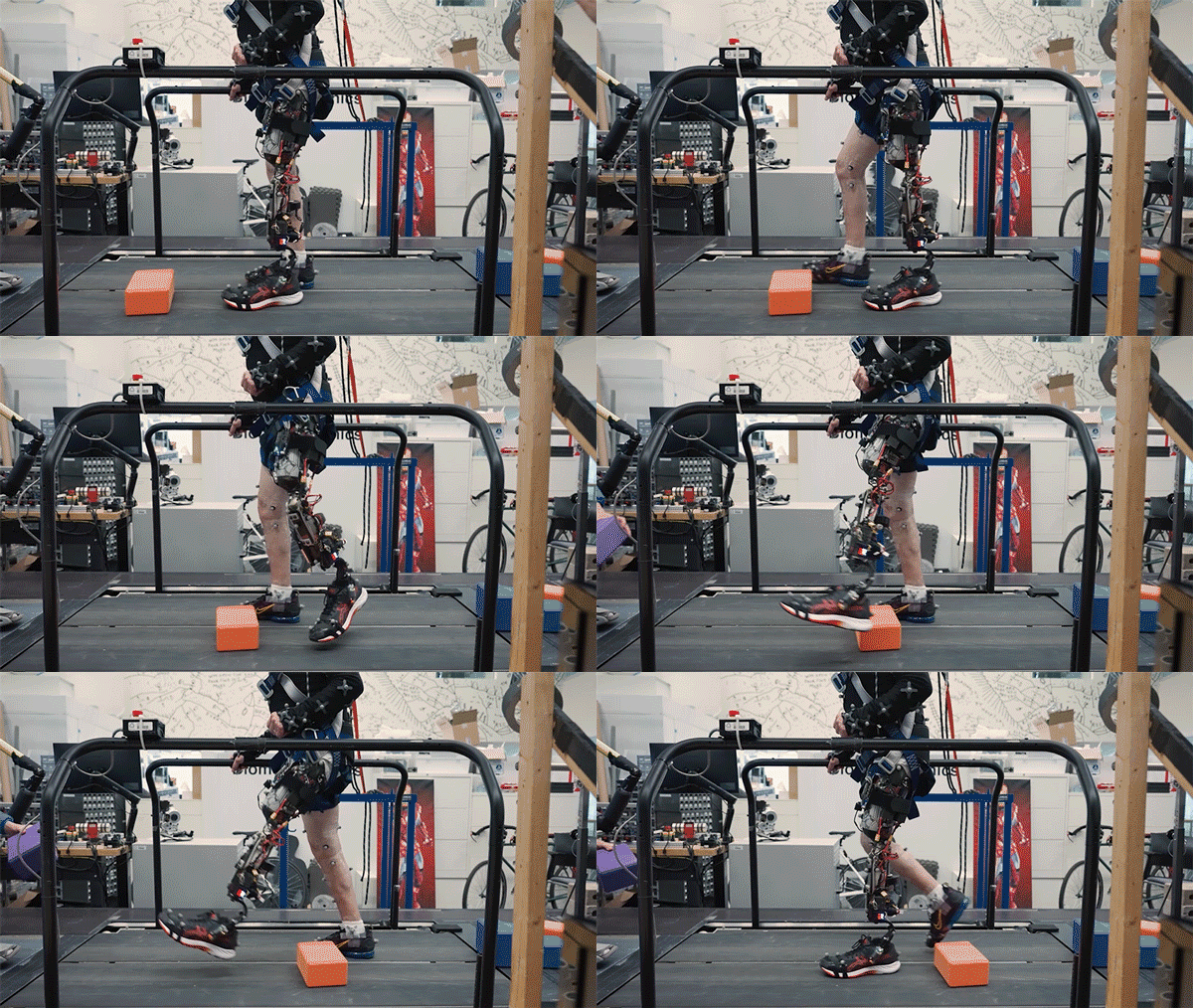
A subject with the osseointegrated mechanoneural prosthesis overcomes an obstacle placed in their walking path by volitionally flexing and extending their phantom knee joint.

Better control

Over the past several years, Herr’s lab has been working on new prostheses that can extract neural information from muscles left behind after an amputation and use that information to help guide a prosthetic limb.

During a traditional amputation, pairs of muscles that take turns stretching and contracting are usually severed, disrupting the normal agonist-antagonist relationship of the muscles. This disruption makes it very difficult for the nervous system to sense the position of a muscle and how fast it’s contracting.

Using the new surgical approach developed by Herr and his colleagues, known as agonist-antagonist myoneuronal interface (AMI), muscle pairs are reconnected during surgery so that they still dynamically communicate with each other within the residual limb. This sensory feedback helps the wearer of the prosthesis to decide how to move the limb, and also generates electrical signals that can be used to control the prosthetic limb.


In a 2024 study, the researchers showed that people with amputations below the knee who received the AMI surgery were able to walk faster and navigate around obstacles much more naturally than people with traditional below-the-knee amputations.

In the new study, the researchers extended the approach to better serve people with amputations above the knee. They wanted to create a system that could not only read out signals from the muscles using AMI but also be integrated into the bone, offering more stability and better sensory feedback.

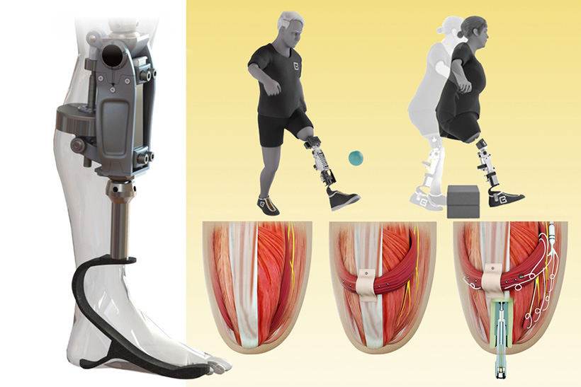
To achieve that, the researchers developed a procedure to insert a titanium rod into the residual femur bone at the amputation site. This implant allows for better mechanical control and load bearing than a traditional prosthesis. Additionally, the implant contains 16 wires that collect information from electrodes located on the AMI muscles inside the body, which enables more accurate transduction of the signals coming from the muscles.

This bone-integrated system, known as e-OPRA, transmits AMI signals to a new robotic controller developed specifically for this study. The controller uses this information to calculate the torque necessary to move the prosthesis the way that the user wants it to move.

The new bionic knee can help people with above-the-knee amputations walk faster, climb stairs, and avoid obstacles more easily than they could with a traditional prosthesis. The new system is directly integrated with the user’s muscle and bone tissue (bottom row right). This enables greater stability and gives the user much more control over the movement of the prosthesis. Image courtesy of the researchers

“All parts work together to better get information into and out of the body and better interface mechanically with the device,” Shu says. “We’re directly loading the skeleton, which is the part of the body that’s supposed to be loaded, as opposed to using sockets, which is uncomfortable and can lead to frequent skin infections.”

In this study, two subjects received the combined AMI and e-OPRA system, known as an osseointegrated mechanoneural prosthesis (OMP). These users were compared with eight who had the AMI surgery but not the e-OPRA implant, and seven users who had neither AMI nor e-OPRA. All subjects took a turn at using an experimental powered knee prosthesis developed by the lab.

The researchers measured the participants’ ability to perform several types of tasks, including bending the knee to a specified angle, climbing stairs, and stepping over obstacles. In most of these tasks, users with the OMP system performed better than the subjects who had the AMI surgery but not the e-OPRA implant, and much better than users of traditional prostheses.

“This paper represents the fulfillment of a vision that the scientific community has had for a long time — the implementation and demonstration of a fully physiologically integrated, volitionally controlled robotic leg,” says Michael Goldfarb, a professor of mechanical engineering and director of the Center for Intelligent Mechatronics at Vanderbilt University, who was not involved in the research. “This is really difficult work, and the authors deserve tremendous credit for their efforts in realizing such a challenging goal.”

A sense of embodiment

In addition to testing gait and other movements, the researchers also asked questions designed to evaluate participants’ sense of embodiment — that is, to what extent their prosthetic limb felt like a part of their own body.

Questions included whether the patients felt as if they had two legs, if they felt as if the prosthesis was part of their body, and if they felt in control of the prosthesis. Each question was designed to evaluate the participants’ feelings of agency, ownership of device, and body representation.

The researchers found that as the study went on, the two participants with the OMP showed much greater increases in their feelings of agency and ownership than the other subjects.

“Another reason this paper is significant is that it looks into these embodiment questions and it shows large improvements in that sensation of embodiment,” Herr says. “No matter how sophisticated you make the AI systems of a robotic prosthesis, it’s still going to feel like a tool to the user, like an external device. But with this tissue-integrated approach, when you ask the human user what is their body, the more it’s integrated, the more they’re going to say the prosthesis is actually part of self.”

The AMI procedure is now done routinely on patients with below-the-knee amputations at Brigham and Women’s Hospital, and Herr expects it will soon become the standard for above-the-knee amputations as well. The combined OMP system will need larger clinical trials to receive FDA approval for commercial use, which Herr expects may take about five years.

The research was funded by the Yang Tan Collective and DARPA.

MIT’s McGovern Institute and Department of Brain and Cognitive Sciences welcome new faculty member Sven Dorkenwald

The McGovern Institute and the Department of Brain and Cognitive Sciences are pleased to announce the appointment of Sven Dorkenwald as an assistant professor starting in January 2026. A trailblazer in the field of computational neuroscience, Dorkenwald is recognized for his leadership in connectomics—an emerging discipline focused on reconstructing and analyzing neural circuitry at unprecedented scale and detail. 

“We are thrilled to welcome Sven to MIT” says McGovern Institute Director Robert Desimone. “He brings visionary science and a collaborative spirit to a rapidly advancing area of brain and cognitive sciences and his appointment strengthens MIT’s position at the forefront of brain research.” 

Dorkenwald’s research is driven by a bold vision: to develop and apply cutting-edge computational methods that reveal how brain circuits are organized and how they give rise to complex computations. His innovative work has led to transformative advances in the reconstruction of connectomes (detailed neural maps) from nanometer-scale electron microscopy images. He has championed open team science and data sharing and played a central role in producing the first connectome of an entire fruit fly brain—a groundbreaking achievement that is reshaping our understanding of sensory processing and brain circuit function. 

Sven is a rising leader in computational neuroscience who has already made significant contributions toward advancing our understanding of the brain,” says Michale Fee, the Glen V. and Phyllis F. Dorflinger Professor of Neuroscience, and Department Head of Brain and Cognitive Sciences. “He brings a combination of technical expertise, a collaborative mindset, and a strong commitment to open science that will be invaluable to our department. I’m pleased to welcome him to our community and look forward to the impact he will have.” 

Dorkenwald earned his BS in physics in 2014 and MS in computer engineering in 2017 from the University of Heidelberg, Germany. He began his research in connectomics as an undergraduate in the group of Winfried Denk at the Max Planck Institute for Medical Research and Max Planck Institute of Neurobiology.  Dorkenwald went on to complete his PhD at Princeton University in 2023, where he studied both computer science and neuroscience under the mentorship of Sebastian Seung and Mala Murthy. 

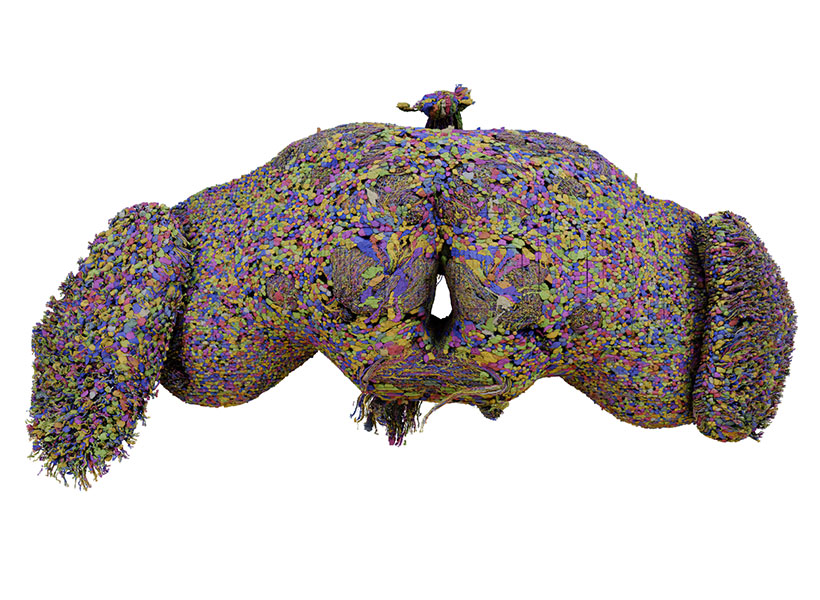
All 139,255 neurons in the brain of an adult fruit fly reconstructed by the FlyWire Consortium, with each neuron uniquely color-coded. Render by Tyler Sloan. Image: Sven Dorkenwald

As a PhD student at Princeton, Dorkenwald spearheaded the FlyWire Consortium, a group of more than 200 scientists, gamers, and proofreaders who combined their skills to create the fruit fly connectome. More than 20 million scientific images of the adult fruit fly brain  were added to an AI model that traced each neuron and synapse in exquisite detail. Members of the consortium then checked the results produced by the AI model and pieced them together into a complete, three-dimensional map. With over 140,000 neurons, it is the most complex brain completely mapped to date. The findings were published in a special issue of Nature in 2024. 

Dorkenwald’s work also played a key role in the MICrONS’ consortium effort to reconstruct a cubic millimeter connectome of the mouse visual cortex. Within the MICrONS effort, he co-lead the development of the software infrastructure, CAVE, that enables scientists to collaboratively edit and analyze large connectomics datasets, including FlyWire’s. The findings of the MICrONS consortium were published in a special issue of Nature in 2025. 

Dorkenwald is currently a Shanahan Fellow at the Allen Institute and the University of Washington. He also serves as a visiting faculty researcher at Google Research, where he has been developing machine learning approaches for the annotation of cell reconstructions as part of the Neuromancer team led by Viren Jain.  

As an investigator at the McGovern Institute and an assistant professor in the department of brain and cognitive sciences at MIT, Dorkenwald  plans to develop computational approaches to overcome challenges in scaling connectomics to whole mammalian brains with the goal of advancing our mechanistic understanding of neuronal circuits and analyzing how they compare across regions and species. 


Feng Zhang elected to EMBO membership

The European Molecular Biology Organization (EMBO), a professional non-profit organization dedicated to promoting international research in life sciences, announced its new members today. Among the 69 new members recognized for their outstanding achievements is Feng Zhang, the James and Patricia Poitras Professor of Neuroscience at MIT and an investigator at the McGovern Institute.

Zhang, who is also a core member of the Broad Institute, a professor of brain and cognitive sciences and biological engineering at MIT, and a Howard Hughes Medical Institute investigator, is a molecular biologist focused on improving human health. He played an integral role in pioneering the use of CRISPR-Cas9 for genome editing in human cells, including working with Stuart Orkin to develop Casgevy, the first CRISPR-based therapeutic approved for clinical use. His team is currently discovering new ways to modify cellular function and activity—including the restoration of diseased, stressed, or aged cells to a more healthful state.

Zhang has been elected to EMBO as an associate member, where he joins a community of more than 2,100 international life scientists that have demonstrated research excellence in their fields.

“A major strength of EMBO lies in the excellence and dedication of its members,” says EMBO Director Fiona Watt. “Science thrives on global collaboration, and the annual election of the new EMBO members and associate members brings fresh energy and inspiration to our community. We are honoured to welcome this remarkable group of scientists to the EMBO Membership. Their ideas and contributions will enrich the organization and help advance the life sciences internationally.”

The 60 new EMBO members in 2025 are based in 18 member states of the EMBC, the intergovernmental organization that funds the main EMBO programs and activities. The nine new EMBO associate members, including Zhang, are based in six countries outside Europe. In total, 29 (42%) of the new members are women and 40 (58%) are men.

The new members will be formally welcomed at the next EMBO Members’ Meeting in Heidelberg, Germany, on 22-24 October 2025.