This winter, may our connections spark new possibilities for the year ahead.
What makes us who we are? How do billions of neurons working together become our thoughts, feelings, and memories? How do they spark imagination and creativity? By tracing these connections, mapping how each neuron links to another, McGovern scientists are carving a path to uncover how these patterns generate the human experience. Because the intricate networks of neurons we’re working to understand, are the very ones that make understanding possible – empowering us to learn, discover, and create. And by exploring them, we see that being human at every level is about connection.
Happy holidays from your friends at the McGovern Institute!
Video credits:
Glass Ink Media and Julie Pryor (video)
Shepherd + Maudsleigh Studio | Megan Cascella (woodcut artist)
When it comes to brain function, neurons get a lot of the glory. But healthy brains depend on the cooperation of many kinds of cells. The most abundant of the brain’s non-neuronal cells are astrocytes, star-shaped cells with a lot of responsibilities. Astrocytes help shape neural circuits, participate in information processing, and provide nutrient and metabolic support to neurons. Individual cells can take on new roles throughout their lifetimes, and at any given time, the astrocytes in one part of the brain will look and behave differently than the astrocytes somewhere else.
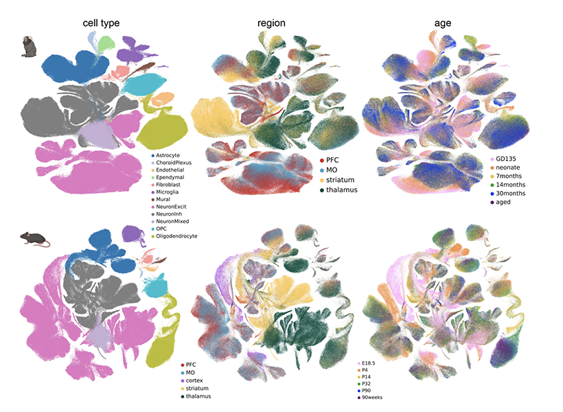
After an extensive analysis by scientists at MIT’s McGovern Institute, neuroscientists now have an atlas detailing astrocytes’ dynamic diversity. Its maps depict the regional specialization of astrocytes across the brains of both mice and marmosets—two powerful models for neuroscience research—and show how their populations shift as brains develop, mature, and age. The study, reported in the November 20 issue of the journal Neuron, was led by Guoping Feng, the James W. (1963) and Patricia T. Poitras Professor of Brain and Cognitive Sciences at MIT. This work was supported by the Hock E. Tan and K. Lisa Yang Center for Autism Research, part of the Yang Tan Collective at MIT, and the National Institutes of Health’s BRAIN Initiative.
Probing the unknown
“It’s really important for us to pay attention to non-neuronal cells’ role in health and disease,” says Feng, who is also the associate director of the McGovern Institute, the director of the Hock E. Tan and K. Lisa Yang Center for Autism Research at MIT, and a member of the Broad Institute of MIT and Harvard. And indeed, these cells—once seen as merely supporting players—have gained more of the spotlight in recent years. Astrocytes are known to play vital roles in the brain’s development and function, and their dysfunction seems to contribute to many psychiatric disorders and neurodegenerative diseases. “But compared to neurons, we know a lot less—especially during development,” Feng adds.
Feng and Margaret Schroeder, a former graduate student in his lab, thought it was important to understand astrocyte diversity across three axes: space, time, and species. They knew from earlier work in the lab, done in collaboration with Steve McCarroll’s lab at Harvard and led by Fenna Krienen in his group, that in adult animals, different parts of the brain have distinctive sets of astrocytes.
“The natural question was, how early in development do we think this regional patterning of astrocytes starts?” Schroeder says.
To find out, she and her colleagues collected brain cells from mice and marmosets at six stages of life, spanning embryonic development to old age. For each animal, they sampled cells from four different brain regions: the prefrontal cortex, the motor cortex, the striatum, and the thalamus.
Then, working with Krienen, who is now an assistant professor at Princeton University, they analyzed the molecular contents of those cells, creating a profile of genetic activity for each one. That profile was based on the mRNA copies of genes found inside the cell, which are known collectively as the cell’s transcriptome. Determining which genes a cell is using and how active those genes are gives researchers insight into a cell’s function and is one way of defining its identity.
Dynamic diversity
After assessing the transcriptomes of about 1.4 million brain cells, the group focused in on the astrocytes, analyzing and comparing their patterns of gene expression. At every life stage, from before birth to old age, the team found regional specialization: Astrocytes from different brain regions had similar patterns of gene expression, which were distinct from those of astrocytes in other brain regions.
This regional specialization was also apparent in the distinct shapes of astrocytes in different parts of the brain, which the team was able to see with expansion microscopy, a high-resolution imaging method developed by McGovern colleague Edward Boyden that reveals fine cellular features.
Notably, the astrocytes in each region changed as animals matured. “When we looked at our late embryonic time point, the astrocytes were already regionally patterned. But when we compare that to the adult profiles, they had completely shifted again,” Schroeder says. “So there’s something happening over postnatal development.” The most dramatic changes the team detected occurred between birth and early adolescence, a period during which brains rapidly rewire as animals begin to interact with the world and learn from their experiences.
Maps generated by Feng’s team depict the regional specialization of astrocytes across the brains of both mice and marmosets—two powerful models for neuroscience research—and show how their populations shift as brains develop, mature, and age.
Feng and Schroeder suspect that the changes they observed may be driven by the neural circuits that are sculpted and refined as the brain matures. “What we think they’re doing is kind of adapting to their local neuronal niche,” Schroeder says. “The types of genes that they are upregulating and changing during development points to their interaction with neurons.” Feng adds that astrocytes may change their genetic programs in response to nearby neurons, or alternatively, they might help direct the development or function of local circuits as they adopt identities best suited to support particular neurons.
Both mouse and marmoset brains exhibited regional specialization of astrocytes and changes in those populations over time. But when the researchers looked at the specific genes whose activity defined various astrocyte populations, the data from the two species diverged. Schroeder calls this a note of caution for scientists who study astrocytes in animal models, and adds that the new atlas will help researchers assess the potential relevance of findings across species.
Beyond astrocytes
With a new understanding of astrocyte diversity, Feng says his team will pay close attention to how these cells are impacted by the disease-related genes they study and how those effects change during development. He also notes that the gene expression data in the atlas can be used to predict interactions between astrocytes and neurons. “This will really guide future experiments: how these cells’ interactions can shift with changes in the neurons or changes in the astrocytes,” he says.
The Feng lab is eager for other researchers to take advantage of the massive amounts of data they generated as they produced their atlas. Schroeder points out that the team analyzed the transcriptomes of all kinds of cells in the brain regions they studied, not just astrocytes. They are sharing their findings so researchers can use them to understand when and where specific genes are used in the brain, or dig in more deeply to further to explore the brain’s cellular diversity.
Large language models (LLMs) like ChatGPT can write an essay or plan a menu almost instantly. But until recently, it was also easy to stump them. The models, which rely on language patterns to respond to users’ queries, often failed at math problems and were not good at complex reasoning. Suddenly, however, they’ve gotten a lot better at these things.
A new generation of LLMs known as reasoning models are being trained to solve complex problems. Like humans, they need some time to think through problems like these—and remarkably, scientists at MIT’s McGovern Institute have found that the kinds of problems that require the most processing from reasoning models are the very same problems that people need take their time with. In other words, they report in the November 18 issue of the journal PNAS, the “cost of thinking” for a reasoning model is similar to the cost of thinking for a human.
The researchers, who were led by McGovern Institute Investigator Evelina Fedorenko, conclude that in at least one important way, reasoning models have a human-like approach to thinking. That, they note, is not by design. “People who build these models don’t care if they do it like humans. They just want a system that will robustly perform under all sorts of conditions and produce correct responses,” Fedorenko says.
“The fact that there’s some convergence is really quite striking.” — Evelina Fedorenko
Reasoning models
Like many forms of artificial intelligence, the new reasoning models are artificial neural networks: computational tools that learn how to process information when they are given data and a problem to solve. Artificial neural networks have been very successful at many of the tasks that the brain’s own neural networks do well—and in some cases, neuroscientists have discovered that those that perform best do share certain aspects of information processing in the brain. Still, some scientists argued that artificial intelligence was not ready to take on more sophisticated aspects of human intelligence.
“Up until recently, I was among the people saying, ‘these models are really good at things like perception and language, but it’s still going to be a long ways off until we have neural network models that can do reasoning,” says Fedorenko, who is also an associate professor of brain and cognitive sciences at MIT. “Then these large reasoning models emerged and they seem to do much better at a lot of these thinking tasks, like solving math problems and writing pieces of computer code.”
Computational neuroscientist Andrea Gregor de Varda is a K. Lisa Yang ICoN Center Fellow and a postdoctoral researcher in Evelina Fedorenko’s lab. Photo: Steph Stevens
Andrea Gregor de Varda, a K. Lisa Yang ICoN Center Fellow and a postdoctoral researcher in Fedorenko’s lab, explains that reasoning models work out problems step by step. “At some point, people realized that models needed to have more space to perform the actual computations that are needed to solve complex problems,” he says. “The performance started becoming way, way stronger if you let the models break down the problems into parts.”
To encourage models to work through complex problems in steps that lead to correct solutions, engineers can use reinforcement learning. During their training, the models are rewarded for correct answers and penalized for wrong ones. “The models explore the problem space themselves,” de Varda says. “The actions that lead to positive rewards are reinforced, so that they produce correct solutions more often.”
Models trained in this way are much more likely than their predecessors to arrive at the same answers a human would when they are given a reasoning task. Their stepwise problem solving does mean reasoning models can take a bit longer to find an answer than the LLMs that came before—but since they’re getting right answers where the previous models would have failed, their responses are worth the wait.
The models’ need to take some time to work through complex problems already hints at a parallel to human thinking: if you demand that a person solve a hard problem instantaneously, they’d probably fail too. De Varda wanted to examine this relationship more systematically. So he gave reasoning models and human volunteers the same set of problems, and tracked not just whether they got the answers right, but also how much time or effort it took them to get there.
Time vs. tokens
This meant measuring how long it took people to respond to each question, down to the millisecond. For the models, Varda used a different metric. It didn’t make sense to measure processing time, since this is more dependent on computer hardware than the effort the model puts into solving a problem. So instead, he tracked tokens, which are part of a model’s internal chain of thought. “They produce tokens that are not meant for the user to see and work on, but just to have some track of the internal computation that they’re doing,” de Varda explains.
“It’s as if they were talking to themselves.” — Andrea Gregor de Varda
Both humans and reasoning models were asked to solve seven different types of problems, like numeric arithmetic and intuitive reasoning. For each problem class, they were given many problems. The harder a given problem was, the longer it took people to solve it—and the longer it took people to solve a problem, the more tokens a reasoning model generated as it came to its own solution.
Likewise, the classes of problems that humans took longest to solve were the same classes of problems that required the most tokens for the models: arithmetic problems were the least demanding, whereas a group of problems called the “ARC challenge,” where pairs of colored grids represent a transformation that must be inferred and then applied to a new object, were the most costly for both people and models.
De Varda and Fedorenko say the striking match in the costs of thinking demonstrates one way in which reasoning models are thinking like humans. That doesn’t mean the models are recreating human intelligence, though. The researchers still want to know whether the models use similar representations of information to the human brain, and how those representations are transformed into solutions to problems. They’re also curious whether the models will be able to handle problems that require world knowledge that is not spelled out in the texts that are used for model training.
The researchers point out that even though reasoning models generate internal monologues as they solve problems, they are not necessarily using language to think. “If you look at the output that these models produce while reasoning, it often contains errors or some nonsensical bits, even if the model ultimately arrives at a correct answer. So the actual internal computations likely take place in an abstract, non-linguistic representation space, similar to how humans don’t use language to think,” he says.
Nidhi Seethapathi is an associate investigator at the McGovern Institute as well as the Frederick A. (1971) and Carole J. Middleton Career Development Assistant Professor in Brain and Cognitive Sciences and Electrical Engineering and Computer Science at MIT.
With every step we take, our brains are already thinking about the next one. If a bump in the terrain or a minor misstep has thrown us off balance, our stride may need to be altered to prevent a fall. Our two-legged posture makes maintaining stability particularly complex, which our brains solve in part by continually monitoring our bodies and adjusting where we place our feet.
Now, scientists at MIT’s McGovern Institute have determined that animals with very different bodies likely use a shared strategy to balance themselves when they walk.
McGovern Associate Investigator Nidhi Seethapathi and K. Lisa Yang ICoN Center Fellow Antoine De Comite found that humans, mice, and fruit flies all use an error-correction process to guide foot placement and maintain stability while walking. Their findings, published October 21, 2025, in the journal PNAS, could inform future studies exploring how the brain achieves stability during locomotion – bridging the gap between animal models and human balance.
Corrective action
Information must be integrated by the brain to keep us upright when we walk or run. Our steps must be continually adjusted according to the terrain, our desired speed, and our body’s current velocity and position in space.
“We rely on a combination of vestibular, proprioceptive, and visual information to build an estimate of our body’s state, determining if we are about to fall. Once we know the body’s state, we can decide which corrective actions to take,” explains Seethapathi, who is also the Frederick A. (1971) and Carole J. Middleton Career Development Assistant Professor in Brain and Cognitive Sciences and Electrical Engineering and Computer Science at MIT.
While humans are known to adjust where they place their feet to correct for errors, it is not known whether animals whose bodies are more stable do this, too.
Antoine DeComite is a K. Lisa Yang ICoN Postdoctoral Fellow in Nidhi Seethapathi’s lab at the McGovern Institute. Photo: Steph Stevens
To find out, Seethapathi and De Comite, who is a postdoctoral research in both Seethapathi’s and Guoping Feng’s labs, turned to locomotion data from mice, fruit flies, and humans shared by other labs, enabling an analysis across species which is otherwise challenging. Importantly, Seethapathi notes, all the animals they studied were walking in everyday natural environments, such as around a room—not on a treadmill or over unusual terrain.
Even in these ordinary circumstances, missteps and minor imbalances are common, and the team’s analysis showed that these errors predicted where all of the animals placed their feet in subsequent steps, regardless of whether they had two, four, or six legs.
By tracking the animals’ bodies and the step-by-step placement of their feet, Seethapathi and De Comite were able to find a measure of error that informs each animal’s next step. “By taking this comparative approach, we’ve forced ourselves to come up with a definition of error that generalizes across species,” Seethapathi says. “An animal moves with an expected body state for a particular speed. If it deviates from that ideal state, that deviation—at any given moment—is the error.”
“It was surprising to find similarities across these three species, which, at first sight, look very different,” says DeComite.
“The methods themselves are surprising because we now have a pipeline to analyze foot placement and locomotion stability in any legged species,” explains DeComite, “which could lead similar analyses in even more species in the future.”
The team’s data suggest that in all of the species in the study, placement of the feet is guided both by an error-correction process and the speed at which an animal is traveling. Steps tend to lengthen and feet spend less time on the ground as animals pick up their pace, while the width of each step seems to change largely to compensate for body-state errors.
Now, Seethapathi says, we can look forward to future studies to explore how the dual control systems might be generated and integrated in the brain to keep moving bodies stable.
Studying how brains help animals move stably may also guide the development of more targeted strategies to help people improve their balance and, ultimately, prevent falls.
“In elderly individuals and individuals with sensorimotor disorders , minimizing fall risk is one of the major functional targets of rehabilitation,” says Seethapathi. “A fundamental understanding of the error correction process that helps us remain stable will provide insight into why this process falls short in populations with neural deficits,” she says.
In most states, schools are required to screen students as they enter kindergarten — a process that is meant to identify students who may need extra help learning to read. However, a new study by MIT researchers suggests that these screenings may not be working as intended in all schools.
The researchers’ survey of about 250 teachers found that many felt they did not receive adequate training to perform the tests, and about half reported that they were not confident that children who need extra instruction in reading end up receiving it.
When performed successfully, these screens can be essential tools to make sure children get the extra help they need to learn to read. However, the new findings suggest that many school districts may need to tweak how they implement the screenings and analyze the results, the researchers say.
“This result demonstrates the need to have a systematic approach for how the basic science on how children learn to read is translated into educational opportunity,” says John Gabrieli, the Grover Hermann Professor of Health Sciences and Technology, a professor of brain and cognitive sciences, and a member of MIT’s McGovern Institute for Brain Research.
Gabrieli is the senior author of the new open-access study, which appears today in Annals of Dyslexia. Ola Ozernov-Palchik, an MIT research scientist who is also a research assistant professor at Boston University Wheelock College of Education and Human Development, is the lead author of the study.
Boosting literacy
Over the past 20 years, national reading proficiency scores in the United States have trended up, but only slightly. In 2022, 33 percent of fourth-graders achieved reading proficiency, compared to 29 percent in 1992, according to the National Assessment of Educational Progress reading report card. (The highest level achieved in the past 20 years was 37 percent, in 2017.)
In hopes of boosting those rates, most states have passed laws requiring students to be screened for potential reading struggles early in elementary school. In most cases, the screenings are required two or three times per year, in kindergarten, first grade, and second grade.
These tests are designed to identify students who have difficulty with skills such as identifying letters and the sounds they make, blending sounds to make words, and recognizing words that rhyme. Students with low scores in these measures can then be offered extra interventions designed to help them catch up.
“The indicators of future reading disability or dyslexia are present as early as within the first few months of kindergarten,” Ozernov-Palchik says. “And there’s also an overwhelming body of evidence showing that interventions are most effective in the earliest grades.”
In the new study, the researchers wanted to evaluate how effectively these screenings are being implemented in schools. With help from the National Center for Improving Literacy, they posted on social media sites seeking classroom teachers and reading specialists who are responsible for administering literacy screening tests.
The survey respondents came from 39 states and represented public and private schools, located in urban, suburban, and rural areas. The researchers asked those teachers dozens of questions about their experience with the literacy screenings, including questions about their training, the testing process itself, and the results of the screenings.
One of the significant challenges reported by the respondents was a lack of training. About 75 percent reported that they received fewer than three hours of training on how to perform the screens, and 44 percent received no training at all or less than an hour of training.
“Under ideal conditions, there is an expert who trains the educators, they provide practice opportunities, they provide feedback, and they observe the educators administer the assessment,” Ozernov-Palchik says. “None of this was done in many of the cases.”
Instead, many educators reported that they spent their own time figuring out how to give the evaluations, sometimes working with colleagues. And, new hires who arrived at a school after the initial training was given were often left on their own to figure it out.
Another major challenge was suboptimal conditions for administering the tests. About 80 percent of teachers reported interruptions during the screenings, and 40 percent had to do the screens in noisy locations such as a school hallway. More than half of the teachers also reported technical difficulties in administering the tests, and that rate was higher among teachers who worked at schools with a higher percentage of students from low socioeconomic (SES) backgrounds.
Teachers also reported difficulties when it came to evaluating students categorized as English language learners (ELL). Many teachers relayed that they hadn’t been trained on how to distinguish students who were having trouble reading from those who struggled on the tests because they didn’t speak English well.
“The study reveals that there’s a lot of difficulty understanding how to handle English language learners in the context of screening,” Ozernov-Palchik says. “Overall, those kids tend to be either over-identified or under-identified as needing help, but they’re not getting the support that they need.”
Unrealized potential
Most concerning, the researchers say, is that in many schools, the results of the screening tests are not being used to get students the extra help that they need. Only 44 percent of the teachers surveyed said that their schools had a formal process for creating intervention plans for students after the screening was performed.
“Even though most educators said they believe that screening is important to do, they’re not feeling that it has the potential to drive change the way that it’s currently implemented,” Ozernov-Palchik says.
In the study, the researchers recommended several steps that state legislatures or individual school districts can take to make the screening process run more smoothly and successfully.
“Implementation is the key here,” Ozernov-Palchik says. “Teachers need more support and professional development. There needs to be systematic support as they administer the screening. They need to have designated spaces for screening, and explicit instruction in how to handle children who are English language learners.”
The researchers also recommend that school districts train an individual to take charge of interpreting the screening results and analyzing the data, to make sure that the screenings are leading to improved success in reading.
In addition to advocating for those changes, the researchers are also working on a technology platform that uses artificial intelligence to provide more individualized instruction in reading, which could help students receive help in the areas where they struggle the most.
The research was funded by Schmidt Futures, the Chan Zuckerberg Initiative for the Reach Every Reader project, and the Halis Family Foundation.
More than 300 million people worldwide are living with rare disorders — many of which have a genetic cause and affect the brain and nervous system — yet the vast majority of these conditions lack an approved therapy. Because each rare disorder affects fewer than 65 out of every 100,000 people, studying these disorders and creating new treatments for them is especially challenging.
Thanks to a generous philanthropic gift from Ana Méndez ’91 and Rajeev Jayavant ’86, EE ’88, SM ’88, MIT is now poised to fill the gaps in this research landscape. By establishing the Rare Brain Disorders Nexus — or RareNet — at MIT’s McGovern Institute, the alumni aim to convene leaders in neuroscience research, clinical medicine, patient advocacy, and industry to streamline the lab-to-clinic pipeline for rare brain disorder treatments.
“Ana and Rajeev’s commitment to MIT will form crucial partnerships to propel the translation of scientific discoveries into promising therapeutics and expand the Institute’s impact on the rare brain disorders community,” says MIT President Sally Kornbluth. “We are deeply grateful for their pivotal role in advancing such critical science and bringing attention to conditions that have long been overlooked.”
Building new coalitions
Several hurdles have slowed the lab-to-clinic pipeline for rare brain disorder research. It is difficult to secure a sufficient number of patients per study, and current research efforts are fragmented since each study typically focuses on a single disorder (there are more than 7,000 known rare disorders, according to the World Health Organization). Pharmaceutical companies are often reluctant to invest in emerging treatments due to a limited market size and the high costs associated with preparing drugs for commercialization.
Méndez and Jayavant envision that RareNet will finally break down these barriers. “Our hope is that RareNet will allow leaders in the field to come together under a shared framework and ignite scientific breakthroughs across multiple conditions. A discovery for one rare brain disorder could unlock new insights that are relevant to another,” says Jayavant. “By congregating the best minds in the field, we are confident that MIT will create the right scientific climate to produce drug candidates that may benefit a spectrum of uncommon conditions.”
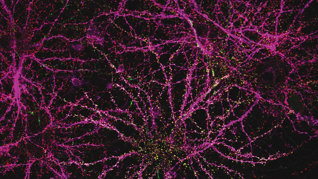
Guoping Feng, the James W. (1963) and Patricia T. Poitras Professor in Neuroscience and associate director of the McGovern Institute for Brain Research at MIT, will serve as RareNet’s inaugural faculty director. Feng holds a strong record of advancing studies on therapies for neurodevelopmental disorders, including autism spectrum disorders, Williams syndrome, and uncommon forms of epilepsy. His team’s gene therapy for Phelan-McDermid syndrome, a rare and profound autism spectrum disorder, has been licensed to Jaguar Gene Therapy and is currently undergoing clinical trials. “RareNet pioneers a unique model for biomedical research — one that is reimagining the role academia can play in developing therapeutics,” says Feng.
An early version of a gene therapy for SHANK3 mutations — linked to a rare brain disorder called Phelan-McDermid syndrome — correctly finds its way to neurons. Image: Feng lab
RareNet plans to deploy two major initiatives: a global consortium and a therapeutic pipeline accelerator. The consortium will form an international network of researchers, clinicians, and patient groups from the outset. It seeks to connect siloed research efforts, secure more patient samples, promote data sharing, and drive a strong sense of trust and goal alignment across the RareNet community. Partnerships within the consortium will support the aim of the therapeutic pipeline accelerator: to de-risk early lab discoveries and expedite their translation to clinic. By fostering more targeted collaborations — especially between academia and industry — the accelerator will prepare potential treatments for clinical use as efficiently as possible.
MIT labs are focusing on four uncommon conditions in the first wave of RareNet projects: Rett syndrome, prion disease, disorders linked to SYNGAP1 mutations, and Sturge-Weber syndrome. The teams are working to develop novel therapies that can slow, halt, or reverse dysfunctions in the brain and nervous system.
These efforts will build new bridges to connect key stakeholders across the rare brain disorders community and disrupt conventional research approaches. “Rajeev and I are motivated to seed powerful collaborations between MIT researchers, clinicians, patients, and industry,” says Méndez. “Guoping Feng clearly understands our goal to create an environment where foundational studies can thrive and seamlessly move toward clinical impact.”
“Patient and caregiver experiences, and our foreseeable impact on their lives, will guide us and remain at the forefront of our work,” Feng adds. “For far too long the rare brain disorders community has been deprived of life-changing treatments — and, importantly, hope. RareNet gives us the opportunity to transform how we study these conditions and to do so at a moment when it’s needed more than ever.”
Which of those sentences are you most likely to remember a few minutes from now? If you guessed the second, you’re probably correct.
According to a new study from MIT cognitive scientists, sentences that stick in your mind longer are those that have distinctive meanings, making them stand out from sentences you’ve previously seen. They found that meaning, not any other trait, is the most important feature when it comes to memorability.
Greta Tuckute, a former graduate student in the Fedorenko lab. Photo: Caitlin Cunningham
“One might have thought that when you remember sentences, maybe it’s all about the visual features of the sentence, but we found that that was not the case. A big contribution of this paper is pinning down that it is the meaning-related space that makes sentences memorable,” says Greta Tuckute PhD ’25, who is now a research fellow at Harvard University’s Kempner Institute.
The findings support the hypothesis that sentences with distinctive meanings — like “Does olive oil work for tanning?” — are stored in brain space that is not cluttered with sentences that mean almost the same thing. Sentences with similar meanings end up densely packed together and are therefore more difficult to recognize confidently later on, the researchers believe.
“When you encode sentences that have a similar meaning, there’s feature overlap in that space. Therefore, a particular sentence you’ve encoded is not linked to a unique set of features, but rather to a whole bunch of features that may overlap with other sentences,” says Evelina Fedorenko, an MIT associate professor of brain and cognitive sciences (BCS), a member of MIT’s McGovern Institute for Brain Research, and the senior author of the study.
Tuckute and Thomas Clark, an MIT graduate student, are the lead authors of the paper, which appears in the Journal of Memory and Language. MIT graduate student Bryan Medina is also an author.
Distinctive sentences
What makes certain things more memorable than others is a longstanding question in cognitive science and neuroscience. In a 2011 study, Aude Oliva, now a senior research scientist at MIT and MIT director of the MIT-IBM Watson AI Lab, showed that not all items are created equal: Some types of images are much easier to remember than others, and people are remarkably consistent in what images they remember best.
In that study, Oliva and her colleagues found that, in general, images with people in them are the most memorable, followed by images of human-scale space and close-ups of objects. Least memorable are natural landscapes.
As a follow-up to that study, Fedorenko and Oliva, along with Ted Gibson, another faculty member in BCS, teamed up to determine if words also vary in their memorability. In a study published earlier this year, co-led by Tuckute and Kyle Mahowald, a former PhD student in BCS, the researchers found that the most memorable words are those that have the most distinctive meanings.
Words are categorized as being more distinctive if they have a single meaning, and few or no synonyms — for example, words like “pineapple” or “avalanche” which were found to be very memorable. On the other hand, words that can have multiple meanings, such as “light,” or words that have many synonyms, like “happy,” were more difficult for people to recognize accurately.
In the new study, the researchers expanded their scope to analyze the memorability of sentences. Just like words, some sentences have very distinctive meanings, while others communicate similar information in slightly different ways.
To do the study, the researchers assembled a collection of 2,500 sentences drawn from publicly available databases that compile text from novels, news articles, movie dialogues, and other sources. Each sentence that they chose contained exactly six words.
The researchers then presented a random selection of about 1,000 of these sentences to each study participant, including repeats of some sentences. Each of the 500 participants in the study was asked to press a button when they saw a sentence that they remembered seeing earlier.
The most memorable sentences — the ones where participants accurately and quickly indicated that they had seen them before — included strings such as “Homer Simpson is hungry, very hungry,” and “These mosquitoes are — well, guinea pigs.”
Those memorable sentences overlapped significantly with sentences that were determined as having distinctive meanings as estimated through the high-dimensional vector space of a large language model (LLM) known as Sentence BERT. That model is able to generate sentence-level representations of sentences, which can be used for tasks like judging meaning similarity between sentences. This model provided researchers with a distinctness score for each sentence based on its semantic similarity to other sentences.
The researchers also evaluated the sentences using a model that predicts memorability based on the average memorability of the individual words in the sentence. This model performed fairly well at predicting overall sentence memorability, but not as well as Sentence BERT. This suggests that the meaning of a sentence as a whole — above and beyond the contributions from individual words — determines how memorable it will be, the researchers say.
Noisy memories
While cognitive scientists have long hypothesized that the brain’s memory banks have a limited capacity, the findings of the new study support an alternative hypothesis that would help to explain how the brain can continue forming new memories without losing old ones.
This alternative, known as the noisy representation hypothesis, says that when the brain encodes a new memory, be it an image, a word, or a sentence, it is represented in a noisy way — that is, this representation is not identical to the stimulus, and some information is lost. For example, for an image, you may not encode the exact viewing angle at which an object is shown, and for a sentence, you may not remember the exact construction used.
Under this theory, a new sentence would be encoded in a similar part of the memory space as sentences that carry a similar meanings, whether they were encountered recently or sometime across a lifetime of language experience. This jumbling of similar meanings together increases the amount of noise and can make it much harder, later on, to remember the exact sentence you have seen before.
“The representation is gradually going to accumulate some noise. As a result, when you see an image or a sentence for a second time, your accuracy at judging whether you’ve seen it before will be affected, and it’ll be less than 100 percent in most cases,” Clark says.
However, if a sentence has a unique meaning that is encoded in a less densely crowded space, it will be easier to pick out later on.
“Your memory may still be noisy, but your ability to make judgments based on the representations is less affected by that noise because the representation is so distinctive to begin with,” Clark says.
The researchers now plan to study whether other features of sentences, such as more vivid and descriptive language, might also contribute to making them more memorable, and how the language system may interact with the hippocampal memory structures during the encoding and retrieval of memories.
The research was funded, in part, by the National Institutes of Health, the McGovern Institute, the Department of Brain and Cognitive Sciences, the Simons Center for the Social Brain, and the MIT Quest Initiative for Intelligence.
In a world full of competing sounds, we often have to filter out a lot of noise to hear what’s most important. This critical skill may come more easily for people with musical training, according to scientists at MIT’s McGovern Institute who used brain imaging to follow what happens when people try to focus their attention on certain sounds.
When Cassia Low Manting, a postdoctoral researcher working in the labs of McGovern Institute Investigators John Gabrieli and Dimitrios Pantazis, asked people to focus on a particular melody while another melody played at the same time, individuals with musical backgrounds were, unsurprisingly, better able to follow the target tune. An analysis of study participants’ brain activity suggests this advantage arises because musical training sharpens neural mechanisms that amplify the sounds they want to listen to while turning down distractions. “This points to the idea that we can train this selective attention ability,” Manting says.
The research team, including senior author Daniel Lundqvist at the Karolinska Institute in Sweden, reported their findings September 17, 2025, in the journal Science Advances.Manting, who is now at the Karolinska Institute, notes that the research is part of an ongoing collaboration between the two institutions.
Overcoming challenges
Participants in the study had vastly difference backgrounds when it came to music. Some were professional musicians with deep training and experience, while others struggled to differentiate between the two tunes they were played, despite each one’s distinct pitch. This disparity allowed the researchers to explore how the brain’s capacity for attention might change with experience. “Musicians are very fun to study because their brains have been morphed in ways based on their training,” Manting says. “It’s a nice model to study these training effects.”
Still, the researchers had significant challenges to overcome. It has been hard to study how the brain manages auditory attention, because when researchers use neuroimaging to monitor brain activity, they see the brain’s response to all sounds: those that the listener cares most about, as well as those the listener is trying to ignore. It is usually difficult to figure out which brain signals were triggered by which sounds.
Manting and her colleagues overcame this challenge with a method called frequency tagging. Rather than playing the melodies in their experiments at a constant volume, the volume of each melody oscillated, rising and falling with a particular frequency. Each melody had its own frequency, creating detectable patterns in the brain signals that responded to it. “When you play these two sounds simultaneously to the subject and you record the brain signal, you can say, this 39-Hertz activity corresponds to the lower pitch sound and the 43-Hertz activity corresponds specifically to the higher pitch sound,” Manting explains. “It is very clean and very clear.”
When they paired frequency tagging with magnetoencephalography, a noninvasive method of monitoring brain activity, the team was able to track how their study participants’ brains responded to each of two melodies during their experiments. While the two tunes played, subjects were instructed to follow either the higher pitched or the lower pitched melody. When the music stopped, they were asked about the final notes of the target tune: did they rise or did they fall? The researchers could make this task harder by making the two tunes closer together in pitch, as well as by altering the timing of the notes.
Manting used a survey that asked about musical experience to score each participant’s musicality, and this measure had an obvious effect on task performance: The more musical a person was, the more successful they were at following the tune they had been asked to track.
To look for differences in brain activity that might explain this, the research team developed a new machine-learning approach to analyze their data. They used it to tease apart what was happening in the brain as participants focused on the target tune—even, in some cases, when the notes of the distracting tune played at the exact same time.
Top-down vs bottom-up attention
What they found was a clear separation of brain activity associated with two kinds of attention, known as top-down and bottom-up attention. Manting explains that top-down attention is goal-oriented, involving a conscious focus—the kind of attention listeners called on as they followed the target tune. Bottom-up attention, on the other hand, is triggered by the nature of the sound itself. A fire alarm would be expected to trigger this kind of attention, both with its volume and its suddenness. The distracting tune in the team’s experiments triggered activity associated with bottom-up attention—but more so in some people than in others.
“The more musical someone is, the better they are at focusing their top-down selective attention, and the less the effect of bottom-up attention is,” Manting explains.
Manting expects that musicians use their heightened capacity for top-down attention in other situations, as well. For example, they might be better than others at following a conversation in a room filled with background chatter. “I would put my bet on it that there is a high chance that they will be great at zooming into sounds,” she says.
She wonders, however, if one kind of distraction might actually be harder for a musician to filter out: the sound of their own instrument. Manting herself plays both the piano and the Chinese harp, and she says hearing those instruments is “like someone calling my name.” It’s one of many questions about how musical training affects cognition that she plans to explore in her future work.
Alan Lightman has spent much of his authorial career writing about scientific discovery, the boundaries of knowledge, and remarkable findings from the world of research. His latest book “The Shape of Wonder,” co-authored with the lauded English astrophysicist Martin Rees and published this month by Penguin Random House, offers both profiles of scientists and an examination of scientific methods, humanizing researchers and making an affirmative case for the value of their work. Lightman is a professor of the practice of the humanities in MIT’s Comparative Media Studies/Writing Program; Rees is a fellow of Trinity College at Cambridge University and the UK’s Astronomer Royal. Lightman talked with MIT News about the new volume.
Q: What is your new book about?
A: The book tries to show who scientists are and how they think. Martin and I wrote it to address several problems. One is mistrust in scientists and their institutions, which is a worldwide problem. We saw this problem illustrated during the pandemic. That mistrust I think is associated with a belief by some people that scientists and their institutions are part of the elite establishment, a belief that is one feature of the populist movement worldwide. In recent years there’s been considerable misinformation about science. And, many people don’t know who scientists are.
Another thing, which is very important, is a lack of understanding about evidence-based critical thinking. When scientists get new data and information, their theories and recommendations change. But this process, part of the scientific method, is not well-understood outside of science. Those are issues we address in the book. We have profiles of a number of scientists and show them as real people, most of whom work for the benefit of society or out of intellectual curiosity, rather than being driven by political or financial interests. We try to humanize scientists while showing how they think.
Q: You profile some well-known figures in the book, as well as some lesser-known scientists. Who are some of the people you feature in it?
A: One person is a young neuroscientist, Lace Riggs, who works at the McGovern Institute for Brain Research at MIT. She grew up in difficult circumstances in southern California, decided to go into science, got a PhD in neuroscience, and works as a postdoc researching the effect of different compounds on the brain and how that might lead to drugs to combat certain mental illnesses. Another very interesting person is Magdalena Lenda, an ecologist in Poland. When she was growing up, her father sold fish for a living, and took her out in the countryside and would identify plants, which got her interested in ecology. She works on stopping invasive species. The intention is to talk about people’s lives and interests, and show them as full people.
While humanizing scientists in the book, we show how critical thinking works in science. By the way, critical thinking is not owned by scientists. Accountants, doctors, and many others use critical thinking. I’ve talked to my car mechanic about what kinds of problems come into the shop. People don’t know what causes the check engine light to go on — the catalytic converter, corroded spark plugs, etc. — so mechanics often start from the simplest and cheapest possibilities and go to the next potential problem, down the list. That’s a perfect example of critical thinking. In science, it is checking your ideas and hypotheses against data, then updating them if needed.
Q: Are there common threads linking together the many scientists you feature in the book?
A: There are common threads, but also no single scientific stereotype. There’s a wide range of personalities in the sciences. But one common thread is that all the scientists I know are passionate about what they’re doing. They’re working for the benefit of society, and out of sheer intellectual curiosity. That links all the people in the book, as well as other scientists I’ve known. I wish more people in America would realize this: Scientists are working for their overall benefit. Science is a great success story. Thanks to scientific advances, since 1900 the expected lifespan in the U.S, has increased from a little more than 45 years to almost 80 years, in just a century, largely due to our ability to combat diseases. What’s more vital than your lifespan?
This book is just a drop in the bucket in terms of what needs to be done. But we all do what we can.
The first comprehensive map of mouse brain activity has been unveiled by a large international collaboration of neuroscientists. Researchers from the International Brain Laboratory (IBL), including McGovern Investigator Ila Fiete, published their findings today in two papers in Nature, revealing insights into how decision-making unfolds across the entire brain in mice at single-cell resolution. This brain-wide activity map challenges the traditional hierarchical view of information processing in the brain and shows that decision-making is distributed across many regions in a highly coordinated way.
“This is the first time anyone has produced a full, brain-wide map of the activity of single neurons during decision-making,” explains Co-Founder of IBL Alexandre Pouget. “The scale is unprecedented as we recorded from over half a million neurons across mice in 12 labs, covering 279 brain areas, which together represent 95% of the mouse brain volume. The decision-making activity, and particularly reward, lit up the brain like a Christmas tree,” adds Pouget, who is also a Group Leader at the University of Geneva.
Brain-wide map showing 75,000 analyzed neurons lighting up during different stages of decision-making. At the beginning of the trial, the activity is quiet. Then it builds up in the visual areas at the back of the brain, followed by a rise in activity spreading across the brain as evidence accumulates towards a decision. Next, motor areas light up as there is movement onset and finally there is a spike in activity everywhere in the brain as the animal is rewarded.
Modeling decision-making
The brain map was made possible by a major international collaboration of neuroscientists from multiple universities, including MIT. Researchers across 12 labs used state-of-the-art silicon electrodes, called Neuropixels probes, for simultaneous neural recordings to measure brain activity while mice were carrying out a decision-making task.
McGovern Associate Investigator Ila Fiete. Photo: Caitlin Cunningham
“Participating in the International Brain Laboratory has added new ways for our group to contribute to science,” says Fiete, who is also a professor of brain and cognitive sciences director of the K. Lisa Yang ICoN Center at MIT. “Our lab has helped standardize methods to analyze and generate robust conclusions from data. As computational neuroscientists interested in building models of how the brain works, access to brainwide recordings is incredible: the traditional approach of recording from one or a few brain areas limited our ability to build and test theories, resulting in fragmented models. Now we have the delightful but formidable task to make sense of how all parts of the brain coordinate to perform a behavior. Surprisingly, having a full view of the brain leads to simplifications in the models of decision making.”
The labs collected data from mice performing a decision-making task with sensory, motor, and cognitive components. In the task, a mouse sits in front of a screen and a light appears on the left or right side. If the mouse then responds by moving a small wheel in the correct direction, it receives a reward.
In some trials, the light is so faint that the animal must guess which way to turn the wheel, for which it can use prior knowledge: the light tends to appear more frequently on one side for a number of trials, before the high-frequency side switches. Well-trained mice learn to use this information to help them make correct guesses. These challenging trials therefore allowed the researchers to study how prior expectations influence perception and decision-making.
Brain-wide results
The first paper, “A brain-wide map of neural activity during complex behaviour,” showed that decision-making signals are surprisingly distributed across the brain, not localized to specific regions. This adds brain-wide evidence to a growing number of studies that challenge the traditional hierarchical model of brain function and emphasizes that there is constant communication across brain areas during decision-making, movement onset, and even reward. This means that neuroscientists will need to take a more holistic, brain-wide approach when studying complex behaviors in future.
Flat maps of the mouse brain showing which areas have significant changes in activity during each of three task intervals. Credit: Michael Schartner & International Brain Laboratory
“The unprecedented breadth of our recordings pulls back the curtain on how the entire brain performs the whole arc of sensory processing, cognitive decision-making, and movement generation,” says Fiete. “Structuring a collaboration that collects a large standardized dataset which single labs could not assemble is a revolutionary new direction for systems neuroscience, initiating the field into the hyper-collaborative mode that has contributed to leaps forward in particle physics and human genetics. Beyond our own conclusions, the dataset and associated technologies, which were released much earlier as part of the IBL mission, have already become a massively used resource for the entire neuroscience community.”
The second paper, “Brain-wide representations of prior information,” showed that prior expectations, our beliefs about what is likely to happen based on our recent experience, are encoded throughout the brain. Surprisingly, these expectations are not only found in cognitive areas, but also brain areas that process sensory information and control actions. For example, expectations are even encoded in early sensory areas such as the thalamus, the brain’s first relay for visual input from the eye. This supports the view that the brain acts as a prediction machine, but with expectations encoded across multiple brain structures playing a central role in guiding behavior responses. These findings could have implications for understanding conditions such as schizophrenia and autism, which are thought to be caused by differences in the way expectations are updated in the brain.
“Much remains to be unpacked: if it is possible to find a signal in a brain area, does it mean that this area is generating the signal, or simply reflecting a signal generated somewhere else? How strongly is our perception of the world is shaped by our expectations? Now we can generate some quantitative answers and begin the next phase experiments to learn about the origins of the expectation signals by intervening to modulate their activity,” says Fiete.
Looking ahead, the team at IBL plan to expand beyond their initial focus on decision-making to explore a broader range of neuroscience questions. With renewed funding in hand, IBL aims to expand its research scope and continue to support large-scale, standardized experiments.
New model of collaborative neuroscience
Officially launched in 2017, IBL introduced a new model of collaboration in neuroscience that uses a standardized set of tools and data processing pipelines shared across multiple labs, enabling the collection of massive datasets while ensuring data alignment and reproducibility. This approach to democratize and accelerate science draws inspiration from large-scale collaborations in physics and biology, such as CERN and the Human Genome Project.
All data from these studies, along with detailed specifications of the tools and protocols used for data collection, are openly accessible to the global scientific community for further analysis and research. Summaries of these resources can be viewed and downloaded on the IBL website under the sections: Data, Tools, Protocols.
This research was supported by grants from Wellcome (209558 and 216324), the Simons Foundation, The National Institutes of Health (NIH U19NS12371601), the National Science Foundation (NSF 1707398), the Gatsby Charitable Foundation (GAT3708), andby the Max Planck Society and the Humboldt Foundation.