Season’s Greetings from the McGovern Institute

This year’s holiday greeting (video above) was inspired by research conducted in John Gabrieli’s lab, which found that practicing mindfulness reduced children’s stress levels and negative emotions during the pandemic. These findings contribute to a growing body of evidence that practicing mindfulness can change patterns of brain activity associated with emotions and mental health.

Coloring is one form of mindfulness, or focusing awareness on the present. Visit our postcard collection to download and color your own brain-themed postcards and may the spirit of mindfulness bring you peace in the year ahead!

Video credits:
Joseph Laney (illustration)
JR Narrows, Space Lute (sound design)
Jacob Pryor (animation)

A mindful McGovern community

Mindfulness is the practice of maintaining a state of complete awareness of one’s thoughts, emotions, or experiences on a moment-to-moment basis. McGovern researchers have shown that practicing mindfulness reduces anxiety and supports emotional resilience.

In a survey distributed to the McGovern Institute community, 57% of the 74 researchers, faculty, and staff who responded, said that they practice mindfulness as a way to reduce anxiety and stress.

Here are a few of their stories.

Fernanda De La Torre

Portrait of a smiling woman leaning back against a railing.
MIT graduate student Fernanda De La Torre. Photo: Steph Stevens

Fernanda De La Torre is a graduate student in MIT’s Department of Brain and Cognitive Sciences, where she is advised by Josh McDermott.

Originally from Mexico, De La Torre took an unconventional path to her education in the United States, where she completed her undergraduate studies in computer science and math at Kansas State University. In 2019, she came to MIT as a postbaccalaureate student in the lab of Tomaso Poggio where she began working on deep-learning theory, an area of machine learning focused on how artificial neural networks modeled on the brain can learn to recognize patterns and learn.

A recent recipient of the prestigious Paul and Daisy Soros Fellowship for New Americans, De La Torre now studies multisensory integration during speech perception using deep learning models in Josh McDermott’s lab.

What kind of mindfulness do you practice, how often, and why?

Metta meditation is the type of meditation I come back to the most. I practice 2-3 times per week. Sometimes by joining Nikki Mirghafori’s Zoom calls or listening to her and other teachers’ recordings on AudioDharma. I practice because when I observe the patterns of my thoughts, I remember the importance of compassion, including self-compassion. In my experience, I find metta meditation is a wonderful way to cultivate the two: observation and compassion. 

When and why did you start practicing mindfulness?

My first meditation practice was as a first-year post-baccalaureate student here at BCS. Gal Raz (also pictured above) carried a lot of peace and attributed it to meditation; this sparked my curiosity. I started practicing more frequently last summer, after realizing my mental health was not in a good place.

How does mindfulness benefit your research at MIT?

This is hard to answer because I think the benefits of meditation are hard to measure. I find that meditation helps me stay centered and healthy, which can indirectly help the research I do. More directly, some of my initial grad school pursuits were fueled by thoughts during meditation but I ended up feeling that a lot of these concepts are hard to explore using non-philosophical approaches. So I think meditation is mainly a practice that helps my health, my relationships with others, and my relationship with work (this last one I find most challenging and personally unresolved). 

Adam Eisen

MIT graduate student Adam Eisen.

Adam Eisen is a graduate student in MIT’s Department of Brain and Cognitive Sciences, where he is co-advised by Ila Fiete (McGovern Institute) and Earl Miller (Picower Institute).

Eisen completed his undergraduate degree in Applied Mathematics & Computer Engineering at Queen’s University in Toronto, Canada. Prior to joining MIT, Eisen built computer vision algorithms at the solar aerial inspection company Heliolytics and worked on developing machine learning tools to predict disease outcomes from genetics at The Hospital for Sick Children.

Today, in the Fiete and Miller labs, Eisen develops tools for analyzing the flow of neural activity, and applies them to understand changes in neural states (such as from consciousness to anesthetic-induced unconsciousness).

What kind of mindfulness do you practice, how often, and why?

I mostly practice simple sitting meditation centered on awareness of senses and breathing. On a good week, I meditate about 3-5 times. The reason I practice are the benefits to my general experience of living. Whenever I’m in a prolonged period of consistent meditation, I’m shocked by how much more awareness I have about thoughts, feelings and sensations that are arising in my mind throughout the day. I’m also amazed by how much easier it is to watch my mind and body react to the context around me, without slipping into the usual patterns and habits. I also find mindful benefits in doing yoga, running and playing music, but the core is really centered on meditation practice.

When and why did you start practicing mindfulness?

I’ve been interested in mindfulness and meditation since undergrad as a path to investigating the nature of mind and thought – an interest which also led me into my PhD. I started practicing meditation more seriously at the start of the pandemic to get more first hand experience with what I had been learning about. I find meditation is one of those things where knowledge and theory can support the practice, but without the experiential component it’s very hard to really start to build an understanding of the core concepts at play.

How does mindfulness benefit your research at MIT?

Mindfulness has definitely informed the kinds of things I’m interested in studying and the questions I’d like to ask – largely in relation to the nature of conscious awareness and the flow of thoughts. Outside of that, I’d like to think that mindfulness benefits my general well-being and spiritual balance, which enables me to do better research.


Sugandha Sharma

Woman clasping hands in a yoga pose, looking directly into the camera.
MIT graduate student Sugandha Sharma. Photo: Steph Stevens

Sugandha (Su) Sharma is a graduate student in MIT’s Department of Brain and Cognitive Sciences (BCS), where she is co-advised by Ila Fiete (McGovern Institute) and Josh Tenenbaum (BCS).

Prior to joining MIT, she studied theoretical neuroscience at the University of Waterloo where she built neural models of context dependent decision making in the prefrontal cortex and spiking neuron models of bayesian inference, based on online learning of priors from life experience.

Today, in the Fiete and Tenenbaum labs, she studies the computational and theoretical principles underlying cognition and intelligence in the human brain.  She is currently exploring the coding principles in the hippocampal circuits implicated in spatial navigation, and their role in cognitive computations like structure learning and relational reasoning.

When did you start practicing mindfulness?

When I first learned to meditate, I was challenged to practice it every day for at least 3 months in a row. I took up the challenge, and by the end of it, the results were profound. My whole perspective towards life changed. It made me more empathetic — I could step in other people’s shoes and be mindful of their situations and feelings;  my focus shifted from myself to the big picture — it made me realize how insignificant my life was on the grand scale of the universe, and how it was worthless to be caught up in small things that I was usually worrying about. It somehow also brought selflessness to me. This experience hooked me to meditation and mindfulness for life!

What kind of mindfulness do you practice and why?

I practice mindfulness because it brings awareness. It helps me to be aware of myself, my thoughts, my actions, and my surroundings at each moment in my life, thus helping me stay in and enjoy the present moment. Awareness is of utmost importance since an aware mind always does the right thing. Imagine that you are angry, in that moment you have lost awareness of yourself. The moment you become aware of yourself; anger goes away. This is why sometimes counting helps to combat anger. If you start counting, that gives you time to think and become aware of yourself and your actions.

Meditating — sitting with my eyes closed and just observing (being aware of) my thoughts — is a yogic technique that helps me clear the noise in my mind and calm it down making it easier for me to be mindful not only while meditating, but also in general after I am done meditating. Over time, the thoughts vanish, and the mind becomes blank (noiseless). For this reason, practicing meditation regularly makes it easier for me to be mindful all the time.

An added advantage of yoga and meditation is that it helps combat stress by relaxing the mind and body. Many people don’t know what to do when they are stressed, but I am grateful to have this toolkit of yoga and meditation to deal with stressful situations in my life. They help me calm my mind in stressful situations and ensure that instead of reacting to a situation, I instead act mindfully and appropriately to make it right.

K. Lisa Yang Postbaccalaureate Program names new scholars

Funded by philanthropist Lisa Yang, the K. Lisa Yang Postbaccalaureate Scholar Program provides two years of paid laboratory experience, mentorship, and education to recent college graduates from backgrounds underrepresented in neuroscience. This year, two young researchers in McGovern Institute labs, Joseph Itiat and Sam Merrow, are the recipients of the Yang postbac program.

Itiat moved to the United States from Nigeria in 2019 to pursue a degree in psychology and cognitive neuroscience at Temple University. Today, he is a Yang postbac in John Gabrieli’s lab studying the relationship between learning and value processes and their influence on future-oriented decision-making. Ultimately, Itiat hopes to develop models that map the underlying mechanisms driving these processes.

“Being African, with limited research experience and little representation in the domain of neuroscience research,” Itiat says, “I chose to pursue a postbaccalaureate
research program to prepare me for a top graduate school and a career in cognitive neuroscience.”

Merrow first fell in love with science while working at the Barrow Neurological Institute in Arizona during high school. After graduating from Simmons University in Boston, Massachusetts, Merrow joined Guoping Feng’s lab as a Yang postbac to pursue research on glial cells and brain disorders. “As a queer, nonbinary, LatinX person, I have not met anyone like me in my field, nor have I had role models that hold a similar identity to myself,” says Merrow.

“My dream is to one day become a professor, where I will be able to show others that science is for anyone.”

Previous Yang postbacs include Alex Negron, Zoe Pearce, Ajani Stewart, and Maya Taliaferro.

Tuning the mind to benefit mental health

This story also appears in the Winter 2024 issue of BrainScan.

___

llustration of woman sitting at end of a dock with head down, arms wrapped around her knees.
Mental health is the defining public health crisis of our time, according to U.S. Surgeon General Vivek Murthy, and the nation’s youth is at the
center of this crisis.

Psychiatrists and pediatricians have sounded an alarm. The mental health of youth in the United States is worsening. Youth visits to emergency departments related to depression, anxiety, and behavioral challenges have been on the rise for years. Suicide rates among young people have escalated, too. Researchers have tracked these trends for more than a decade, and the Covid-19 pandemic only exacerbated the situation.

“It’s all over the news, how shockingly common mental health difficulties are,” says John Gabrieli, the Grover Hermann Professor of Health Sciences and Technology at MIT and an investigator at the McGovern Institute. “It’s worsening by every measure.”

Experts worry that our mental health systems are inadequate to meet the growing need. “This has gone from bad to catastrophic, from my perspective,” says Susan Whitfeld-Gabrieli, a professor of psychology at Northeastern University and a research affiliate at the McGovern Institute.

“We really need to come up with novel interventions that target the neural mechanisms that we believe potentiate depression and anxiety.”

Training the brain

One approach may be to help young people learn to modulate some of the relevant brain circuitry themselves. Evidence is accumulating that practicing mindfulness — focusing awareness on the present, typically through meditation — can change patterns of brain activity associated with emotions and mental health.

“There’s been a steady flow of moderate-size studies showing that when you help people gain mindfulness through training programs, you get all kinds of benefits in terms of people feeling less stress, less anxiety, fewer negative emotions, and sometimes more positive ones as well,” says Gabrieli, who is also a professor of brain and cognitive sciences at MIT. “Those are the things you wish for people.”

“If there were a medicine with as much evidence of its effectiveness as mindfulness, it would be flying off the shelves of every pharmacy.”
– John Gabrieli

Researchers have even begun testing mindfulness-based interventions head-to-head against standard treatments for psychiatric disorders. The results of recent studies involving hundreds of adults with anxiety disorders or depression are encouraging. “It’s just as good as the best medicines and the best behavioral treatments that we know a ton about,” Gabrieli says.

Much mindfulness research has focused on adults, but promising data about the benefits of mindfulness training for children and adolescents is emerging as well. In studies supported by the McGovern Institute’s Poitras Center for Psychiatric Disorders Research in 2019 and 2020, Gabrieli and Whitfield-Gabrieli found that sixth-graders in a Boston middle school who participated in eight weeks of mindfulness training experienced reductions in feelings of stress and increases in sustained attention. More recently, Gabrieli and Whitfeld-Gabrieli’s teams have shown how new tools can support mindfulness training and make it accessible to more children and their families — from a smartphone app that can be used anywhere to real-time neurofeedback inside an MRI scanner.

Three people practicing mindfulness in MIT Building 46. Woman on left is leaning on a railing, wearing headphones with eyes closed. Man seated in the center holds a bowl and a wooden spoon. Woman on right is seated with legs crossed and eyes closed.
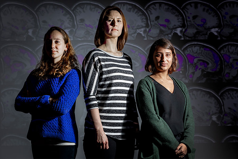
Isaac Treves (center), a PhD student in the lab of John Gabrieli, is the lead author of two studies which found that mindfulness training may improve children’s mental health. Treves and his co-authors Kimberly Wang (left) and Cindy Li (right) also practice mindfulness in their daily lives. Photo: Steph Stevens

Mindfulness and mental health

Mindfulness is not just a practice, it is a trait — an open, non-judgmental way of attending to experiences that some people exhibit more than others. By assessing individuals’ mindfulness with questionnaires that ask about attention and awareness, researchers have found the trait associates with many measures of mental health. Gabrieli and his team measured mindfulness in children between the ages of eight and ten and found it was highest in those who were most emotionally resilient to the stress they experienced during the Covid-19 pandemic. As the team reported this year in the journal PLOS One, children who were more mindful rated the impact of the pandemic on their own lives lower than other participants in the study. They also reported lower levels of stress, anxiety, and depression.

Illustration of a finger tracing the outline of a hand. There is a circle next to the hand with text that says, "Breathe In, Breathe Out. Children enrolled in John Gabrieli’s mindfulness study learned to trace the outline of their fingers in rhythm with their in-andout breathing pattern. This multisensory breathing technique has been shown to relieve anxiety and relax the body."

Mindfulness doesn’t come naturally to everyone, but brains are malleable, and both children and adults can cultivate mindfulness with training and practice. In their studies of middle schoolers, Gabrieli and Whitfeld-Gabrieli showed that the emotional effects of mindfulness training corresponded to measurable changes in the brain: Functional MRI scans revealed changes in regions involved in stress, negative feelings, and focused attention.

Whitfeld-Gabrieli says if mindfulness training makes kids more resilient, it could be a valuable tool for managing symptoms of anxiety and depression before they become severe. “I think it should be part of the standard school day,” she says. “I think we would have a much happier, healthier society if we could be doing this from the ground up.”

Data from Gabrieli’s lab suggests broadly implementing mindfulness training might even pay off in terms of academic achievement. His team found in a 2019 study that middle school students who reported greater levels of mindfulness had, on average, better grades, better scores on standardized tests, fewer absences, and fewer school suspensions than their peers.

Some schools have begun making mindfulness programs available to their students. But those programs don’t reach everyone, and their type and quality vary tremendously. Indeed, not every study of mindfulness training in schools has found the program to significantly benefit participants, which may be because not every approach to mindfulness training is equally effective.

“This is where I think the science matters,” Gabrieli says. “You have to find out what kinds of supports really work and you have to execute them reasonably. A recent report from Gabrieli’s lab offers encouraging news: mindfulness training doesn’t have to be in-person. Gabrieli and his team found that children can benefit from practicing mindfulness at home with the help of an app.

When the pandemic closed schools in 2020, school-based mindfulness programs came to an abrupt halt. Soon thereafter, a group called Inner Explorer had developed a smartphone app that could teach children mindfulness at home. Gabrieli and his team were eager to find out if this easy-access tool could effectively support children’s emotional well-being.

In October of this year, they reported in the journal Mindfulness that after 40 days of app use, children between the ages of eight and ten reported less stress than they had before beginning mindfulness training. Parents reported that their children were also experiencing fewer negative emotions, such as loneliness and fear.

The outcomes suggest a path toward making evidence-based mindfulness training for children broadly accessible. “Tons of people could do this,” says Gabrieli. “It’s super scalable. It doesn’t cost money; you don’t have to go somewhere. We’re very excited about that.”

Visualizing healthy minds

Mindfulness training may be even more effective when practitioners can visualize what’s happening in their brains. In Whitfeld-Gabrieli’s lab, teenagers have had a chance to slide inside an MRI scanner and watch their brain activity shift in real time as they practiced mindfulness meditation. The visualization they see focuses on the brain’s default mode network (DMN), which is most active when attention is not focused on a particular task. Certain patterns of activity in the DMN have been linked to depression, anxiety, and other psychiatric conditions, and mindfulness training may help break these patterns.

McGovern research affiliate Susan Whitfield-Gabrieli in the Martinos Imaging Center. Photo: Caitlin Cunningham

Whitfeld-Gabrieli explains that when the mind is free to wander, two hubs of the DMN become active. “Typically, that means we’re engaged in some kind of mental time travel,” she says. That might mean reminiscing about the past or planning for the future, but can be more distressing when it turns into obsessive rumination or worry. In people with anxiety, depression, and psychosis, these network hubs are often hyperconnected.

“It’s almost as if they’re hijacked,” Whitfeld-Gabrieli says. “The more they’re correlated, the more psychopathology one might be experiencing. We wanted to unlock that hyperconnectivity for kids who are suffering from depression and anxiety.” She hoped that by replacing thoughts of the past and the future with focus on the present, mindfulness meditation would rein in overactive DMNs, and she wanted a way to encourage kids to do exactly that.

The neurofeedback tool that she and her colleagues created focuses on the DMN as well as separate brain region that is called on during attention-demanding tasks. Activity in those regions is monitored with functional MRI and displayed to users in a game-like visualization. Inside the scanner, participants see how that activity changes as they focus on a meditation or when their mind wanders. As their mind becomes more focused on the present moment, changes in brain activity move a ball toward a target.

Whitfeld-Gabrieli says the real-time feedback was motivating for adolescents who participated in a recent study, who all had histories of anxiety or depression. “They’re training their brain to tune their mind, and they love it,” she says.

MRI images of two brains, one showing an active DMN and the other showing a healthy DMN.
The default mode network (DMN) is a large-scale brain network that is active when a person is not focused on the outside world and the brain is at wakeful rest. The DMN is often over-engaged in adolescents with depression and anxiety, as well as teens at risk for these affective disorders (left). DMN activation and connectivity can be “tuned” to a healthier state through the practice of mindfulness (right).

In March, she and her team reported in Molecular Psychiatry that the neurofeedback tool helped those study participants reduce connectivity in the DMN and engage a more desirable brain state. It’s not the first success the team has had with the approach. Previously, they found that the decreases in DMN connectivity brought about by mindfulness meditation with neurofeedback were associated with reduced hallucinations for patients with schizophrenia. Testing the clinical benefits of the approach in teens is on the horizon; Whitfeld-Gabrieli and her collaborators plan to investigate how mindfulness meditation with real-time neurofeedback affects depression symptoms in an upcoming clinical trial.

Whitfeld-Gabrieli emphasizes that the neurofeedback is a training tool, helping users improve mindfulness techniques they can later call on anytime, anywhere. While that training currently requires time inside an MRI scanner, she says it may be possible create an EEG-based version of the approach, which could be deployed in doctors’ offices and other more accessible settings.

Both Gabrieli and Whitfeld-Gabrieli continue to explore how mindfulness training impacts different aspects of mental health, in both children and adults and with a range of psychiatric conditions. Whitfeld-Gabrieli expects it will be one powerful tool for combating a youth mental health crisis for which there will be no single solution. “I think it’s going to take a village,” she says. “We are all going to have to work together, and we’ll have to come up some really innovative ways to help.”

Search algorithm reveals nearly 200 new kinds of CRISPR systems

Microbial sequence databases contain a wealth of information about enzymes and other molecules that could be adapted for biotechnology. But these databases have grown so large in recent years that they’ve become difficult to search efficiently for enzymes of interest.

Now, scientists at the Broad Institute of MIT and Harvard, the McGovern Institute for Brain Research at MIT, and the National Center for Biotechnology Information (NCBI) at the National Institutes of Health have developed a new search algorithm that has identified 188 kinds of new rare CRISPR systems in bacterial genomes, encompassing thousands of individual systems. The work appears today in Science.

The algorithm, which comes from the lab of CRISPR pioneer Feng Zhang, uses big-data clustering approaches to rapidly search massive amounts of genomic data. The team used their algorithm, called Fast Locality-Sensitive Hashing-based clustering (FLSHclust) to mine three major public databases that contain data from a wide range of unusual bacteria, including ones found in coal mines, breweries, Antarctic lakes, and dog saliva. The scientists found a surprising number and diversity of CRISPR systems, including ones that could make edits to DNA in human cells, others that can target RNA, and many with a variety of other functions.

The new systems could potentially be harnessed to edit mammalian cells with fewer off-target effects than current Cas9 systems. They could also one day be used as diagnostics or serve as molecular records of activity inside cells.

The researchers say their search highlights an unprecedented level of diversity and flexibility of CRISPR and that there are likely many more rare systems yet to be discovered as databases continue to grow.

“Biodiversity is such a treasure trove, and as we continue to sequence more genomes and metagenomic samples, there is a growing need for better tools, like FLSHclust, to search that sequence space to find the molecular gems,” said Zhang, a co-senior author on the study and a core institute member at the Broad.

Zhang is also an investigator at the McGovern Institute for Brain Research at MIT, the James and Patricia Poitras Professor of Neuroscience at MIT with joint appointments in the departments of Brain and Cognitive Sciences and Biological Engineering, and an investigator at the Howard Hughes Medical Institute. Eugene Koonin, a distinguished investigator at the NCBI, is co-senior author on the study as well.

Searching for CRISPR

CRISPR, which stands for Clustered Regularly Interspaced Short Palindromic Repeats, is a bacterial defense system that has been engineered into many tools for genome editing and diagnostics.

To mine databases of protein and nucleic acid sequences for novel CRISPR systems, the researchers developed an algorithm based on an approach borrowed from the big data community. This technique, called locality-sensitive hashing, clusters together objects that are similar but not exactly identical. Using this approach allowed the team to probe billions of protein and DNA sequences — from the NCBI, its Whole Genome Shotgun database, and the Joint Genome Institute — in weeks, whereas previous methods that look for identical objects would have taken months. They designed their algorithm to look for genes associated with CRISPR.

“This new algorithm allows us to parse through data in a time frame that’s short enough that we can actually recover results and make biological hypotheses,” said Soumya Kannan, who is a co-first author on the study. Kannan was a graduate student in Zhang’s lab when the study began and is currently a postdoctoral researcher and Junior Fellow at Harvard University. Han Altae-Tran, a graduate student in Zhang’s lab during the study and currently a postdoctoral researcher at the University of Washington, was the study’s other co-first author.

“This is a testament to what you can do when you improve on the methods for exploration and use as much data as possible,” said Altae-Tran. “It’s really exciting to be able to improve the scale at which we search.”

New systems

In their analysis, Altae-Tran, Kannan, and their colleagues noticed that the thousands of CRISPR systems they found fell into a few existing and many new categories. They studied several of the new systems in greater detail in the lab.

They found several new variants of known Type I CRISPR systems, which use a guide RNA that is 32 base pairs long rather than the 20-nucleotide guide of Cas9. Because of their longer guide RNAs, these Type I systems could potentially be used to develop more precise gene-editing technology that is less prone to off-target editing. Zhang’s team showed that two of these systems could make short edits in the DNA of human cells. And because these Type I systems are similar in size to CRISPR-Cas9, they could likely be delivered to cells in animals or humans using the same gene-delivery technologies being used today for CRISPR.

One of the Type I systems also showed “collateral activity” — broad degradation of nucleic acids after the CRISPR protein binds its target. Scientists have used similar systems to make infectious disease diagnostics such as SHERLOCK, a tool capable of rapidly sensing a single molecule of DNA or RNA. Zhang’s team thinks the new systems could be adapted for diagnostic technologies as well.

The researchers also uncovered new mechanisms of action for some Type IV CRISPR systems, and a Type VII system that precisely targets RNA, which could potentially be used in RNA editing. Other systems could potentially be used as recording tools — a molecular document of when a gene was expressed — or as sensors of specific activity in a living cell.

Mining data

The scientists say their algorithm could aid in the search for other biochemical systems. “This search algorithm could be used by anyone who wants to work with these large databases for studying how proteins evolve or discovering new genes,” Altae-Tran said.

The researchers add that their findings illustrate not only how diverse CRISPR systems are, but also that most are rare and only found in unusual bacteria. “Some of these microbial systems were exclusively found in water from coal mines,” Kannan said. “If someone hadn’t been interested in that, we may never have seen those systems. Broadening our sampling diversity is really important to continue expanding the diversity of what we can discover.”

This work was supported by the Howard Hughes Medical Institute; K. Lisa Yang and Hock E. Tan Molecular Therapeutics Center at MIT; Broad Institute Programmable Therapeutics Gift Donors; The Pershing Square Foundation, William Ackman and Neri Oxman; James and Patricia Poitras; BT Charitable Foundation; Asness Family Foundation; Kenneth C. Griffin; the Phillips family; David Cheng; and Robert Metcalfe.

A multifunctional tool for cognitive neuroscience

A team of researchers at MIT’s McGovern and Picower Institutes has advanced the clinical potential of a thin, flexible fiber designed to simultaneously monitor and manipulate neural activity at targeted sites in the brain. The collaborative team improved upon an earlier model of the multifunctional fiber, developed in the lab of McGovern Institute Associate Investigator Polina Anikeeva, to explore dynamic changes to neural signaling as large animals engage in a working memory task. The results appear Oct. 6 in Science Advances.

The new device, developed by Indie Garwood, who recently received her PhD in the Harvard-MIT Program in Health Sciences and Technology, includes four microelectrodes for detecting neural activity and two microfluidic channels through which drugs can be delivered. This means scientists can deliver a drug that alters neural signaling within a particular part of the brain, then monitor the consequences for local brain activity. This technology was a collaborative effort between Anikeeva, who is also the Matoula S. Salapatas Professor in Materials Science and Engineering and a professor of brain and cognitive sciences, and Picower Institute Investigators Emery Brown and Earl Miller, who jointly supervised Garwood to develop a multifunctional neurotechnology for larger and translational animal models, which are necessary to investigate the neural circuits that underlie high-level cognitive functions.  With further development and testing, similar devices might one day be deployed to diagnose or treat brain disorders in human patients.

Brown is the Edward Hood Taplin Professor of Medical Engineering and Computational Neuroscience in the Picower Institute, the Institute for Medical Engineering and Science, and the Department of Brain and Cognitive Sciences, as well as an anesthesiologist at Massachusetts General Hospital and Harvard Medical School. Miller is the Picower Professor of Neuroscience and a professor of brain and cognitive sciences at MIT.

The new multifunctional fiber is not the first produced by Anikeeva and her team. An earlier model engineered in their lab has already reached the neuroscience community, whose members use it to simultaneously monitor and manipulate neural activity in the brains of mice and rats. But for studies in larger animals, the existing tools for delivering drugs to the brains were rigid, bulky devices, which were both fragile and prone to causing tissue damage. A better tool was needed, both to advance cognitive neuroscience research and to set the stage for developing devices that can deliver drugs directly to the brains of patients and monitor the effects.

Like the devices that Anikeeva’s team designed for rodent studies, the new tool is created by first assembling a larger version of the fiber—a preform cylinder with multiple channels that is then heated and stretched until it is thin and long. As the channels narrow, microelectrodes are incorporated into to the fiber. The final step is to link the electrodes in the fiber to a connector that will relay data collected inside the brain to a unit in the lab.

The final device is long enough to access areas deep in the brain of a large animal. It is built to withstand rigorous sterilization procedures and to stay in place even in an active animal. And it integrates directly with experimental systems that cognitive neuroscientists already use in their labs. “We really wanted this to be something that we could easily hand somebody and they’re going to know how to implement it in their system,” says Garwood, who led development of the device as a graduate student in Anikeeva’s lab.

Once the new device was developed, Garwood and colleagues in the Miller and Brown labs put it to work.  They used the tool to study changes in neural activity as an animal completed a task requiring working memory. The fluid channels in the fiber were used to deliver small amounts of GABA, a neurotransmitter that dampens neuronal activity, to the animal’s premotor cortex, a part of the brain that helps plan movement. At the same time, the device recorded electrical activity from individual neurons, as well as broader patterns of activity in this part of the brain. By monitoring these signals over time, the team learned how neural circuits adapted to the local inhibition they had applied. In another experiment, the team used the device to record neural activity from the putamen, a region deep in the brain involved in reward processing and motivation.

The data collected by the device was extensive and complex, tracking changes that unfolded in the brain over seconds to hours. Interpreting those data required the team to devise new methods of data analysis, which Garwood worked on closely with the Brown lab. Garwood says these methods will be shared with users of the new devices, providing “a roadmap for extracting all of these rich dynamics that you can get out of them.”

These successes, the researchers say, are an important step toward the development of tools to modulate and manipulate neuronal activity in the human brain to benefit patients. For example, they say, a multifunctional fiber might one day be used to more accurately pinpoint the origin of seizures in people with epilepsy, by testing the effects of activating or inhibiting specific brain cells.


New cellular census maps the complexity of a primate brain

A new atlas developed by researchers at MIT’s McGovern Institute and Harvard Medical School catalogs a diverse array of brain cells throughout the marmoset brain. The atlas helps establish marmosets—small monkeys whose brains share many functional and structural features with the human brain—as a valuable model for neuroscience research.

Data from more than two million brain cells are included in the atlas, which spans 18 regions of the marmoset brain. A research team led by Guoping Feng, associate director of the McGovern Institute and member of the Broad Institute of Harvard and MIT, Harvard biologist and member of the Broad Institute of Harvard and MIT Steven McCarroll, and Princeton neurobiologist Fenna Krienen classified each cell according to its particular pattern of genetic activity, providing an important reference for studies of the marmoset brain. The team’s analysis, reported October 13, 2023, in the journal Science Advances, also reveals the profound influence of a cell’s developmental origin on its identity in the primate brain.

Regional variation in neocortical cell types and expression patterns. Image courtesy of the researchers.

Cellular diversity

Brains are made up of a tremendous diversity of cells. Neurons with dramatically different gene expression, shapes, and activities work together to process information and drive behavior, supported by an assortment of immune cells and other cell types. Scientists have only recently begun to catalog this cellular diversity—first in mice, and now in primates.

The marmoset is a quick-breeding monkey whose small brain has many of features similar to those that enable higher cognitive processes in humans. Feng says neuroscientists have begun turning to marmosets as a research model in recent years because new gene editing technology has made it easier to modify the animal’s DNA, so scientists can now study the genetic factors that shape marmosets’ brains and behavior. Feng, McCarroll, Krienen and others hope these animals will offer insights into how primate brains handle complex decision-making, social interactions, and other higher brain functions that are difficult to study in mice. Likewise, Feng says, the monkeys will help scientists investigate the impact of genetic mutations associated with brain disorders and explore potential therapeutic strategies.

To make marmosets a practical model for neuroscience, scientists need to understand the fundamental composition of their brains. Feng and McCarroll’s team have begun that characterization with their cell census, which was supported by the National Institutes of Health’s Brain Research Through Advancing Innovative Neurotechnologies (BRAIN) Initiative’s Cell Census Network (BICCN), as part a larger effort to map cellular features in the brains of mice, non-human primates, and humans. It is an essential first step in the creation of a comprehensive atlas charting the molecular, anatomical, and functional features of cells in the marmoset brain.

“Hopefully, when the BRAIN Initiative is complete, we will have a very complete map of these cells: where they are located, their abundance, their functional properties,” says Feng. “This not only gives you knowledge of the normal brain, but you can also look at what aspects change in diseases of the brain. So it’s a really powerful database.”

To catalog the diversity of cells in the marmoset brain, the researchers undertook an expansive analysis of the molecular contents of 2.4 million brain cells from adult marmosets. For each of these cells, they analyzed the complete set of RNA copies of its genes that the cell had produced, known as the cell’s transcriptome. Because the transcriptome captures patterns of genetic activity inside a cell, it is an indication of the cell’s function and can be used to assess cellular identity.

Gene expression across neural populations. Image courtesy of the researchers.

The team’s analysis is one of the first to compare patterns of gene activity in cells from disparate regions of the marmoset brain. Doing so yielded surprising insights into the factors that shape brain cells’ transcriptomic identities. “What we found is that the cell’s transcriptome contains breadcrumbs that link back to the developmental origin of that cell type,” says Krienen, who led the cellular census as a postdoctoral researcher in McCarroll’s lab. That suggests that comparing cells’ transcriptomes can help scientists figure out how primate brains are assembled, which might lead to insights into neurodevelopmental disorders, she says.

The team also learned that a cell’s location in the brain was critical to shaping its transcriptomic identity. For example, Krienen says, “it turns out that an inhibitory neuron in the cortex doesn’t look very anything like an inhibitory neuron in the thalamus, probably because they have distinct embryonic origins.”

Expanding the cell census

This new picture of cellular diversity in the marmoset brain will help researchers understand how genetic perturbations affect different brain cells and interpret the results of future experiments. Importantly, Krienen says, it could help researchers pinpoint exactly which cells are affected in brain disorders, and how the effects of a disease might localize to specific brain regions.

Krienen, McCarroll, and Feng went beyond their initial survey of cellular diversity with analyses of specific subsets of cells, charting the spatial distribution of interneurons in a key region of the prefrontal cortex and visualizing the shapes of several molecularly-defined cell types. Now, they have begun expanding their cell census beyond the 18 brain structures represented in the reported work. As part of the BRAIN Initiative’s Brain Cell Atlas Network (BICAN), the team will profile cells throughout the entire adult marmoset brain, including multiple data types in their analysis. Building on cell census data, NIH BRAIN Initiative has also launched BRAIN CONNECTS projects to map cellular connectivity in the brain.

This work was supported by the National Institutes of Health, the National Science Foundation, MathWorks, MIT, Harvard Medical School, the Broad Institute’s Stanley Center for Psychiatric Research, the Hock E. Tan and K. Lisa Yang Center for Autism Research at MIT, the Poitras Center for Psychiatric Disorders Research at MIT, and the McGovern Institute for Brain Research at MIT.

Thousands of programmable DNA-cutters found in algae, snails, and other organisms

A diverse set of species, from snails to algae to amoebas, make programmable DNA-cutting enzymes called Fanzors—and a new study from scientists at MIT’s McGovern Institute has identified thousands of them. Fanzors are RNA-guided enzymes that can be programmed to cut DNA at specific sites, much like the bacterial enzymes that power the widely used gene-editing system known as CRISPR. The newly recognized diversity of natural Fanzor enzymes, reported September 27, 2023, in the journal Science Advances, gives scientists an extensive set of programmable enzymes that might be adapted into new tools for research or medicine.

“RNA-guided biology is what lets you make programmable tools that are really easy to use. So the more we can find, the better,” says McGovern fellow Omar Abudayyeh, who led the research with McGovern fellow Jonathan Gootenberg.

CRISPR, an ancient bacterial defense system, has made it clear how useful RNA-guided enzymes can be when they are adapted for use in the lab. CRISPR-based genome editing tools developed by McGovern investigator Feng Zhang, Abudayyeh, Gootenberg and others have changed the way scientists modify DNA, accelerating research and enabling the development of many experimental gene therapies.

Researchers have since uncovered other RNA-guide enzymes throughout the bacterial world, many with features that make them valuable in the lab. The discovery of Fanzors, whose ability to cut DNA in an RNA-guided manner was reported by Zhang’s group earlier this year, opens a new frontier of RNA-guided biology. Fanzors were the first such enzymes to be found in eukaryotic organisms—a wide group of lifeforms, including plants, animals, and fungi, defined by the membrane-bound nucleus that holds each cell’s genetic material. (Bacteria, which lack nuclei, belong to a group known as prokaryotes.)

Structural illustration of Fanzors.
Predicted structural image of Fanzors. Image: Jonathan Gootenberg and Omar Abudayyeh

“People have been searching for interesting tools in prokaryotic systems for a long time, and I think that that has been incredibly fruitful,” says Gootenberg. “Eukaryotic systems are really just a whole new kind of playground to work in.”

One hope, Abudayyeh and Gootenberg say, is that enzymes that naturally evolved in eukaryotic organisms might be better suited to function safely and efficiently in the cells of other eukaryotic organisms, including humans. Zhang’s group has shown that Fanzor enzymes can be engineered to precisely cut specific DNA sequences in human cells. In the new work, Abudayyeh and Gootenberg discovered that some Fanzors can target DNA sequences in human cells even without optimization. “The fact that they work quite efficiently in mammalian cells was really fantastic to see,” Gootenberg says.

Prior to the current study, hundreds of Fanzors had been found among eukaryotic organisms. Through an extensive search of genetic databases led by lab member Justin Lim, Gootenberg and Abudayyeh’s team has now expanded the known diversity of these enzymes by an order of magnitude.

Among the more than 3,600 Fanzors that the team found in eukaryotes and the viruses that infect them, the researchers were able to identify five different families of the enzymes. By comparing these enzymes’ precise makeup, they found evidence of a long evolutionary history.

Fanzors likely evolved from RNA-guided DNA-cutting bacterial enzymes called TnpBs. In fact, it was Fanzors’ genetic similarities to these bacterial enzymes that first caught the attention of both Zhang’s group and Gootenberg and Abudayyeh’s team.

The evolutionary connections that Gootenberg and Abudayyeh traced suggest that these bacterial predecessors of Fanzors probably entered eukaryotic cells, initiating their evolution, more than once. Some were likely transmitted by viruses, while others may have been introduced by symbiotic bacteria. The research also suggests that after they were taken up by eukaryotes, the enzymes evolved features suited to their new environment, such as a signal that allows them to enter a cell nucleus, where they have access to DNA.

Through genetic and biochemical experiments led by graduate student Kaiyi Jiang, the team determined that Fanzors have evolved a DNA-cutting active site that is distinct from that of their bacterial predecessors. This seems to allow the enzyme to cut its target sequence more precisely the ancestors of TnpB, when targeted to a sequence of DNA in a test tube, become activated and cut other sequences in the tube; Fanzors lack this promiscuous activity. When they used an RNA guide to direct the enzymes to cut specific sites in the genome of human cells, they found that certain Fanzors were able to cut these target sequences with about 10 to 20 percent efficiency.

With further research, Abudayyeh and Gootenberg hope that a variety of sophisticated genome editing tools can be developed from Fanzors. “It’s a new platform, and they have many capabilities,” says Gootenberg. “Opening up the whole eukaryotic world to these types of RNA-guided systems is going to give us a lot to work on,” Abudayyeh adds.

Four McGovern Investigators receive NIH BRAIN Initiative grants

In the human brain, 86 billion neurons form more than 100 trillion connections with other neurons at junctions called synapses. Scientists at the McGovern Institute are working with their collaborators to develop technologies to map these connections across the brain, from mice to humans.

Today, the National Institutes of Health (NIH) announced a new program to support research projects that have the potential to reveal an unprecedented and dynamic picture of the connected networks in the brain. Four of these NIH-funded research projects will take place in McGovern labs.

BRAIN Initiative

In 2013, the Obama administration announced the Brain Research Through Advancing Innovative Neurotechnologies® (BRAIN) Initiative, a public-private research effort to support the development and application of new technologies to understand brain function.

Today, the NIH announced its third project supported by the BRAIN Initiative, called BRAIN Initiative Connectivity Across Scales (BRAIN CONNECTS). The new project complements two previous large-scale projects, which together aim to transform neuroscience research by generating wiring diagrams that can span entire brains across multiple species. These detailed wiring diagrams can help uncover the logic of the brain’s neural code, leading to a better understanding of how this circuitry makes us who we are and how it could be rewired to treat brain diseases.

BRAIN CONNECTS at McGovern

The initial round of BRAIN CONNECTS awards will support researchers at more than 40 university and research institutions across the globe with 11 grants totaling $150 million over five years. Four of these grants have been awarded to McGovern researchers Guoping Feng, Ila Fiete, Satra Ghosh, and Ian Wickersham, whose projects are outlined below:

BRAIN CONNECTS: Comprehensive regional projection map of marmoset with single axon and cell type resolution
Team: Guoping Feng (McGovern Institute, MIT), Partha Mitra (Cold Spring Harbor Laboratory), Xiao Wang (Broad Institute), Ian Wickersham (McGovern Institute, MIT)

Summary: This project will establish an integrated experimental-computational platform to create the first comprehensive brain-wide mesoscale connectivity map in a non-human primate (NHP), the common marmoset (Callithrix jacchus). It will do so by tracing axonal projections of RNA barcode-identified neurons brain-wide in the marmoset, utilizing a sequencing-based imaging method that also permits simultaneous transcriptomic cell typing of the identified neurons. This work will help bridge the gap between brain-wide mesoscale connectivity data available for the mouse from a decade of mapping efforts using modern techniques and the absence of comparable data in humans and NHPs.

BRAIN CONNECTS: A center for high-throughput integrative mouse connectomics
Team: Jeff Lichtman (Harvard University), Ila Fiete (McGovern Institute, MIT), Sebastian Seung (Princeton University), David Tank (Princeton University), Hongkui Zeng (Allen Institute), Viren Jain (Google), Greg Jeffries (Oxford University)

Summary: This project aims to produce a large-scale synapse-level brain map (connectome) that includes all the main areas of the mouse hippocampus. This region is of clinical interest because it is an essential part of the circuit underlying spatial navigation and memory and the earliest impairments and degeneration related to Alzheimer’s disease.

BRAIN CONNECTS: The center for Large-scale Imaging of Neural Circuits (LINC)
Team: Anastasia Yendiki (MGH), Satra Ghosh (McGovern, MIT), Suzanne Haber (University of Rochester), Elizabeth Hillman (Columbia University)

Summary: This project will generate connectional diagrams of the monkey and human brain at unprecedented resolutions. These diagrams will be linked both to the neuroanatomic literature and to in vivo neuroimaging techniques, bridging between the rigor of the former and the clinical relevance of the latter. The data to be generated by this project will advance our understanding of brain circuits that are implicated in motor and psychiatric disorders, and that are targeted by deep-brain stimulation to treat these disorders.

BRAIN CONNECTS: Mapping brain-wide connectivity of neuronal types using barcoded connectomics
Team: Xiaoyin Chen (Allen Institute), Ian Wickersham (McGovern Institute, MIT), and Justus Kebschull of JHU

Summary: This project aims to optimize and develop barcode sequencing-based neuroanatomical techniques to achieve brain-wide, high-throughput, highly multiplexed mapping of axonal projections and synaptic connectivity of neuronal types at cellular resolution in primate brains. The team will work together to apply these techniques to generate an unprecedented multi-resolution map of brain-wide projections and synaptic inputs of neurons in the macaque visual cortex at cellular resolution.


Nuevo podcast de neurociencia en español celebra su tercera temporada

Sylvia Abente, neuróloga clínica de la Universidad Nacional de Asunción (Paraguay), investiga la variedad de síntomas que son característicos de la epilepsia. Trabaja con los pueblos indígenas de Paraguay, y su dominio del español y el guaraní, los dos idiomas oficiales de Paraguay, le permite ayudar a los pacientes a encontrar las palabras que ayuden a describir sus síntomas de epilepsia para poder tratarlos.

Juan Carlos Caicedo Mera, neurocientífico de la Universidad Externado de Colombia, utiliza modelos de roedores para investigar los efectos neurobiológicos del estrés en los primeros años de vida. Ha desempeñado un papel decisivo en despertar la conciencia pública sobre los efectos biológicos y conductuales del castigo físico a edades tempranas, lo que ha propiciado cambios políticos encaminados a reducir su prevalencia como práctica cultural en Colombia.

Woman interviews a man at a table with a camera recording the interview in the foreground.
Jessica Chomik-Morales (right) interviews Pedro Maldonado at the Biomedical Neuroscience Institute of Chile at the University of Chile. Photo: Jessica Chomik-Morales

Estos son solo dos de los 33 neurocientíficos de siete países latinoamericanos que Jessica Chomik-Morales entrevistó durante 37 días para la tercera temporada de su podcast en español “Mi Última Neurona,” que se estrenará el 18 de septiembre a las 5:00 p. m. en YouTube. Cada episodio dura entre 45 y 90 minutos.

“Quise destacar sus historias para disipar la idea errónea de que la ciencia de primer nivel solo puede hacerse en Estados Unidos y Europa,” dice Chomik-Morales, “o que no se consigue en Sudamérica debido a barreras financieras y de otro tipo.”

Chomik-Morales, graduada universitaria de primera generación que creció en Asunción (Paraguay) y Boca Ratón (Florida), es ahora investigadora académica de post licenciatura en el MIT. Aquí trabaja con Laura Schulz, profesora de Ciencia Cognitiva, y Nancy Kanwisher, investigadora del McGovern Institute y la profesora Walter A. Rosenblith de Neurociencia Cognitiva, utilizando imágenes cerebrales funcionales para investigar de qué forma el cerebro explica el pasado, predice el futuro e interviene sobre el presente a traves del razonamiento causal.

“El podcast está dirigido al público en general y es apto para todas las edades,” afirma. “Se explica la neurociencia de forma fácil para inspirar a los jóvenes en el sentido de que ellos también pueden llegar a ser científicos y para mostrar la amplia variedad de investigaciones que se realizan en los países de origen de los escuchas.”

El viaje de toda una vida

“Mi Última Neurona” comenzó como una idea en 2021 y creció rápidamente hasta convertirse en una serie de conversaciones con destacados científicos hispanos, entre ellos L. Rafael Reif, ingeniero electricista venezolano-estadounidense y 17.º presidente del MIT.

Woman interviews man at a table while another man adjusts microphone.
Jessica Chomik-Morales (left) interviews the 17th president of MIT, L. Rafael Reif (right), for her podcast while Héctor De Jesús-Cortés (center) adjusts the microphone. Photo: Steph Stevens

Con las relaciones profesionales que estableció en las temporadas uno y dos, Chomik-Morales amplió su visión y reunió una lista de posibles invitados en América Latina para la tercera temporada. Con la ayuda de su asesor científico, Héctor De Jesús-Cortés, un investigador Boricua de posdoctorado del MIT, y el apoyo financiero del McGovern Institute, el Picower Institute for Learning and Memory, el Departamento de Ciencias Cerebrales y Cognitivas, y las Iniciativas Internacionales de Ciencia y Tecnología del MIT, Chomik-Morales organizó entrevistas con científicos en México, Perú, Colombia, Chile, Argentina, Uruguay y Paraguay durante el verano de 2023.

Viajando en avión cada cuatro o cinco días, y consiguiendo más posibles participantes de una etapa del viaje a la siguiente por recomendación, Chomik-Morales recorrió más de 10,000 millas y recopiló 33 historias para su tercera temporada. Las áreas de especialización de los científicos abarcan toda una variedad de temas, desde los aspectos sociales de los ciclos de sueño y vigilia hasta los trastornos del estado de ánimo y la personalidad, pasando por la lingüística y el lenguaje en el cerebro o el modelado por computadoras como herramienta de investigación.

“Si alguien estudia la depresión y la ansiedad, quiero hablar sobre sus opiniones con respecto a diversas terapias, incluidos los fármacos y también las microdosis con alucinógenos,” dice Chomik-Morales. “Estas son las cosas de las que habla la gente.” No le teme a abordar temas delicados, como la relación entre las hormonas y la orientación sexual, porque “es importante que la gente escuche a los expertos hablar de estas cosas,” comenta.

El tono de las entrevistas va de lo informal (“el investigador y yo somos como amigos”, dice) a lo pedagógico (“de profesor a alumno”). Lo que no cambia es la accesibilidad (se evitan términos técnicos) y las preguntas iniciales y finales en cada entrevista. Para empezar: “¿Cómo ha llegado hasta aquí? ¿Qué le atrajo de la neurociencia?”. Para terminar: “¿Qué consejo le daría a un joven estudiante latino interesado en Ciencias, Ingeniería, Tecnología y Matemáticas[1]?

Permite que el marco de referencia de sus escuchas sea lo que la guíe. “Si no entendiera algo o pensara que se podría explicar mejor, diría: ‘Hagamos una pausa’. ¿Qué significa esta palabra?”, aunque ella conociera la definición. Pone el ejemplo de la palabra “MEG” (magnetoencefalografía): la medición del campo magnético generado por la actividad eléctrica de las neuronas, que suele combinarse con la resonancia magnética para producir imágenes de fuentes magnéticas. Para aterrizar el concepto, preguntaría: “¿Cómo funciona? ¿Este tipo de exploración hace daño al paciente?”.

Allanar el camino para la creación de redes globales

El equipo de Chomik-Morales era escaso: tres micrófonos Yeti y una cámara de video Canon conectada a su computadora portátil. Las entrevistas se realizaban en salones de clase, oficinas universitarias, en la casa de los investigadores e incluso al aire libre, ya que no había estudios insonorizados disponibles. Ha estado trabajando con el ingeniero de sonido David Samuel Torres, de Puerto Rico, para obtener un sonido más claro.

Ninguna limitación tecnológica podía ocultar la importancia del proyecto para los científicos participantes.

Two women talking at a table in front of a camera.
Jessica Chomik-Morales (left) interviews Josefina Cruzat (right) at Adolfo Ibañez University in Chile. Photo: Jessica Chomik-Morales

“Mi Última Neurona” muestra nuestro conocimiento diverso en un escenario global, proporcionando un retrato más preciso del panorama científico en América Latina,” dice Constanza Baquedano, originaria de Chile. “Es un avance hacia la creación de una representación más inclusiva en la ciencia”. Baquendano es profesora adjunta de psicología en la Universidad Adolfo Ibáñez, en donde utiliza electrofisiología y mediciones electroencefalográficas y conductuales para investigar la meditación y otros estados contemplativos. “Estaba ansiosa por ser parte de un proyecto que buscara brindar reconocimiento a nuestras experiencias compartidas como mujeres latinoamericanas en el campo de la neurociencia.”

“Comprender los retos y las oportunidades de los neurocientíficos que trabajan en América Latina es primordial,” afirma Agustín Ibáñez, profesor y director del Instituto Latinoamericano de Salud Cerebral (BrainLat) de la Universidad Adolfo Ibáñez de Chile. “Esta región, que se caracteriza por tener importantes desigualdades que afectan la salud cerebral, también presenta desafíos únicos en el campo de la neurociencia,” afirma Ibáñez, quien se interesa principalmente en la intersección de la neurociencia social, cognitiva y afectiva. “Al centrarse en América Latina, el podcast da a conocer las historias que frecuentemente no se cuentan en la mayoría de los medios. Eso tiende puentes y allana el camino para la creación de redes globales.”

Por su parte, Chomik-Morales confía en que su podcast generará un gran número de seguidores en América Latina. “Estoy muy agradecida por el espléndido patrocinio del MIT,” dice Chomik-Morales. “Este es el proyecto más gratificante que he hecho en mi vida.”

__

[1] En inglés Science, Technology, Engineering and Mathematics (STEM)