Author: Julie Pryor
Seminar: Peixin Zhu, PhD
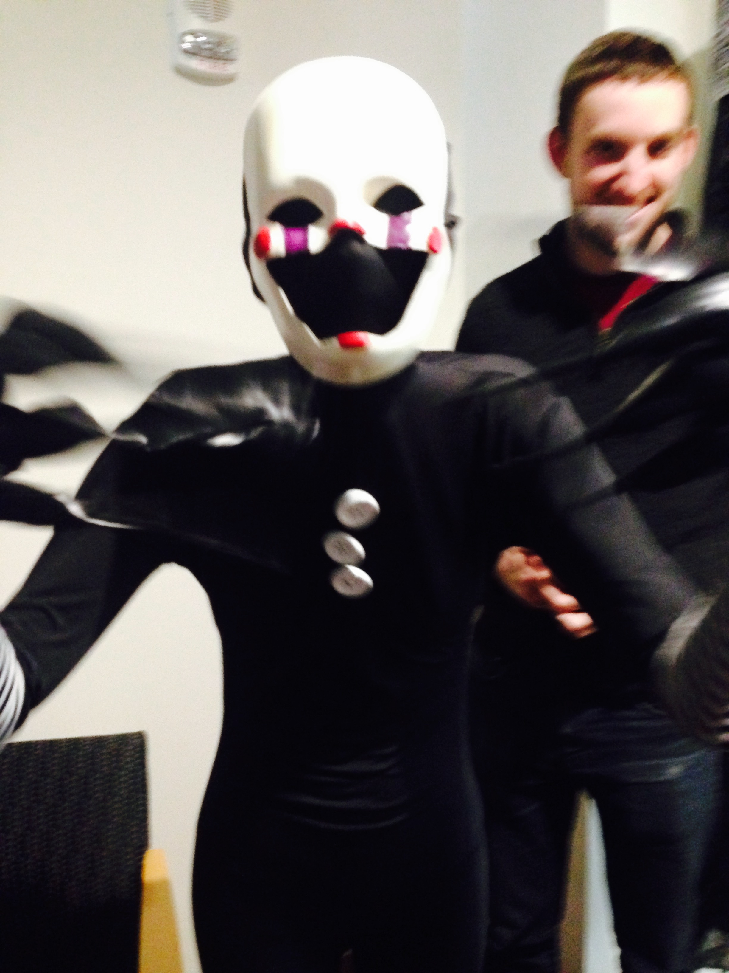
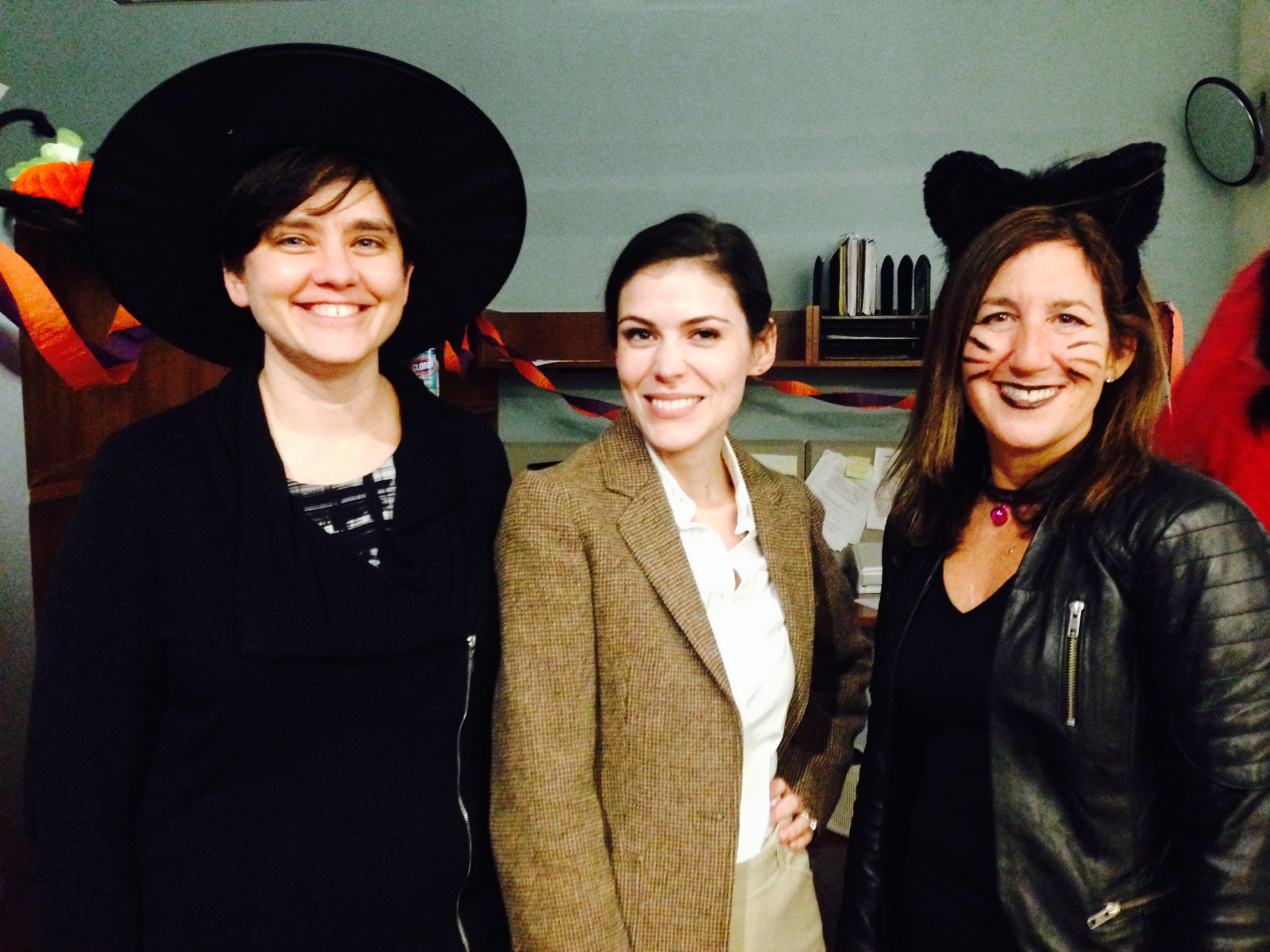
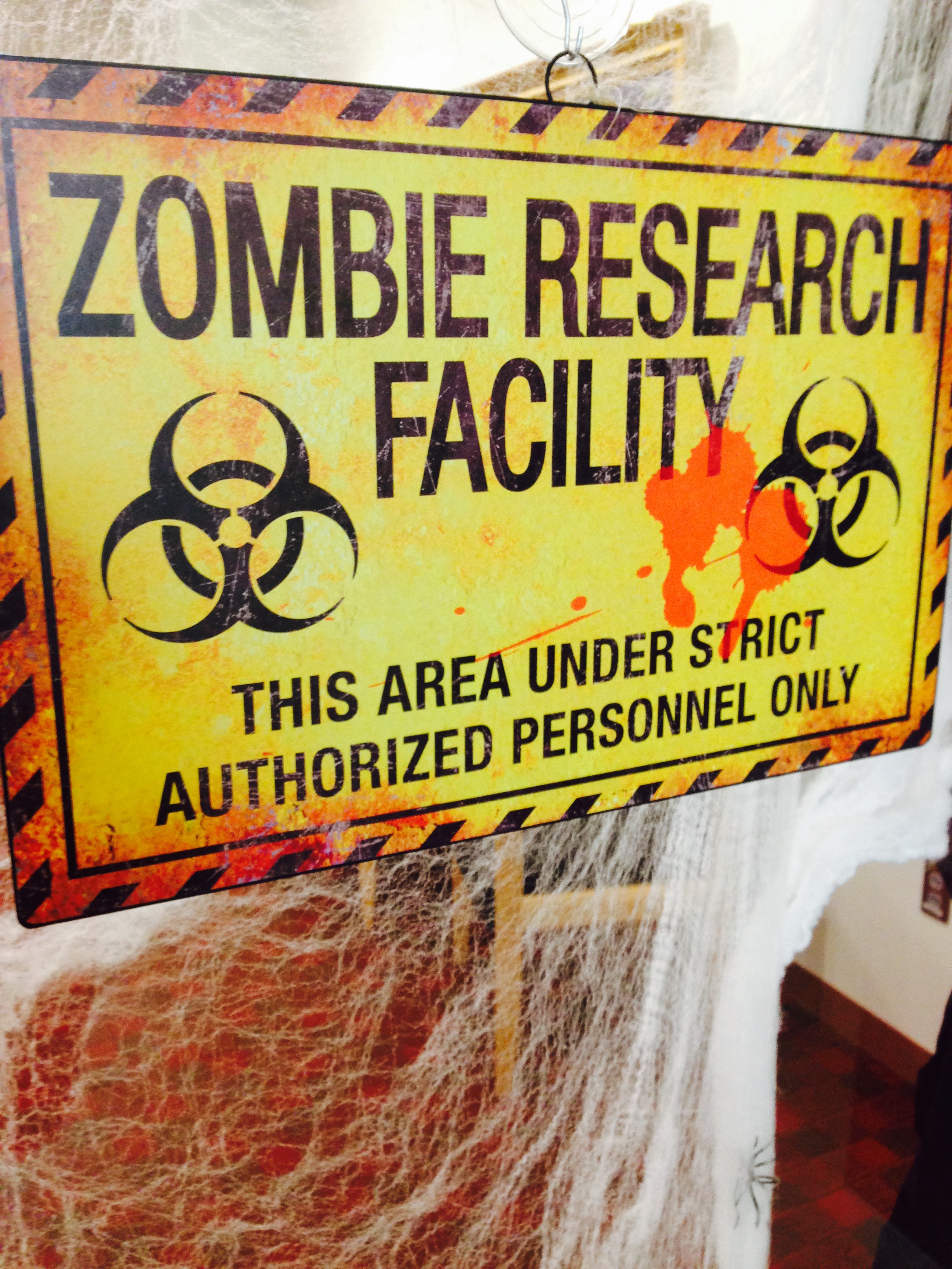
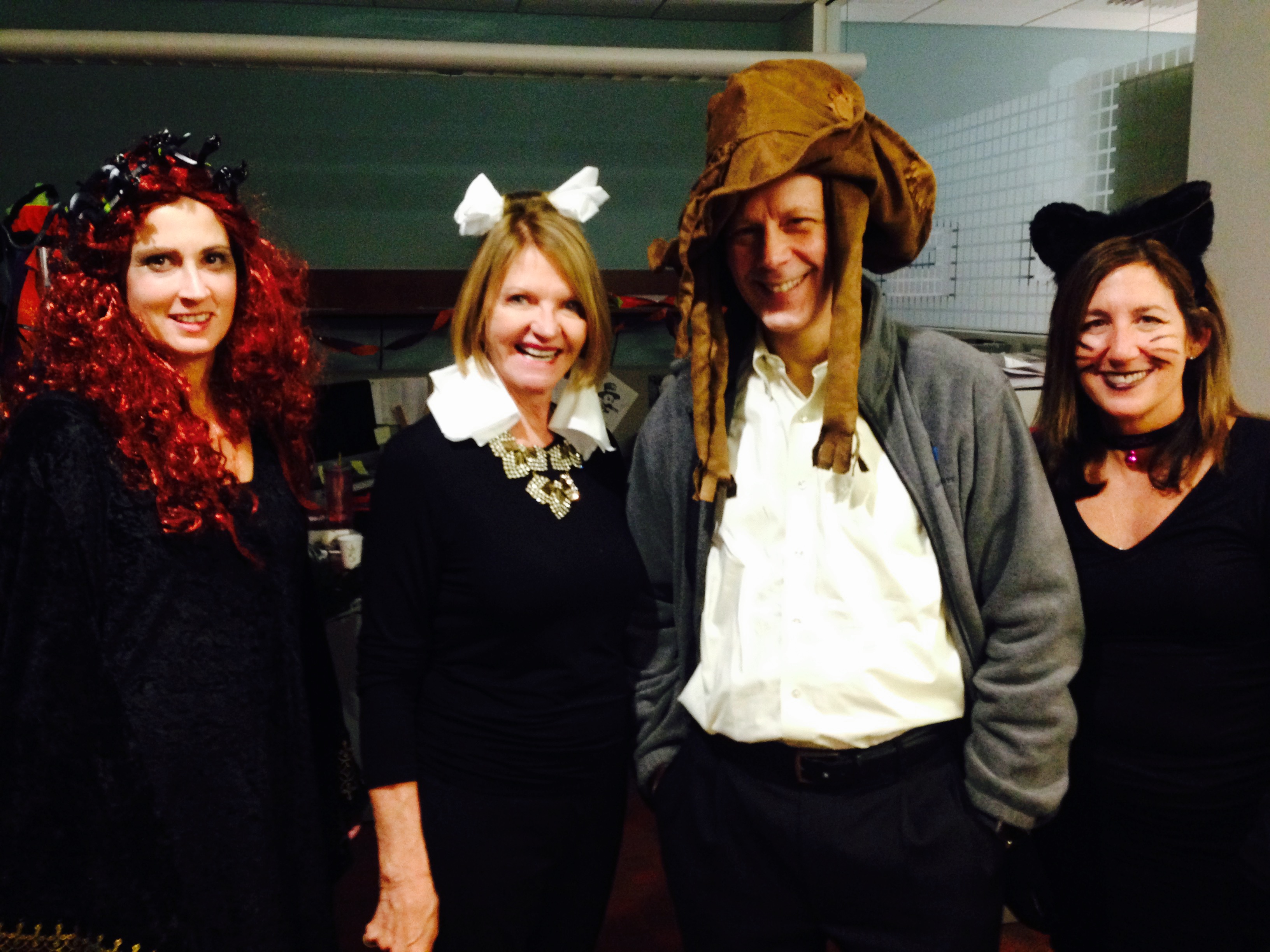
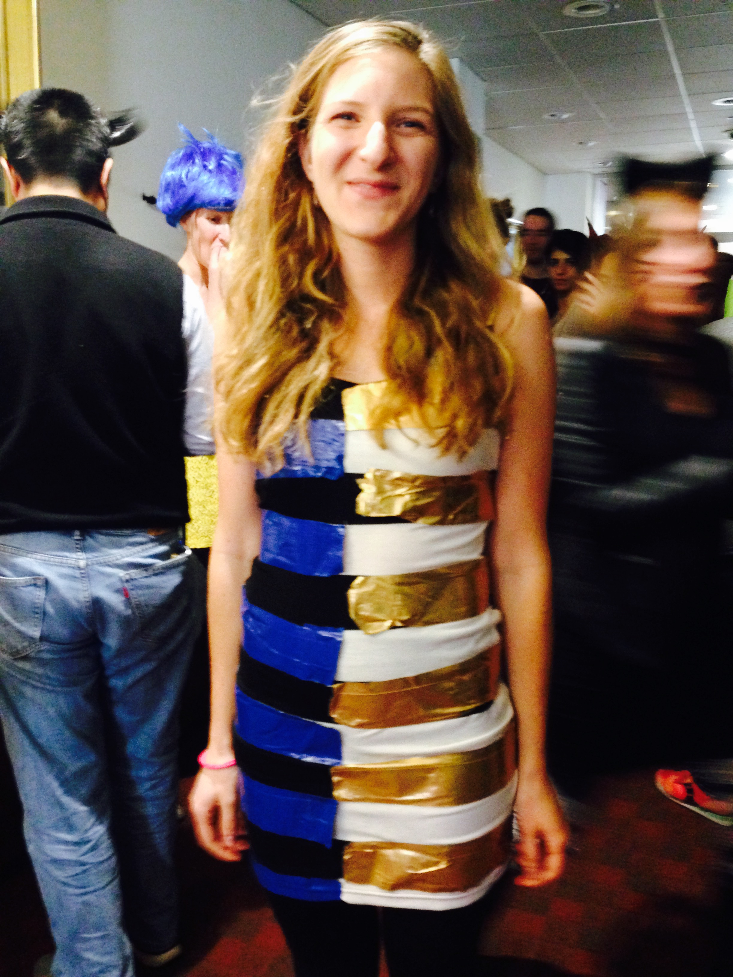
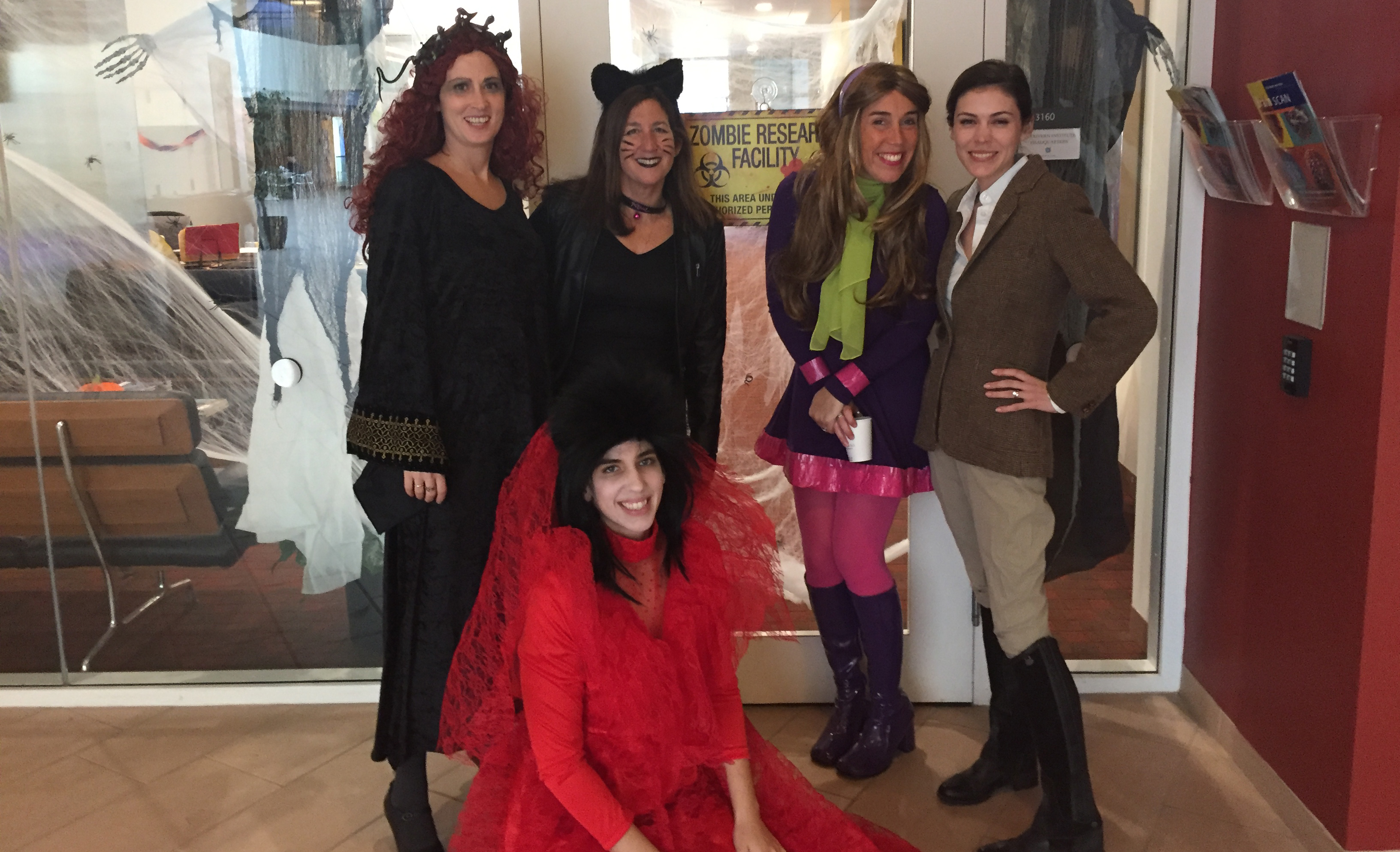
2015 McGovern Institute Halloween Party
To view the 2015 McGovern Institute Halloween Party gallery, please click on one of the thumbnail images below.


Used Book Fair
Stanley Center & Poitras Center Joint Translational Neuroscience Seminar Series: Dr. Steven Hyman
Abstract:
The genetic analysis of schizophrenia, bipolar disorder, and autism spectrum disorders has achieved early success. Much work remains: increasing the size and diversity of cohorts, fine mapping GWAS loci, and improving tools to implicate variants too rare to allow statistical certainty. However, the greater challenges lie in transforming gene lists into biological insights and therapies. The genetic architecture of neuropsychiatric disorders creates special difficulties for biology including polygenicity, low penetrance alleles and sharing across multiple disorders. These difficulties are heightened by the challenges posed by the human brain, with its diversity of cells and circuits, its inaccessibility in life, and by recent evolutionary changes that often limits the utility of animal models. I will review progress in genetics and discuss why the Stanley Center is pursing genetic analysis to “diminishing returns”. I will then argue that to exploit genetics we must (1) significantly humanize our model systems and commit to using the “right” cell types; (2) enhance molecular tools to interrogate human neurons and glia at the single cell level; (3) eschew overreliance on approaches that have worked for the investigation of highly penetrant alleles; and (4) develop ethical and practical frameworks so that compounds, once shown to be safe, can be studied in patients without attempting to gain false reassurance of efficacy from animal behavior.
Dr. Steve Hyman Joint Translational Neuroscience Seminar
Halloween Party
A new way to see the brain
Neurotech 2015
The Neurotech 2015 symposium presents talks by neurotechnology pioneers whose cutting-edge innovations are changing the face of neurobiological research from molecules to cognition. The symposium is open to the public, and registration is required, but seating is limited.
For more information and to register for this event, visit neurotech.mit.edu