What if we could peer into the brain to determine guilt or innocence? Could advances in neuroscience help reform our criminal justice system?
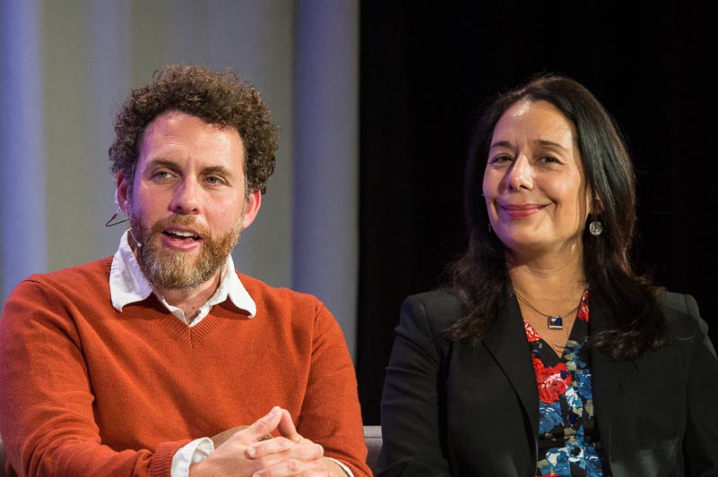
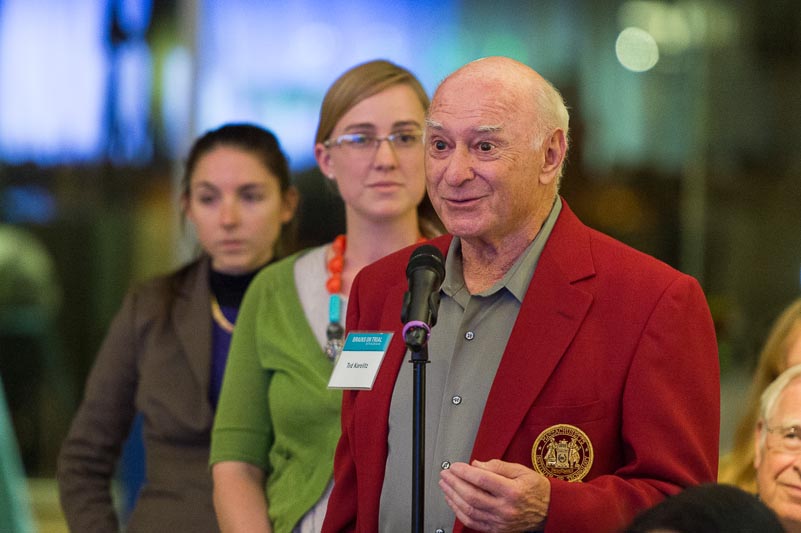
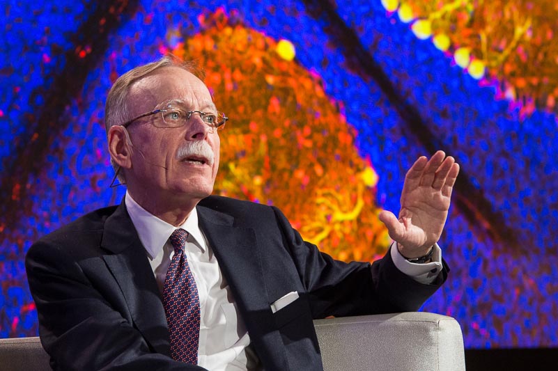
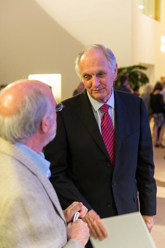
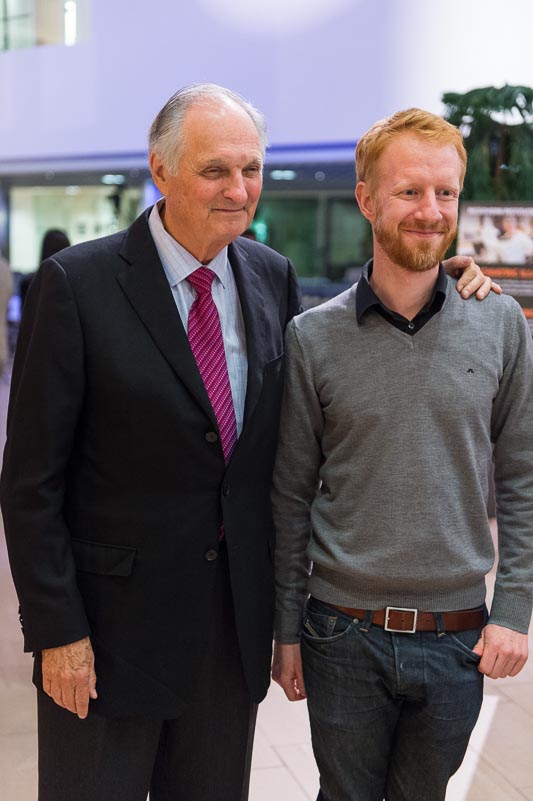
On Tuesday, September 17th, the McGovern Institute hosted a discussion with a distinguished group of legal and neuroscience experts who debated these and related questions. Alan Alda moderated the panel of experts, showed clips from his 2-part PBS special, “Brains on Trial,” and engaged the audience in a Q&A session.
See below for a photo gallery of the event. All photos courtesy of Justin Knight.
Alan Alda moderates a panel of experts on the role of neuroscience in the criminal justice system.
Alan Alda moderates a panel of experts on the role of neuroscience in the criminal justice system. Photo: Justin Knight
What if we could peer into the brain to determine guilt or innocence? Could advances in neuroscience help reform our criminal justice system?
On Tuesday, September 17th, the McGovern Institute hosted a discussion with a distinguished group of legal and neuroscience experts who debated these and related questions. Alan Alda moderated the panel of experts, showed clips from his 2-part PBS special, “Brains on Trial,” and engaged the audience in a Q&A session.
Popular Science magazine has named two MIT junior faculty members — Pedro Reis and Feng Zhang — to its 2013 Brilliant 10 list of young stars in science and technology. The list will appear in the magazine’s October issue.
“Popular Science prides itself on revealing the innovations and ideas that are laying today’s groundwork for tomorrow’s breakthroughs, and the Brilliant 10 is one of the most exciting ways we do that,” says Jake Ward, editor-in-chief. “This collection of 10 brilliant young researchers is our chance to honor the most promising work — and the most hardworking people — in science and technology today. This year’s winners are particularly distinguished and I’m proud to welcome them all as members of the 2013 Brilliant 10.”
Pedro Reis, the Esther and Harold E. Edgerton Assistant Professor of Civil and Environmental Engineering and Mechanical Engineering, studies the mechanics of slender structures, with a particular focus on devising new ways of turning mechanical failure into functionality.
Over the past few years, Reis, 35, has published a number of eclectic and impactful papers in prominent journals. In 2009 he reported on the delamination of thin films adhered to soft foundations, which is relevant for stretchable electronics. He explained why adhesive films tear into triangular shapes, a problem that applies to both the everyday peeling of adhesive tape from a roll and the manufacturing of tapered graphene nanoribbons. Motivated by the closing of aquatic flowers, he recently discovered a new mechanism for passively pipetting liquids using a petal-shaped object. And last year inspired by a toy, Reis introduced the Buckliball, a new class of structures that uses buckling to provide origami-like folding capabilities to curved structures with potential uses for encapsulation and soft robotics.
In other work undertaken just for fun, Reis and colleagues reported in 2010 that when cats lap fluids (milk or water, for example), they take advantage of a perfect balance between gravity and inertia.
Feng Zhang, 31, is the W.M. Keck Career Development Professor in Biomedical Engineering, an assistant professor in the department of Brain and Cognitive Sciences, a member of the McGovern Institute for Brain Research and a core member of the Broad Institute. He received the award for his work on genome editing. Earlier this year he reported a powerful new way to make targeted mutations in genomic DNA, based on a bacterial system known as CRISPR. The new method will greatly accelerate the development of animal models of human genetic diseases, and may eventually make it possible to correct genetic mutations in patients. Zhang, a pioneer in optogenetics, has also recently invented a new method for controlling gene expression with light, in which light-sensitive plant proteins are engineered to create an “optical switch” that can turn other genes on or off at will.
This is the 12th annual Brilliant 10 list. Ten MIT researchers were included on previous lists.
Zeynep Saygin, a postdoc in Nancy Kanwisher’s lab uses a technology known as diffusion-weighted MR imaging to reveal long-range connections in the brain.
As anyone who has traveled with young children knows, maintaining focus on distant goals can be a challenge. A new study from MIT suggests how the brain achieves this task, and indicates that the neurotransmitter dopamine may signal the value of long-term rewards. The findings may also explain why patients with Parkinson’s disease — in which dopamine signaling is impaired — often have difficulty in sustaining motivation to finish tasks.
The work is described this week in the journal Nature.
Previous studies have linked dopamine to rewards, and have shown that dopamine neurons show brief bursts of activity when animals receive an unexpected reward. These dopamine signals are believed to be important for reinforcement learning, the process by which an animal learns to perform actions that lead to reward.
Taking the long view
In most studies, that reward has been delivered within a few seconds. In real life, though, gratification is not always immediate: Animals must often travel in search of food, and must maintain motivation for a distant goal while also responding to more immediate cues. The same is true for humans: A driver on a long road trip must remain focused on reaching a final destination while also reacting to traffic, stopping for snacks, and entertaining children in the back seat.
The MIT team, led by Institute Professor Ann Graybiel — who is also an investigator at MIT’s McGovern Institute for Brain Research — decided to study how dopamine changes during a maze task approximating work for delayed gratification. The researchers trained rats to navigate a maze to reach a reward. During each trial a rat would hear a tone instructing it to turn either right or left at an intersection to find a chocolate milk reward.
Rather than simply measuring the activity of dopamine-containing neurons, the MIT researchers wanted to measure how much dopamine was released in the striatum, a brain structure known to be important in reinforcement learning. They teamed up with Paul Phillips of the University of Washington, who has developed a technology called fast-scan cyclic voltammetry (FSCV) in which tiny, implanted, carbon-fiber electrodes allow continuous measurements of dopamine concentration based on its electrochemical fingerprint.
“We adapted the FSCV method so that we could measure dopamine at up to four different sites in the brain simultaneously, as animals moved freely through the maze,” explains first author Mark Howe, a former graduate student with Graybiel who is now a postdoc in the Department of Neurobiology at Northwestern University. “Each probe measures the concentration of extracellular dopamine within a tiny volume of brain tissue, and probably reflects the activity of thousands of nerve terminals.”
Gradual increase in dopamine
From previous work, the researchers expected that they might see pulses of dopamine released at different times in the trial, “but in fact we found something much more surprising,” Graybiel says: The level of dopamine increased steadily throughout each trial, peaking as the animal approached its goal — as if in anticipation of a reward.
The rats’ behavior varied from trial to trial — some runs were faster than others, and sometimes the animals would stop briefly — but the dopamine signal did not vary with running speed or trial duration. Nor did it depend on the probability of getting a reward, something that had been suggested by previous studies.
“Instead, the dopamine signal seems to reflect how far away the rat is from its goal,” Graybiel explains. “The closer it gets, the stronger the signal becomes.” The researchers also found that the size of the signal was related to the size of the expected reward: When rats were trained to anticipate a larger gulp of chocolate milk, the dopamine signal rose more steeply to a higher final concentration.
In some trials the T-shaped maze was extended to a more complex shape, requiring animals to run further and to make extra turns before reaching a reward. During these trials, the dopamine signal ramped up more gradually, eventually reaching the same level as in the shorter maze. “It’s as if the animal were adjusting its expectations, knowing that it had further to go,” Graybiel says.
The traces represent brain activity in rats as they navigate through different mazes to receive a chocolate milk reward.
An ‘internal guidance system’
“This means that dopamine levels could be used to help an animal make choices on the way to the goal and to estimate the distance to the goal,” says Terrence Sejnowski of the Salk Institute, a computational neuroscientist who is familiar with the findings but who was not involved with the study. “This ‘internal guidance system’ could also be useful for humans, who also have to make choices along the way to what may be a distant goal.”
One question that Graybiel hopes to examine in future research is how the signal arises within the brain. Rats and other animals form cognitive maps of their spatial environment, with so-called “place cells” that are active when the animal is in a specific location. “As our rats run the maze repeatedly,” she says, “we suspect they learn to associate each point in the maze with its distance from the reward that they experienced on previous runs.”
As for the relevance of this research to humans, Graybiel says, “I’d be shocked if something similar were not happening in our own brains.” It’s known that Parkinson’s patients, in whom dopamine signaling is impaired, often appear to be apathetic, and have difficulty in sustaining motivation to complete a long task. “Maybe that’s because they can’t produce this slow ramping dopamine signal,” Graybiel says.
Patrick Tierney at MIT and Stefan Sandberg at the University of Washington also contributed to the study, which was funded by the National Institutes of Health, the National Parkinson Foundation, the CHDI Foundation, the Sydney family and Mark Gorenberg.
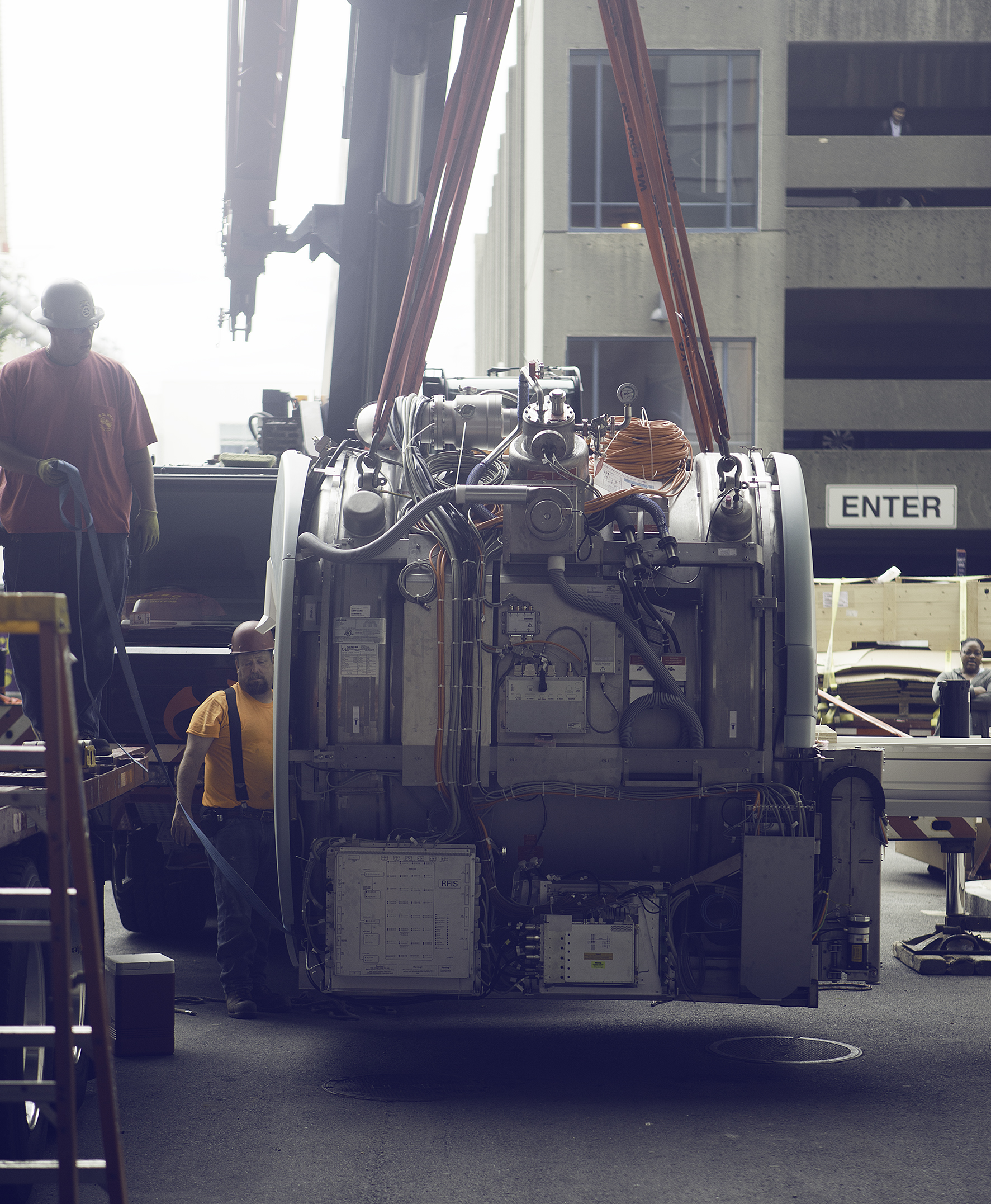
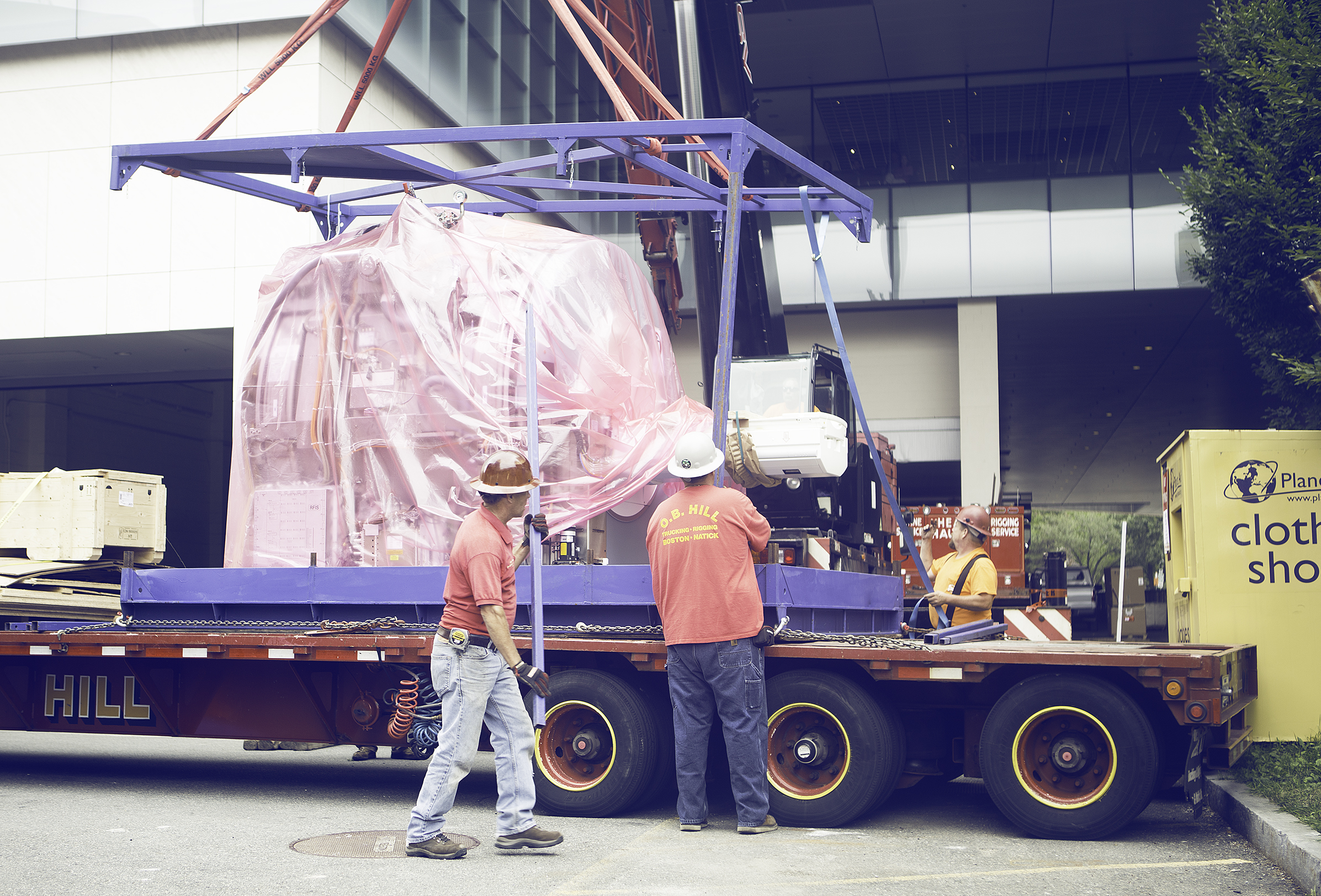
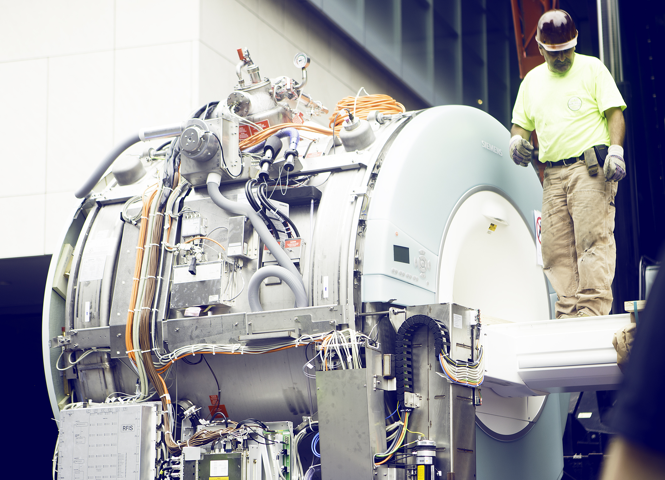
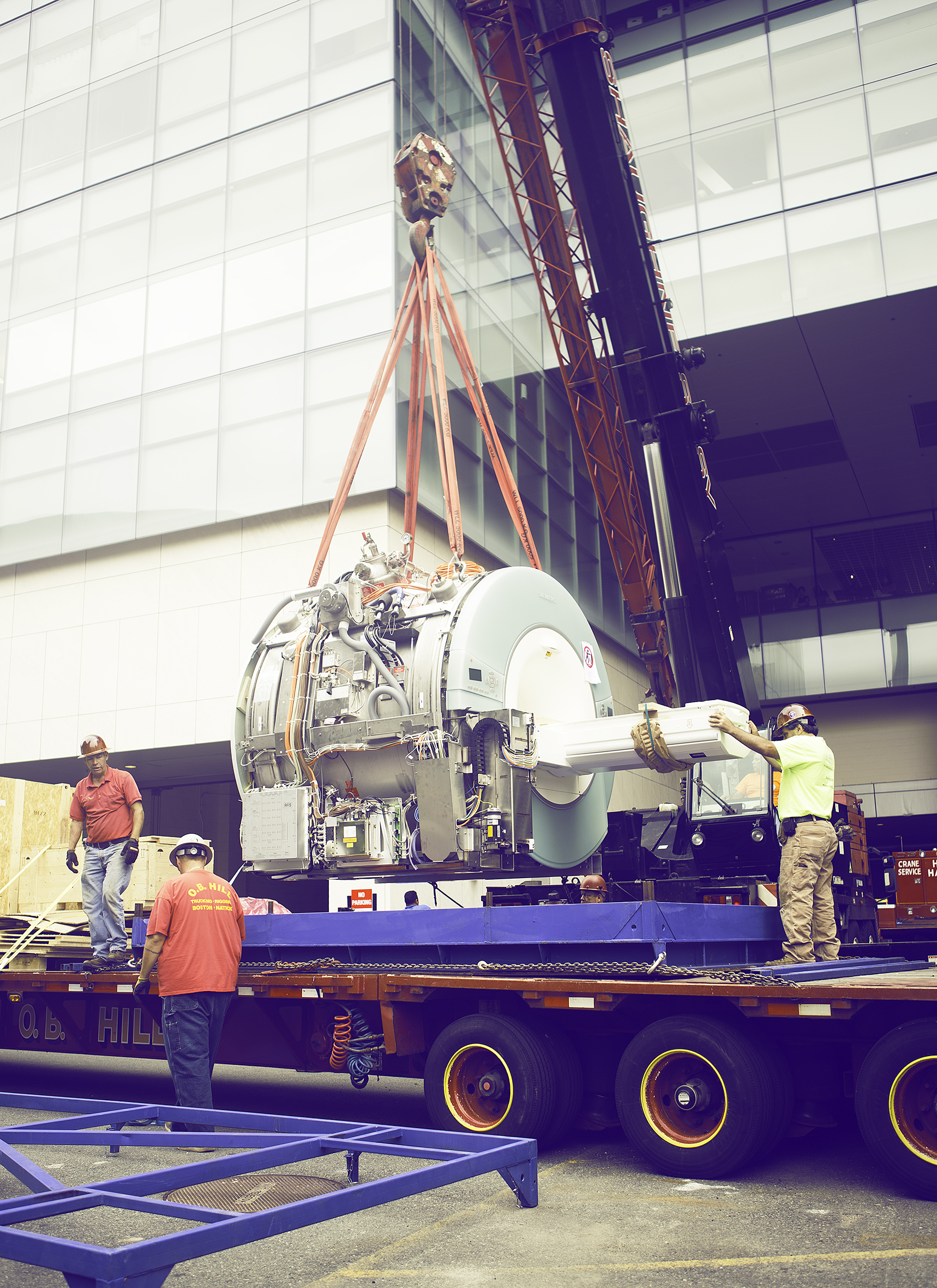
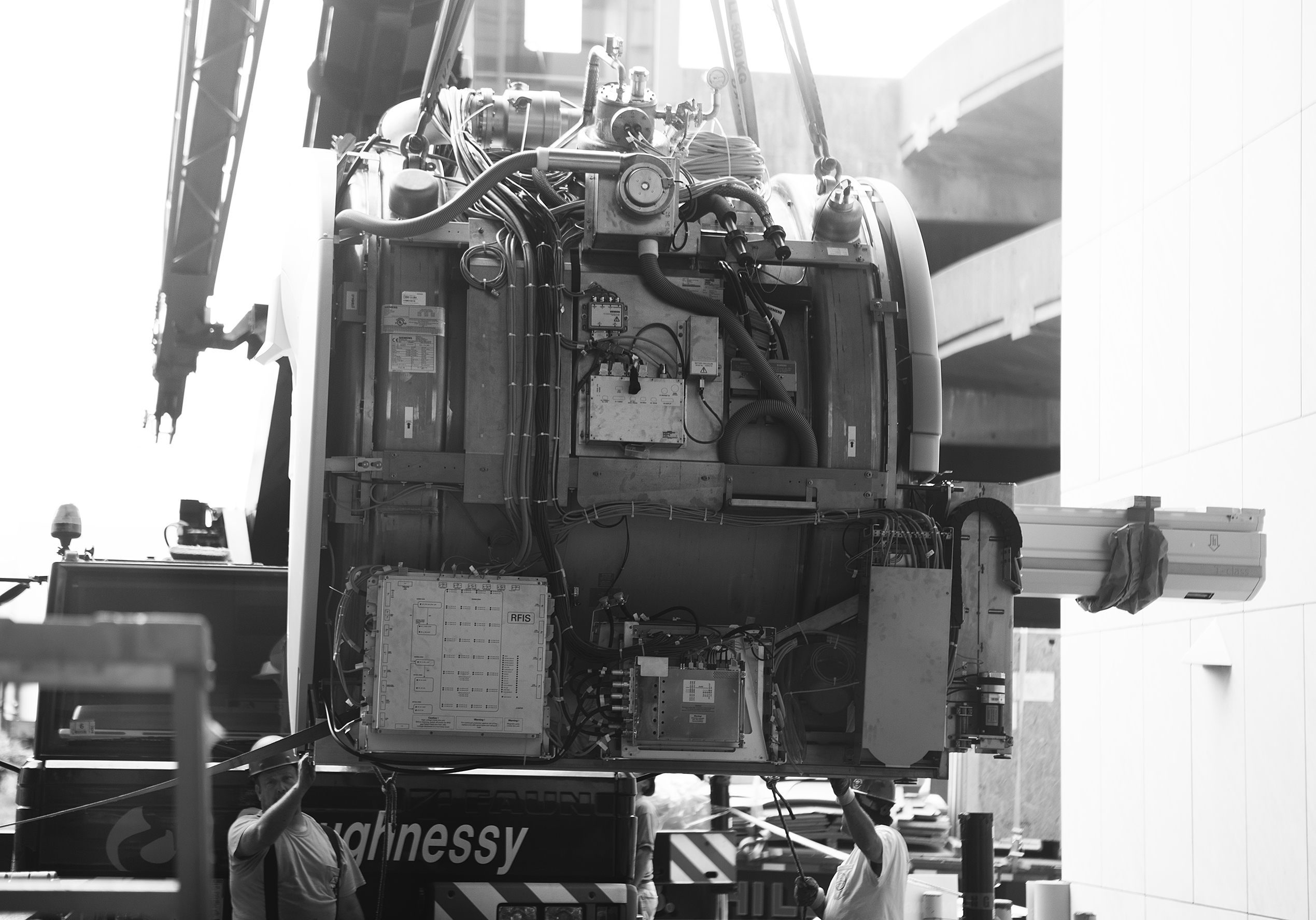
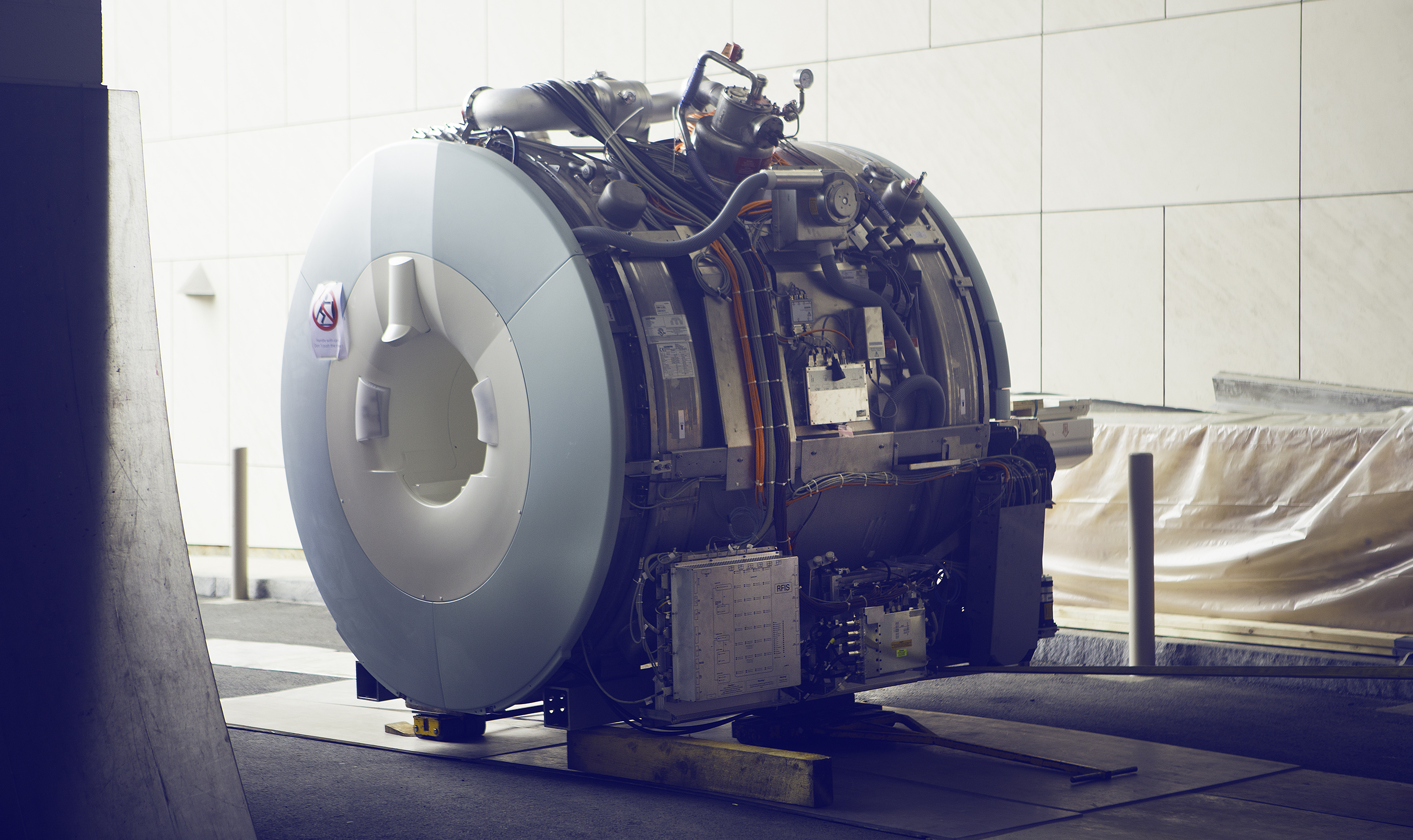
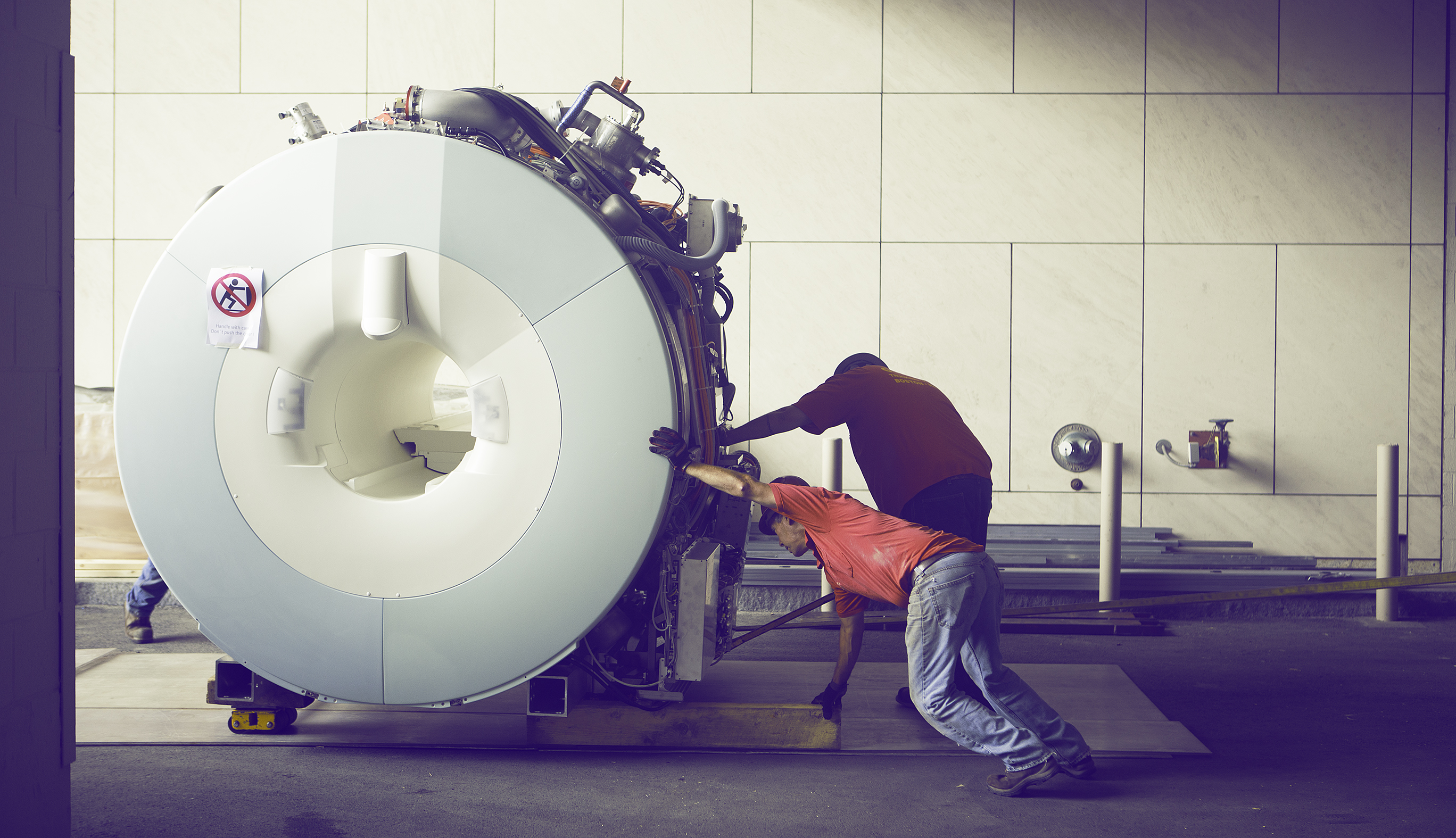
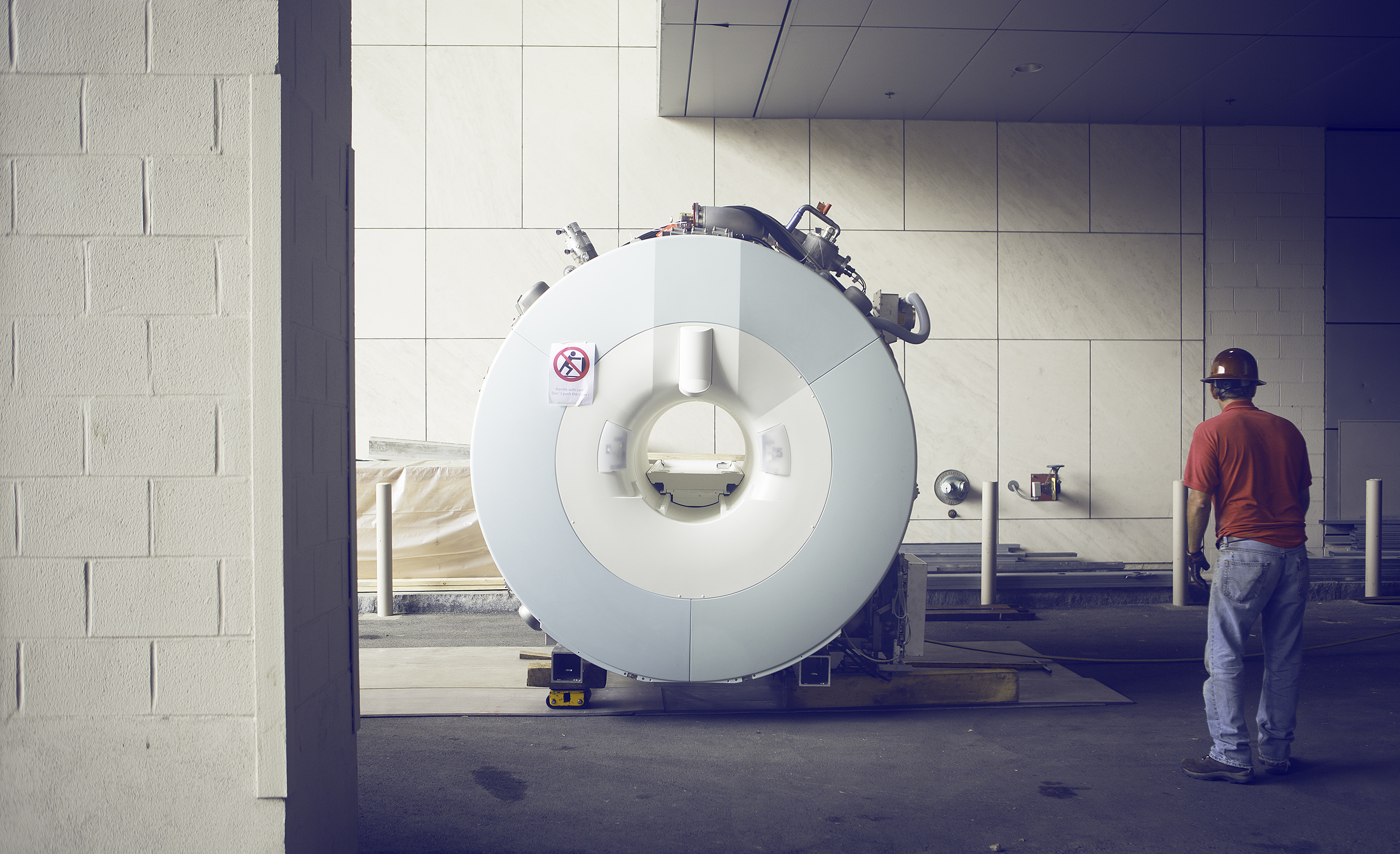
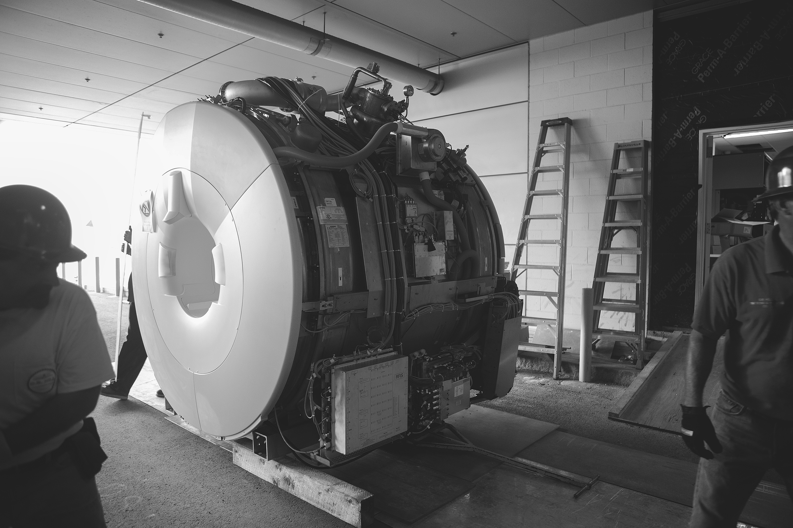
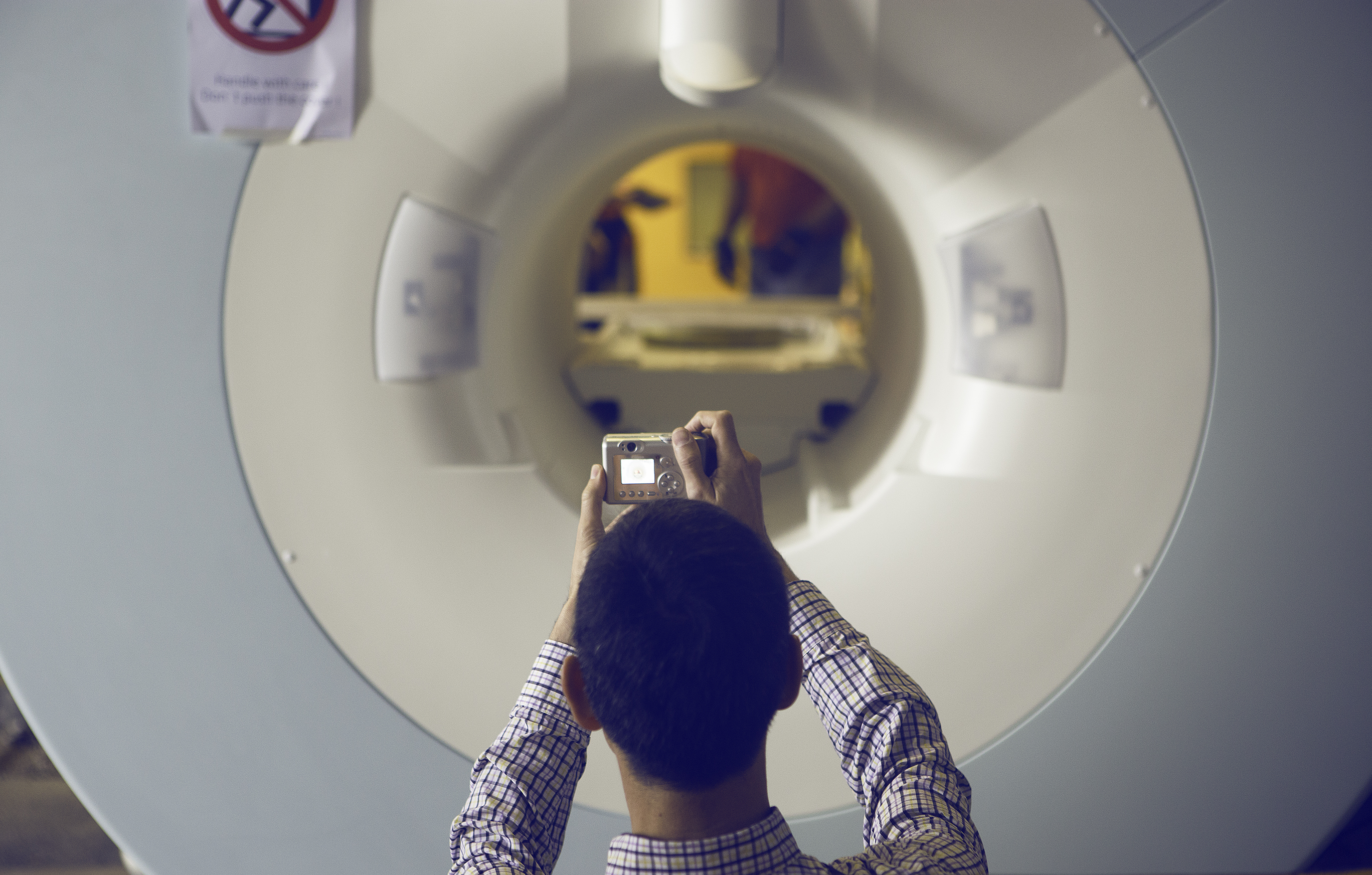
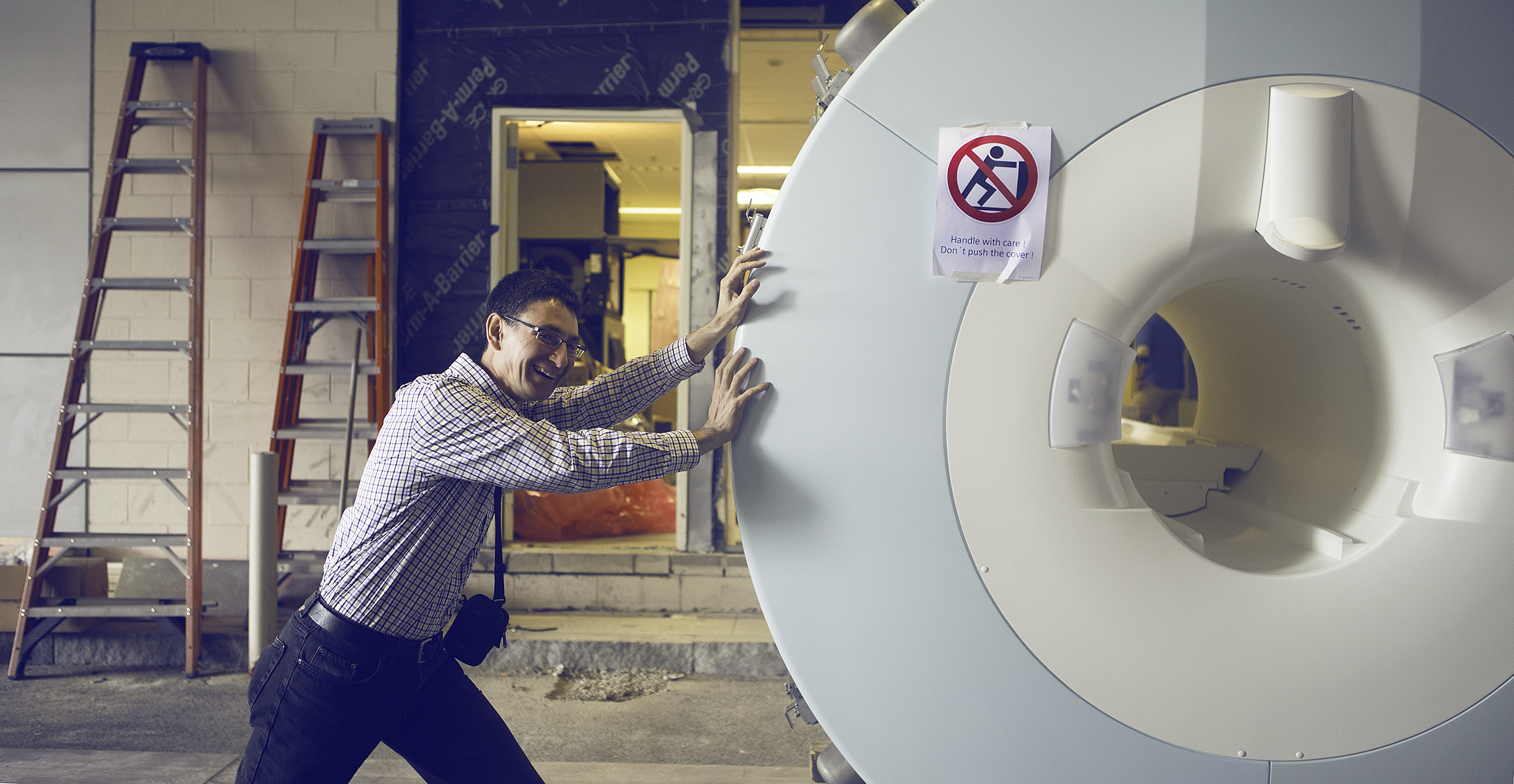
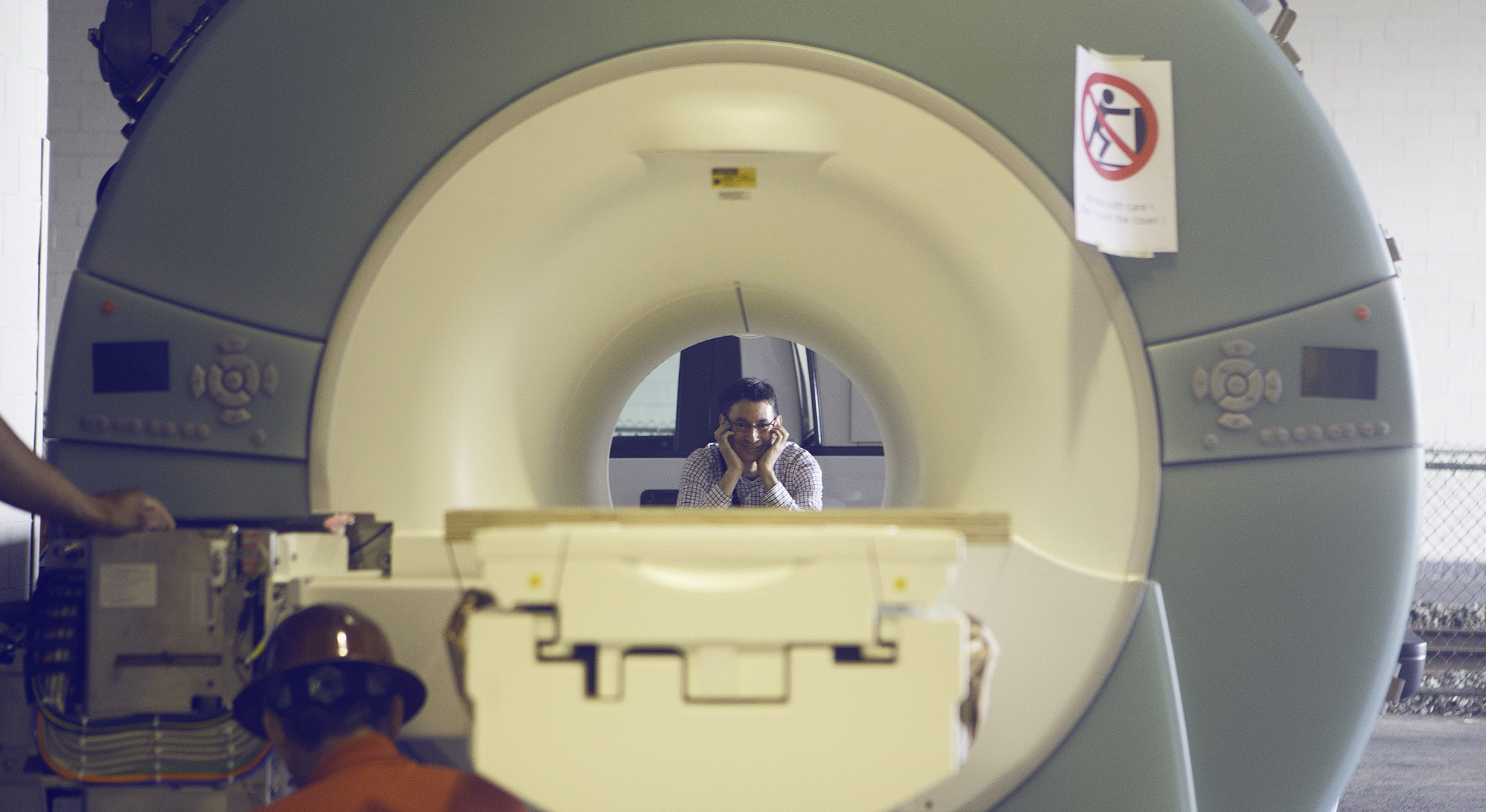
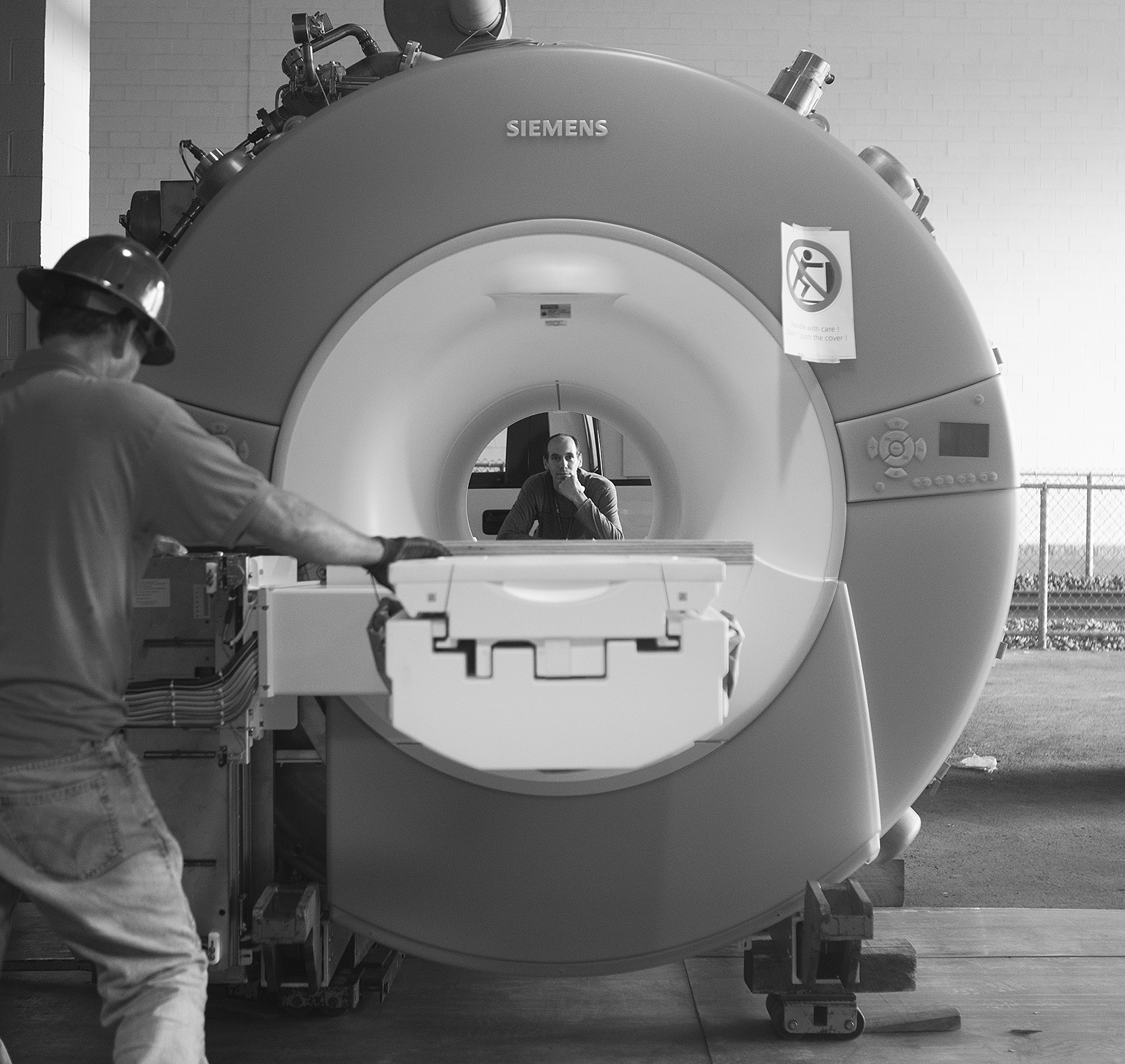
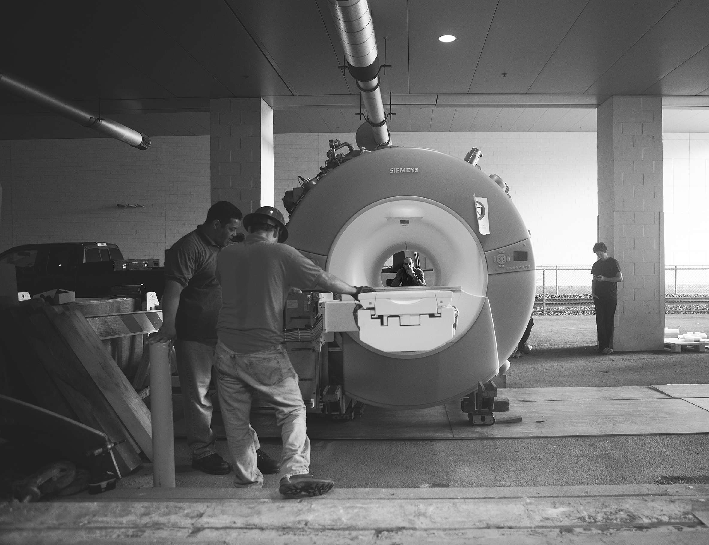
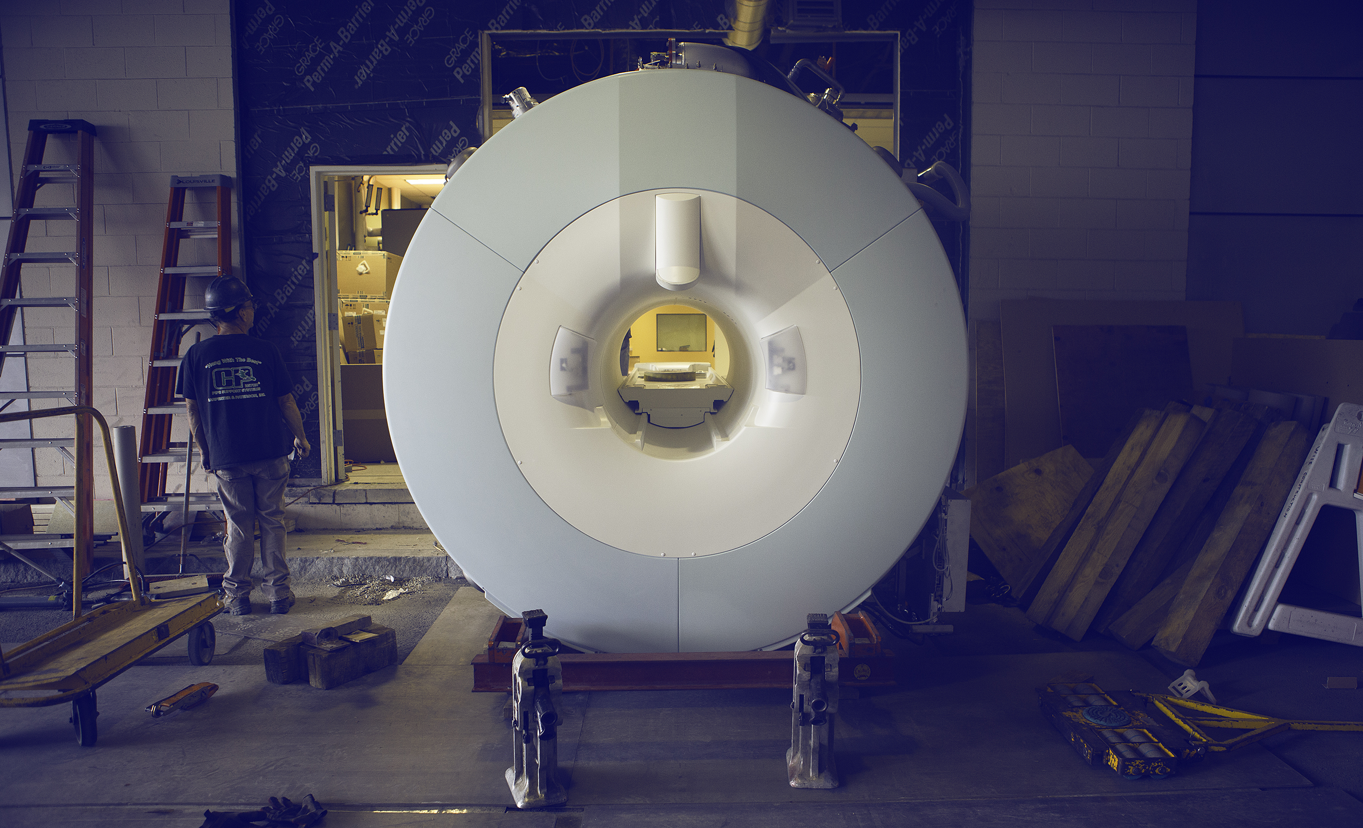
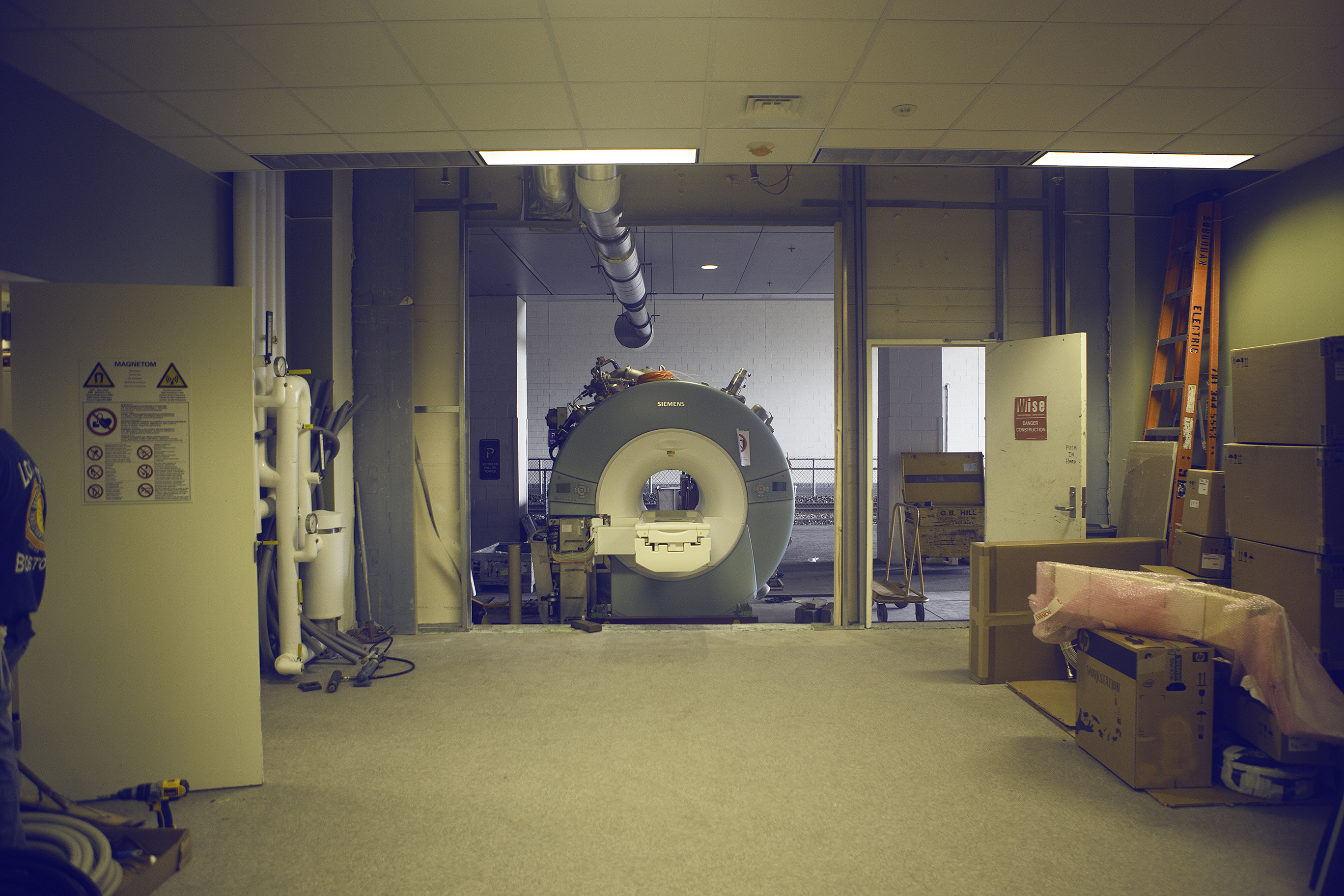
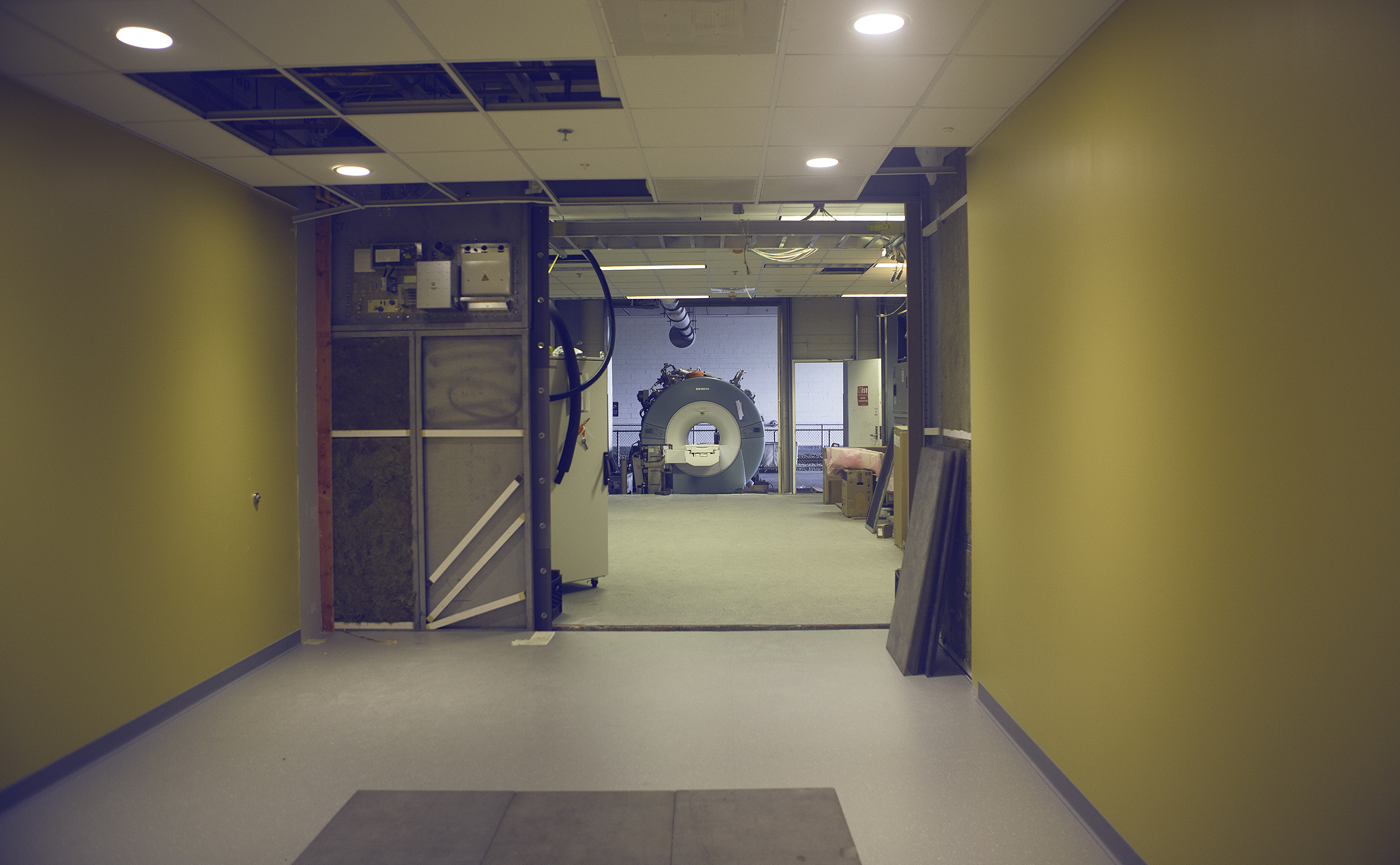
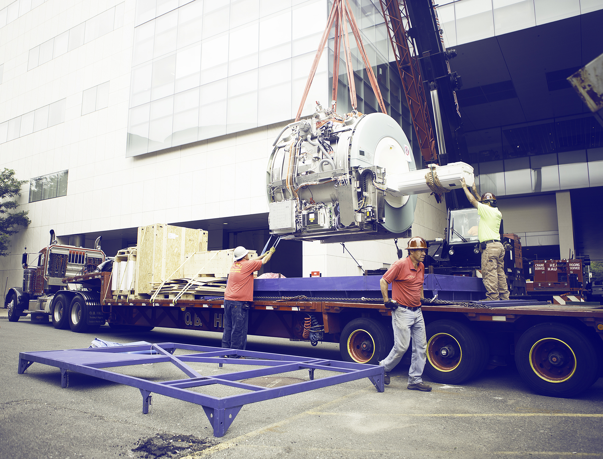
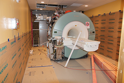
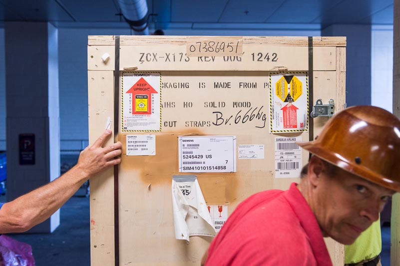
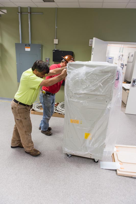
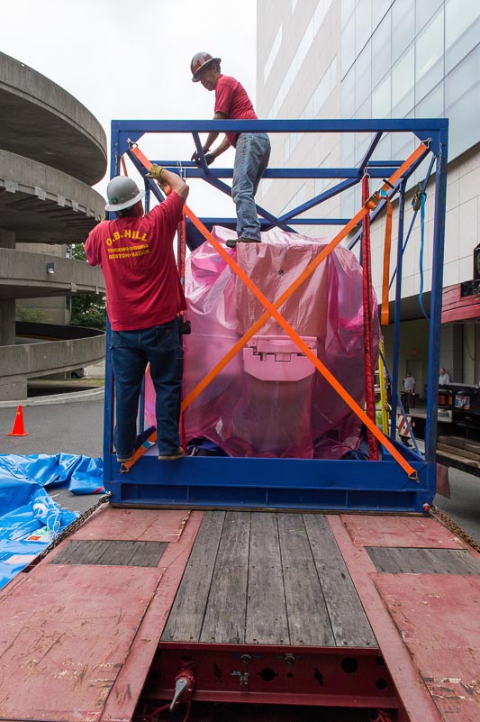
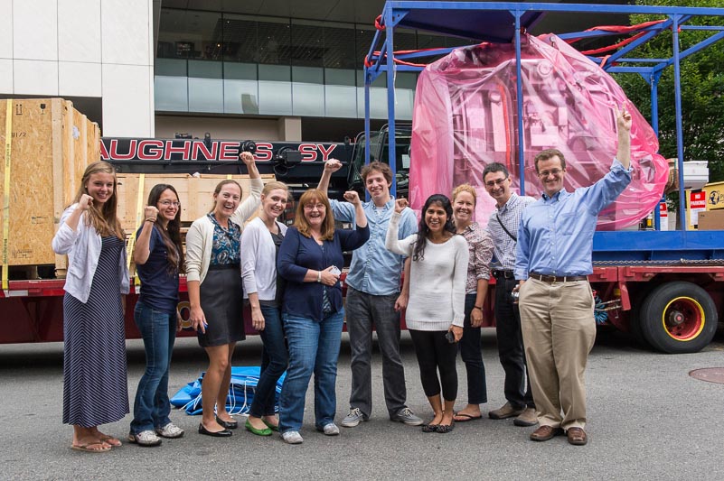
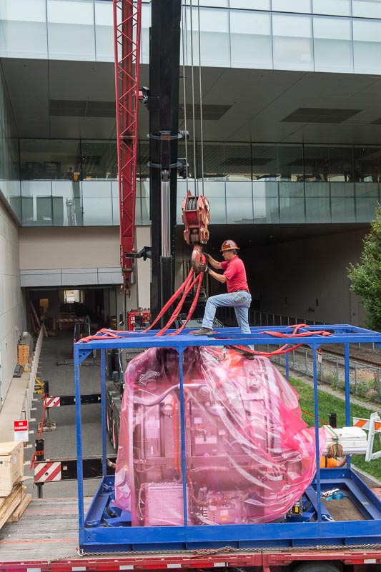
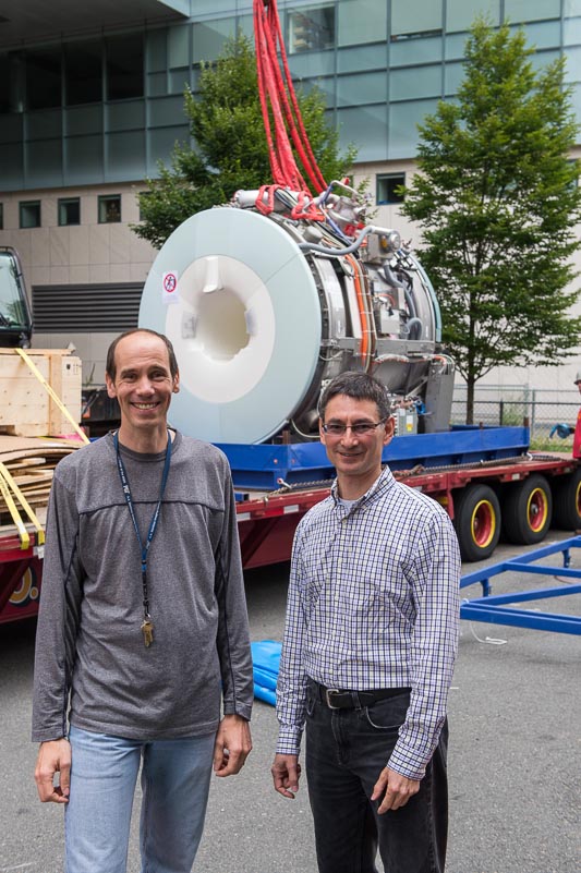
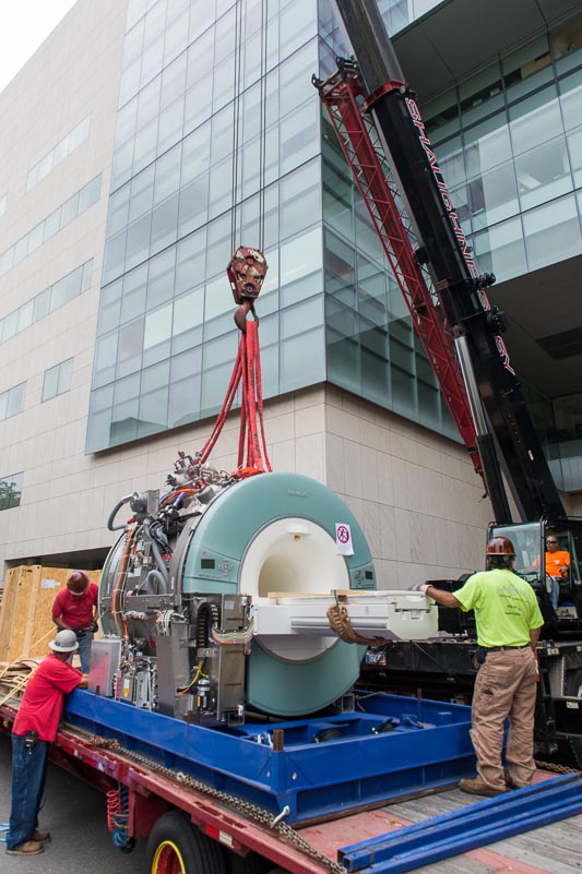
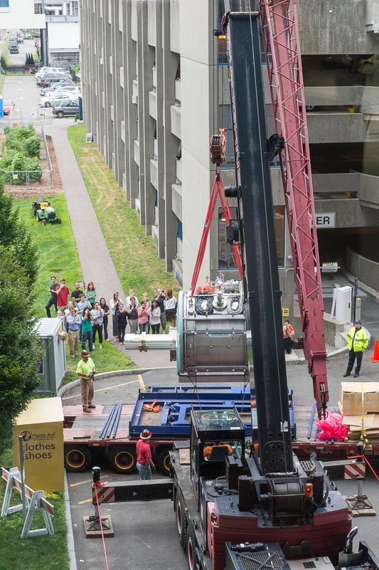
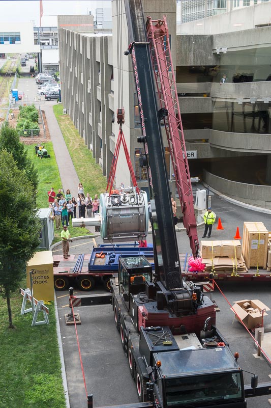
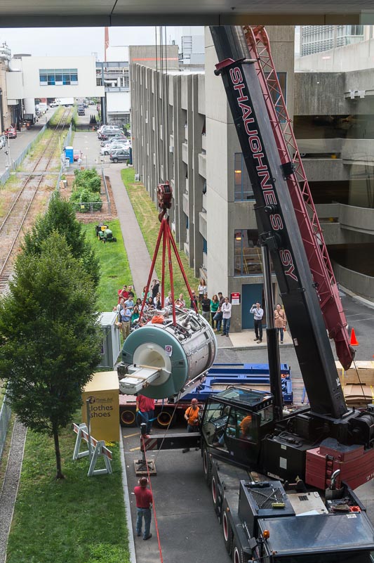
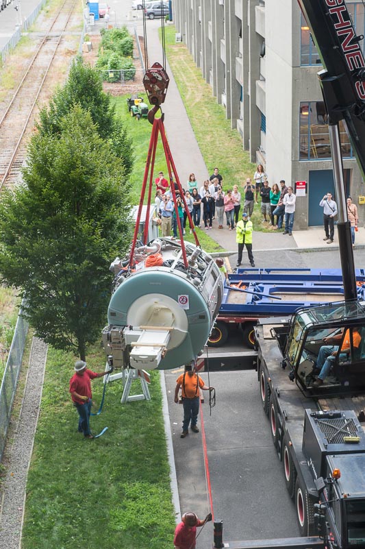
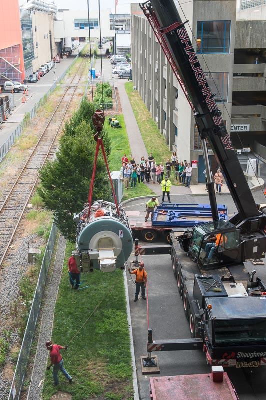
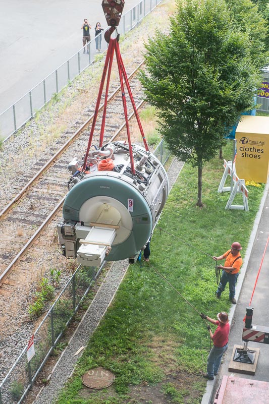
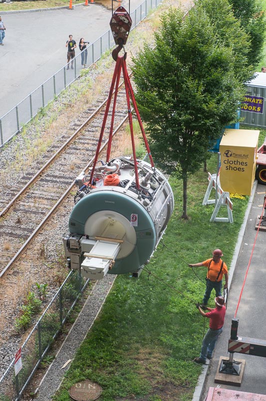
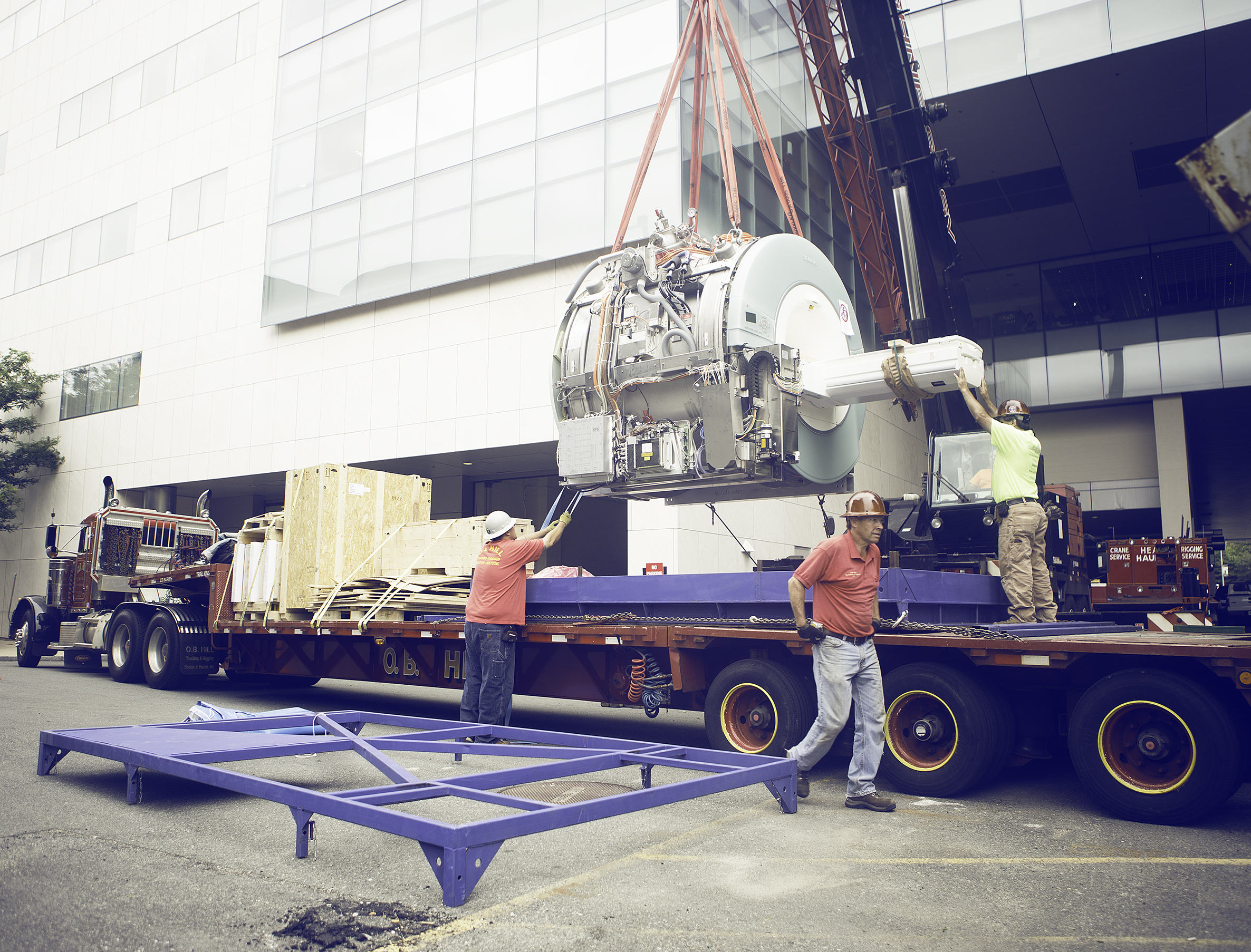
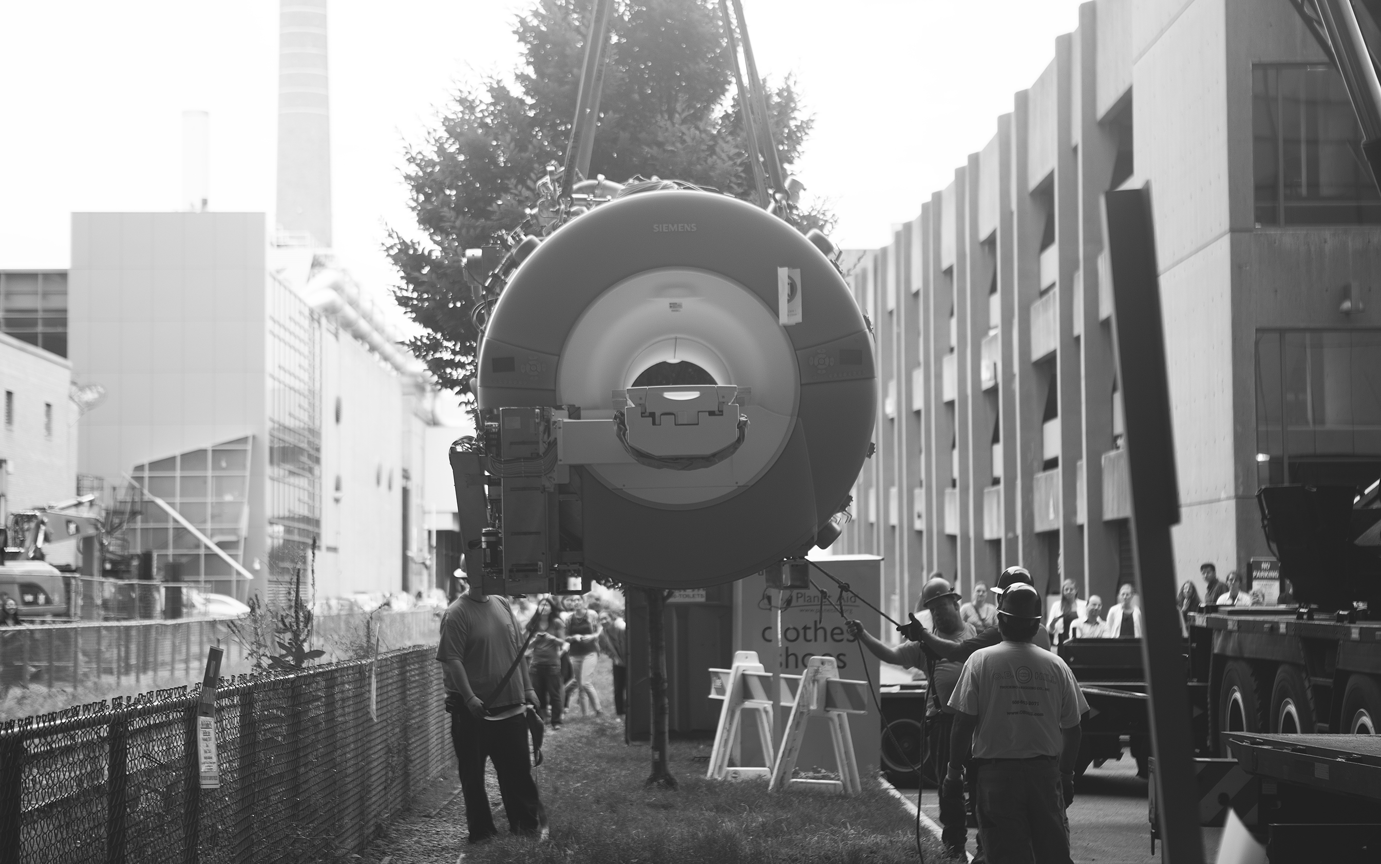
After months of planning and construction, we are delighted to report that we have installed a new 3-tesla MRI scanner for human neuroimaging. The $2M scanner, a Siemens Magnetom Trio, was delivered to the Martinos Imaging Center on July 25, 2013.
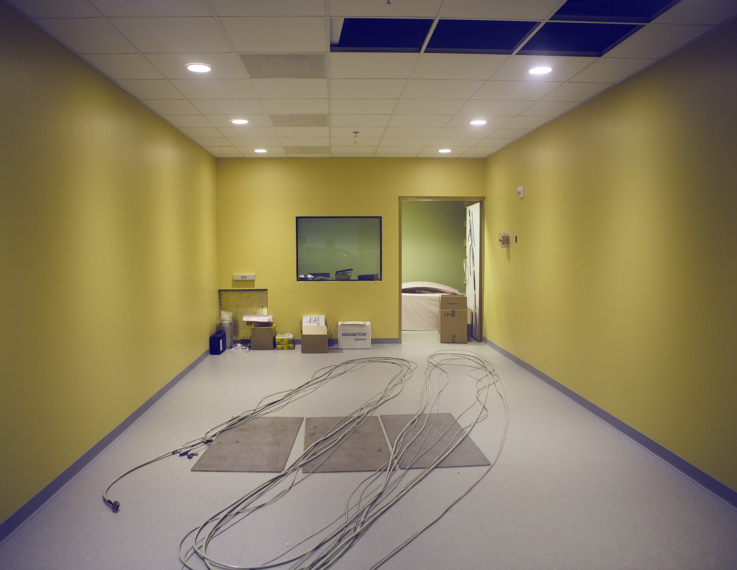
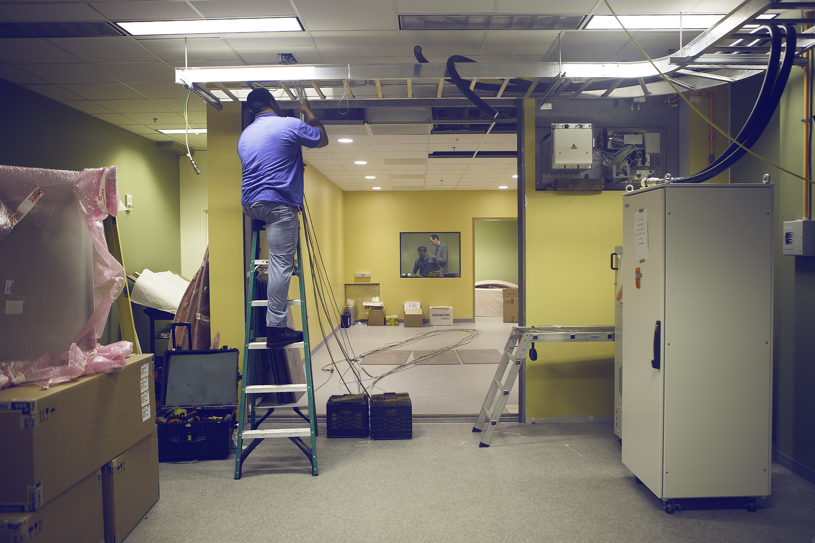
The core of the scanner is a large electromagnet, weighing around 13 tons and containing superconducting coils that are chilled in liquid helium to within a few degrees of absolute zero. It is housed in a custom-built room, with a specially reinforced floor to support the scanner’s weight, and with some 5000 steel panels to shield the system from RF interference.
The acquisition of the new scanner was made possible by Bruce Dayton, Jeffrey and Nancy Halis, the Simons Foundation, and an anonymous donor. The scanner is expected to be fully operational by the fall, and will be used for a wide range of studies on brain function, in both children and adults.
Click here to view a photo album of the MRI installation.