Mapping the brain’s sensory gatekeeper

Many people with autism experience sensory hypersensitivity, attention deficits, and sleep disruption. One brain region that has been implicated in these symptoms is the thalamic reticular nucleus (TRN), which is believed to act as a gatekeeper for sensory information flowing to the cortex.

A team of researchers from MIT and the Broad Institute of MIT and Harvard has now mapped the TRN in unprecedented detail, revealing that the region contains two distinct subnetworks of neurons with different functions. The findings could offer researchers more specific targets for designing drugs that could alleviate some of the sensory, sleep, and attention symptoms of autism, says Guoping Feng, one of the leaders of the research team.
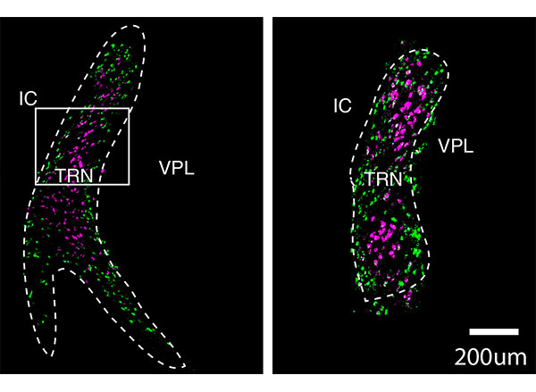
These cross-sections of the thalamic reticular nucleus (TRN) show two distinct populations of neurons, labeled in purple and green. A team of researchers from MIT and the Broad Institute of MIT and Harvard has now mapped the TRN in unprecedented detail.
Image: courtesy of the researchers

“The idea is that you could very specifically target one group of neurons, without affecting the whole brain and other cognitive functions,” says Feng, the James W. and Patricia Poitras Professor of Neuroscience at MIT and a member of MIT’s McGovern Institute for Brain Research.

Feng; Zhanyan Fu, associate director of neurobiology at the Broad Institute’s Stanley Center for Psychiatric Research; and Joshua Levin, a senior group leader at the Broad Institute, are the senior authors of the study, which appears today in Nature. The paper’s lead authors are former MIT postdoc Yinqing Li, former Broad Institute postdoc Violeta Lopez-Huerta, and Broad Institute research scientist Xian Adiconis.

Distinct populations

When sensory input from the eyes, ears, or other sensory organs arrives in our brains, it goes first to the thalamus, which then relays it to the cortex for higher-level processing. Impairments of these thalamo-cortical circuits can lead to attention deficits, hypersensitivity to noise and other stimuli, and sleep problems.

One of the major pathways that controls information flow between the thalamus and the cortex is the TRN, which is responsible for blocking out distracting sensory input. In 2016, Feng and MIT Assistant Professor Michael Halassa, who is also an author of the new Nature paper, discovered that loss of a gene called Ptchd1 significantly affects TRN function. In boys, loss of this gene, which is carried on the X chromosome, can lead to attention deficits, hyperactivity, aggression, intellectual disability, and autism spectrum disorders.

In that study, the researchers found that when the Ptchd1 gene was knocked out in mice, the animals showed many of the same behavioral defects seen in human patients. When it was knocked out only in the TRN, the mice showed only hyperactivity, attention deficits, and sleep disruption, suggesting that the TRN is responsible for those symptoms.

In the new study, the researchers wanted to try to learn more about the specific types of neurons found in the TRN, in hopes of finding new ways to treat hyperactivity and attention deficits. Currently, those symptoms are most often treated with stimulant drugs such as Ritalin, which have widespread effects throughout the brain.

“Our goal was to find some specific ways to modulate the function of thalamo-cortical output and relate it to neurodevelopmental disorders,” Feng says. “We decided to try using single-cell technology to dissect out what cell types are there, and what genes are expressed. Are there specific genes that are druggable as a target?”

To explore that possibility, the researchers sequenced the messenger RNA molecules found in neurons of the TRN, which reveals genes that are being expressed in those cells. This allowed them to identify hundreds of genes that could be used to differentiate the cells into two subpopulations, based on how strongly they express those particular genes.

They found that one of these cell populations is located in the core of the TRN, while the other forms a very thin layer surrounding the core. These two populations also form connections to different parts of the thalamus, the researchers found. Based on those connections, the researchers hypothesize that cells in the core are involved in relaying sensory information to the brain’s cortex, while cells in the outer layer appear to help coordinate information that comes in through different senses, such as vision and hearing.

“Druggable targets”

The researchers now plan to study the varying roles that these two populations of neurons may have in a variety of neurological symptoms, including attention deficits, hypersensitivity, and sleep disruption. Using genetic and optogenetic techniques, they hope to determine the effects of activating or inhibiting different TRN cell types, or genes expressed in those cells.

“That can help us in the future really develop specific druggable targets that can potentially modulate different functions,” Feng says. “Thalamo-cortical circuits control many different things, such as sensory perception, sleep, attention, and cognition, and it may be that these can be targeted more specifically.”

This approach could also be useful for treating attention or hypersensitivity disorders even when they aren’t caused by defects in TRN function, the researchers say.

“TRN is a target where if you enhance its function, you might be able to correct problems caused by impairments of the thalamo-cortical circuits,” Feng says. “Of course we are far away from the development of any kind of treatment, but the potential is that we can use single-cell technology to not only understand how the brain organizes itself, but also how brain functions can be segregated, allowing you to identify much more specific targets that modulate specific functions.”

The research was funded by the Simons Center for the Social Brain at MIT, the Hock E. Tan and K. Lisa Yang Center for Autism Research at MIT, the James and Patricia Poitras Center for Psychiatric Disorders Research at MIT, the Stanley Center for Psychiatric Research at the Broad Institute, the National Institutes of Health/National Institute for Mental Health, the Klarman Cell Observatory at the Broad Institute, the Pew Foundation, and the Human Frontiers Science Program.
A mechanical way to stimulate neurons

In addition to responding to electrical and chemical stimuli, many of the body’s neural cells can also respond to mechanical effects, such as pressure or vibration. But these responses have been more difficult for researchers to study, because there has been no easily controllable method for inducing such mechanical stimulation of the cells. Now, researchers at MIT and elsewhere have found a new method for doing just that.

The finding might offer a step toward new kinds of therapeutic treatments, similar to electrically based neurostimulation that has been used to treat Parkinson’s disease and other conditions. Unlike those systems, which require an external wire connection, the new system would be completely contact-free after an initial injection of particles, and could be reactivated at will through an externally applied magnetic field.

The finding is reported in the journal ACS Nano, in a paper by former MIT postdoc Danijela Gregurec, Alexander Senko PhD ’19, Associate Professor Polina Anikeeva, and nine others at MIT, at Boston’s Brigham and Women’s Hospital, and in Spain.

The new method opens a new pathway for the stimulation of nerve cells within the body, which has so far almost entirely relied on either chemical pathways, through the use of pharmaceuticals, or on electrical pathways, which require invasive wires to deliver voltage into the body. This mechanical stimulation, which activates entirely different signaling pathways within the neurons themselves, could provide a significant area of study, the researchers say.

“An interesting thing about the nervous system is that neurons can actually detect forces,” Senko says. “That’s how your sense of touch works, and also your sense of hearing and balance.” The team targeted a particular group of neurons within a structure known as the dorsal root ganglion, which forms an interface between the central and peripheral nervous systems, because these cells are particularly sensitive to mechanical forces.

The applications of the technique could be similar to those being developed in the field of bioelectronic medicines, Senko says, but those require electrodes that are typically much bigger and stiffer than the neurons being stimulated, limiting their precision and sometimes damaging cells.

The key to the new process was developing minuscule discs with an unusual magnetic property, which can cause them to start fluttering when subjected to a certain kind of varying magnetic field. Though the particles themselves are only 100 or so nanometers across, roughly a hundredth of the size of the neurons they are trying to stimulate, they can be made and injected in great quantities, so that collectively their effect is strong enough to activate the cell’s pressure receptors. “We made nanoparticles that actually produce forces that cells can detect and respond to,” Senko says.

Anikeeva says that conventional magnetic nanoparticles would have required impractically large magnetic fields to be activated, so finding materials that could provide sufficient force with just moderate magnetic activation was “a very hard problem.” The solution proved to be a new kind of magnetic nanodiscs.

These discs, which are hundreds of nanometers in diameter, contain a vortex configuration of atomic spins when there are no external magnetic fields applied. This makes the particles behave as if they were not magnetic at all, making them exceptionally stable in solutions. When these discs are subjected to a very weak varying magnetic field of a few millitesla, with a low frequency of just several hertz, they switch to a state where the internal spins are all aligned in the disc plane. This allows these nanodiscs to act as levers — wiggling up and down with the direction of the field.

Anikeeva, who is an associate professor in the departments of Materials Science and Engineering and Brain and Cognitive Sciences, says this work combines several disciplines, including new chemistry that led to development of these nanodiscs, along with electromagnetic effects and work on the biology of neurostimulation.

The team first considered using particles of a magnetic metal alloy that could provide the necessary forces, but these were not biocompatible materials, and they were prohibitively expensive. The researchers found a way to use particles made from hematite, a benign iron oxide, which can form the required disc shapes. The hematite was then converted into magnetite, which has the magnetic properties they needed and is known to be benign in the body. This chemical transformation from hematite to magnetite dramatically turns a blood-red tube of particles to jet black.

“We had to confirm that these particles indeed supported this really unusual spin state, this vortex,” Gregurec says. They first tried out the newly developed nanoparticles and proved, using holographic imaging systems provided by colleagues in Spain, that the particles really did react as expected, providing the necessary forces to elicit responses from neurons. The results came in late December and “everyone thought that was a Christmas present,” Anikeeva recalls, “when we got our first holograms, and we could really see that what we have theoretically predicted and chemically suspected actually was physically true.”

The work is still in its infancy, she says. “This is a very first demonstration that it is possible to use these particles to transduce large forces to membranes of neurons in order to stimulate them.”

She adds “that opens an entire field of possibilities. … This means that anywhere in the nervous system where cells are sensitive to mechanical forces, and that’s essentially any organ, we can now modulate the function of that organ.” That brings science a step closer, she says, to the goal of bioelectronic medicine that can provide stimulation at the level of individual organs or parts of the body, without the need for drugs or electrodes.

The work was supported by the U.S. Defense Advanced Research Projects Agency, the National Institute of Mental Health, the Department of Defense, the Air Force Office of Scientific Research, and the National Defense Science and Engineering Graduate Fellowship.

Full paper at ACS Nano

Signs of COVID19 may be hidden in speech signals

It’s often easy to tell when colleagues are struggling with a cold — they sound sick. Maybe their voices are lower or have a nasally tone. Infections change the quality of our voices in various ways. But MIT Lincoln Laboratory researchers are detecting these changes in Covid-19 patients even when these changes are too subtle for people to hear or even notice in themselves.

By processing speech recordings of people infected with Covid-19 but not yet showing symptoms, these researchers found evidence of vocal biomarkers, or measurable indicators, of the disease. These biomarkers stem from disruptions the infection causes in the movement of muscles across the respiratory, laryngeal, and articulatory systems. A technology letter describing this research was recently published in IEEE Open Journal of Engineering in Medicine and Biology.

While this research is still in its early stages, the initial findings lay a framework for studying these vocal changes in greater detail. This work may also hold promise for using mobile apps to screen people for the disease, particularly those who are asymptomatic.

Talking heads

“I had this ‘aha’ moment while I was watching the news,” says Thomas Quatieri, a senior staff member in the laboratory’s Human Health and Performance Systems Group. Quatieri has been leading the group’s research in vocal biomarkers for the past decade; their focus has been on discovering vocal biomarkers of neurological disorders such as amyotrophic lateral sclerosis (ALS) and Parkinson’s disease. These diseases, and many others, change the brain’s ability to turn thoughts into words, and those changes can be detected by processing speech signals.

He and his team wondered whether vocal biomarkers might also exist for COVID19. The symptoms led them to think so. When symptoms manifest, a person typically has difficulty breathing. Inflammation in the respiratory system affects the intensity with which air is exhaled when a person talks. This air interacts with hundreds of other potentially inflamed muscles on its journey to speech production. These interactions impact the loudness, pitch, steadiness, and resonance of the voice — measurable qualities that form the basis of their biomarkers.

While watching the news, Quatieri realized there were speech samples in front of him of people who had tested positive for COVID19. He and his colleagues combed YouTube for clips of celebrities or TV hosts who had given interviews while they were COVID19 positive but asymptomatic. They identified five subjects. Then, they downloaded interviews of those people from before they had COVID19, matching audio conditions as best they could.

They then used algorithms to extract features from the vocal signals in each audio sample. “These vocal features serve as proxies for the underlying movements of the speech production systems,” says Tanya Talkar, a PhD candidate in the Speech and Hearing Bioscience and Technology program at Harvard University.

The signal’s amplitude, or loudness, was extracted as a proxy for movement in the respiratory system. For studying movements in the larynx, they measured pitch and the steadiness of pitch, two indicators of how stable the vocal cords are. As a proxy for articulator movements — like those of the tongue, lips, jaw, and more — they extracted speech formants. Speech formants are frequency measurements that correspond to how the mouth shapes sound waves to create a sequence of phonemes (vowels and consonants) and to contribute to a certain vocal quality (nasally versus warm, for example).

They hypothesized that Covid19 inflammation causes muscles across these systems to become overly coupled, resulting in a less complex movement. “Picture these speech subsystems as if they are the wrist and fingers of a skilled pianist; normally, the movements are independent and highly complex,” Quatieri says. Now, picture if the wrist and finger movements were to become stuck together, moving as one. This coupling would force the pianist to play a much simpler tune.

The researchers looked for evidence of coupling in their features, measuring how each feature changed in relation to another in 10 millisecond increments as the subject spoke. These values were then plotted on an eigenspectrum; the shape of this eigenspectrum plot indicates the complexity of the signals. “If the eigenspace of the values forms a sphere, the signals are complex. If there is less complexity, it might look more like a flat oval,” Talkar says.

In the end, they found a decreased complexity of movement in the Covid-19 interviews as compared to the pre-Covid-19 interviews. “The coupling was less prominent between larynx and articulator motion, but we’re seeing a reduction in complexity between respiratory and larynx motion,” Talkar says.

Early detections

These preliminary results hint that biomarkers derived from vocal system coordination can indicate the presence of Covid-19. However, the researchers note that it’s still early to draw conclusions, and more data are needed to validate their findings. They’re working now with a publicly released dataset from Carnegie Mellon University that contains audio samples from individuals who have tested positive for COVID19.

Beyond collecting more data to fuel this research, the team is looking at using mobile apps to implement it. A partnership is underway with Satra Ghosh at the MIT McGovern Institute for Brain Research to integrate vocal screening for Covid-19 into its VoiceUp app, which was initially developed to study the link between voice and depression. A follow-on effort could add this vocal screening into the How We Feel app. This app asks users questions about their daily health status and demographics, with the aim to use these data to pinpoint hotspots and predict the percentage of people who have the disease in different regions of the country. Asking users to also submit a daily voice memo to screen for biomarkers of Covid-19 could potentially help scientists catch on to an outbreak.

“A sensing system integrated into a mobile app could pick up on infections early, before people feel sick or, especially, for these subsets of people who don’t ever feel sick or show symptoms,” says Jeffrey Palmer, who leads the research group. “This is also something the U.S. Army is interested in as part of a holistic Covid-19 monitoring system.” Even after a diagnosis, this sensing ability could help doctors remotely monitor their patients’ progress or monitor the effects of a vaccine or drug treatment.

As the team continues their research, they plan to do more to address potential confounders that could cause inaccuracies in their results, such as different recording environments, the emotional status of the subjects, or other illnesses causing vocal changes. They’re also supporting similar research. The Mass General Brigham Center for COVID Innovation has connected them to international scientists who are following the team’s framework to analyze coughs.

“There are a lot of other interesting areas to look at. Here, we looked at the physiological impacts on the vocal tract. We’re also looking to expand our biomarkers to consider neurophysiological impacts linked to Covid-19, like the loss of taste and smell,” Quatieri says. “Those symptoms can affect speaking, too.”

Nine MIT School of Science professors receive tenure for 2020

Beginning July 1, nine faculty members in the MIT School of Science have been granted tenure by MIT. They are appointed in the departments of Brain and Cognitive Sciences, Chemistry, Mathematics, and Physics.

Physicist Ibrahim Cisse investigates living cells to reveal and study collective behaviors and biomolecular phase transitions at the resolution of single molecules. The results of his work help determine how disruptions in genes can cause diseases like cancer. Cisse joined the Department of Physics in 2014 and now holds a joint appointment with the Department of Biology. His education includes a bachelor’s degree in physics from North Carolina Central University, concluded in 2004, and a doctoral degree in physics from the University of Illinois at Urbana-Champaign, achieved in 2009. He followed his PhD with a postdoc at the École Normale Supérieure of Paris and a research specialist appointment at the Howard Hughes Medical Institute’s Janelia Research Campus.

Jörn Dunkel is a physical applied mathematician. His research focuses on the mathematical description of complex nonlinear phenomena in a variety of fields, especially biophysics. The models he develops help predict dynamical behaviors and structure formation processes in developmental biology, fluid dynamics, and even knot strengths for sailing, rock climbing and construction. He joined the Department of Mathematics in 2013 after completing postdoctoral appointments at Oxford University and Cambridge University. He received diplomas in physics and mathematics from Humboldt University of Berlin in 2004 and 2005, respectively. The University of Augsburg awarded Dunkel a PhD in statistical physics in 2008.

A cognitive neuroscientist, Mehrdad Jazayeri studies the neurobiological underpinnings of mental functions such as planning, inference, and learning by analyzing brain signals in the lab and using theoretical and computational models, including artificial neural networks. He joined the Department of Brain and Cognitive Sciences in 2013. He achieved a BS in electrical engineering from the Sharif University of Technology in 1994, an MS in physiology at the University of Toronto in 2001, and a PhD in neuroscience from New York University in 2007. Prior to joining MIT, he was a postdoc at the University of Washington. Jazayeri is also an investigator at the McGovern Institute for Brain Research.

Yen-Jie Lee is an experimental particle physicist in the field of proton-proton and heavy-ion physics. Utilizing the Large Hadron Colliders, Lee explores matter in extreme conditions, providing new insight into strong interactions and what might have existed and occurred at the beginning of the universe and in distant star cores. His work on jets and heavy flavor particle production in nuclei collisions improves understanding of the quark-gluon plasma, predicted by quantum chromodynamics (QCD) calculations, and the structure of heavy nuclei. He also pioneered studies of high-density QCD with electron-position annihilation data. Lee joined the Department of Physics in 2013 after a fellowship at CERN and postdoc research at the Laboratory for Nuclear Science at MIT. His bachelor’s and master’s degrees were awarded by the National Taiwan University in 2002 and 2004, respectively, and his doctoral degree by MIT in 2011. Lee is a member of the Laboratory for Nuclear Science.

Josh McDermott investigates the sense of hearing. His research addresses both human and machine audition using tools from experimental psychology, engineering, and neuroscience. McDermott hopes to better understand the neural computation underlying human hearing, to improve devices to assist hearing impaired, and to enhance machine interpretation of sounds. Prior to joining MIT’s Department of Brain and Cognitive Sciences, he was awarded a BA in 1998 in brain and cognitive sciences by Harvard University, a master’s degree in computational neuroscience in 2000 by University College London, and a PhD in brain and cognitive sciences in 2006 by MIT. Between his doctoral time at MIT and returning as a faculty member, he was a postdoc at the University of Minnesota and New York University, and a visiting scientist at Oxford University. McDermott is also an associate investigator at the McGovern Institute for Brain Research and an investigator in the Center for Brains, Minds and Machines.

Solving environmental challenges by studying and manipulating chemical reactions is the focus of Yogesh Surendranath’s research. Using chemistry, he works at the molecular level to understand how to efficiently interconvert chemical and electrical energy. His fundamental studies aim to improve energy storage technologies, such as batteries, fuel cells, and electrolyzers, that can be used to meet future energy demand with reduced carbon emissions. Surendranath joined the Department of Chemistry in 2013 after a postdoc at the University of California at Berkeley. His PhD was completed in 2011 at MIT, and BS in 2006 at the University of Virginia. Suendranath is also a collaborator in the MIT Energy Initiative.

A theoretical astrophysicist, Mark Vogelsberger is interested in large-scale structures of the universe, such as galaxy formation. He combines observational data, theoretical models, and simulations that require high-performance supercomputers to improve and develop detailed models that simulate galaxy diversity, clustering, and their properties, including a plethora of physical effects like magnetic fields, cosmic dust, and thermal conduction. Vogelsberger also uses simulations to generate scenarios involving alternative forms of dark matter. He joined the Department of Physics in 2014 after a postdoc at the Harvard-Smithsonian Center for Astrophysics. Vogelsberger is a 2006 graduate of the University of Mainz undergraduate program in physics, and a 2010 doctoral graduate of the University of Munich and the Max Plank Institute for Astrophysics. He is also a principal investigator in the MIT Kavli Institute for Astrophysics and Space Research.

Adam Willard is a theoretical chemist with research interests that fall across molecular biology, renewable energy, and material science. He uses theory, modeling, and molecular simulation to study the disorder that is inherent to systems over nanometer-length scales. His recent work has highlighted the fundamental and unexpected role that such disorder plays in phenomena such as microscopic energy transport in semiconducting plastics, ion transport in batteries, and protein hydration. Joining the Department of Chemistry in 2013, Willard was formerly a postdoc at Lawrence Berkeley National Laboratory and then the University of Texas at Austin. He holds a PhD in chemistry from the University of California at Berkeley, achieved in 2009, and a BS in chemistry and mathematics from the University of Puget Sound, granted in 2003.

Lindley Winslow seeks to understand the fundamental particles shaped the evolution of our universe. As an experimental particle and nuclear physicist, she develops novel detection technology to search for axion dark matter and a proposed nuclear decay that makes more matter than antimatter. She started her faculty position in the Department of Physics in 2015 following a postdoc at MIT and a subsequent faculty position at the University of California at Los Angeles. Winslow achieved her BA in physics and astronomy in 2001 and PhD in physics in 2008, both at the University of California at Berkeley. She is also a member of the Laboratory for Nuclear Science.

Producing a gaseous messenger molecule inside the body, on demand

Nitric oxide is an important signaling molecule in the body, with a role in building nervous system connections that contribute to learning and memory. It also functions as a messenger in the cardiovascular and immune systems.

But it has been difficult for researchers to study exactly what its role is in these systems and how it functions. Because it is a gas, there has been no practical way to direct it to specific individual cells in order to observe its effects. Now, a team of scientists and engineers at MIT and elsewhere has found a way of generating the gas at precisely targeted locations inside the body, potentially opening new lines of research on this essential molecule’s effects.

The findings are reported today in the journal Nature Nanotechnology, in a paper by MIT professors Polina Anikeeva, Karthish Manthiram, and Yoel Fink; graduate student Jimin Park; postdoc Kyoungsuk Jin; and 10 others at MIT and in Taiwan, Japan, and Israel.

“It’s a very important compound,” says Anikeeva, who is also an Investigator at the McGovern Institute. But figuring out the relationships between the delivery of nitric oxide to particular cells and synapses, and the resulting higher-level effects on the learning process has been difficult. So far, most studies have resorted to looking at systemic effects, by knocking out genes responsible for the production of enzymes the body uses to produce nitric oxide where it’s needed as a messenger.

But that approach, she says, is “very brute force. This is a hammer to the system because you’re knocking it out not just from one specific region, let’s say in the brain, but you essentially knock it out from the entire organism, and this can have other side effects.”

Others have tried introducing compounds into the body that release nitric oxide as they decompose, which can produce somewhat more localized effects, but these still spread out, and it is a very slow and uncontrolled process.

The team’s solution uses an electric voltage to drive the reaction that produces nitric oxide. This is similar to what is happening on a much larger scale with some industrial electrochemical production processes, which are relatively modular and controllable, enabling local and on-demand chemical synthesis. “We’ve taken that concept and said, you know what? You can be so local and so modular with an electrochemical process that you can even do this at the level of the cell,” Manthiram says. “And I think what’s even more exciting about this is that if you use electric potential, you have the ability to start production and stop production in a heartbeat.”

The team’s key achievement was finding a way for this kind of electrochemically controlled reaction to be operated efficiently and selectively at the nanoscale. That required finding a suitable catalyst material that could generate nitric oxide from a benign precursor material. They found that nitrite offered a promising precursor for electrochemical nitric oxide generation.

“We came up with the idea of making a tailored nanoparticle to catalyze the reaction,” Jin says. They found that the enzymes that catalyze nitric oxide generation in nature contain iron-sulfur centers. Drawing inspiration from these enzymes, they devised a catalyst that consisted of nanoparticles of iron sulfide, which activates the nitric oxide-producing reaction in the presence of an electric field and nitrite. By further doping these nanoparticles with platinum, the team was able to enhance their electrocatalytic efficiency.

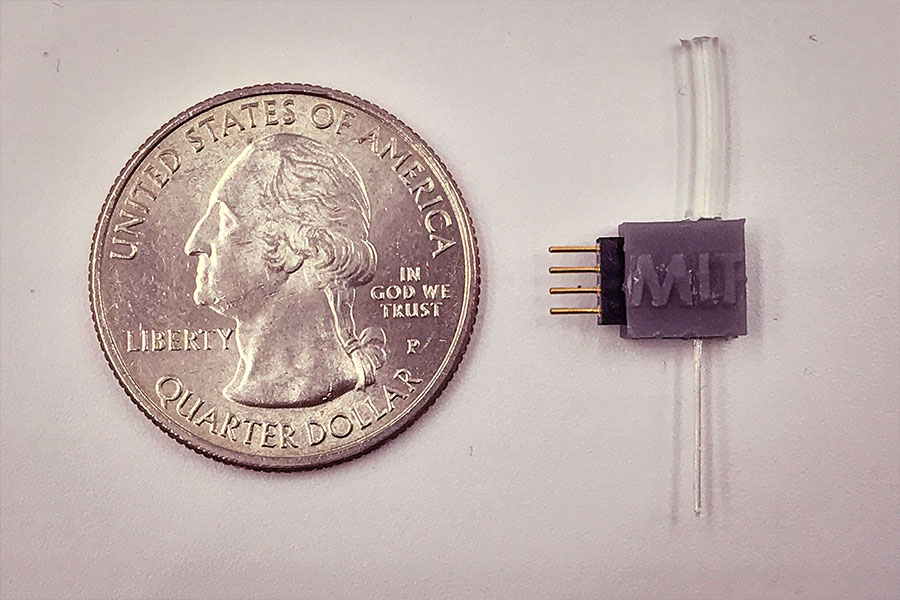
To miniaturize the electrocatalytic cell to the scale of biological cells, the team has created custom fibers containing the positive and negative microelectrodes, which are coated with the iron sulfide nanoparticles, and a microfluidic channel for the delivery of sodium nitrite, the precursor material. When implanted in the brain, these fibers direct the precursor to the specific neurons. Then the reaction can be activated at will electrochemically, through the electrodes in the same fiber, producing an instant burst of nitric oxide right at that spot so that its effects can be recorded in real-time.

Device created by the Anikeeva lab. The tube at top is connected to a supply of the precursor material, sodium nitrite, which then passes through a channel in the fiber at the bottom and into the body, which also contains the electrodes to stimulate the release of nitric oxide. The electrodes are connected through the four-pin connector on the left.
Photo: Anikeeva Lab

As a test, they used the system in a rodent model to activate a brain region that is known to be a reward center for motivation and social interaction, and that plays a role in addiction. They showed that it did indeed provoke the expected signaling responses, demonstrating its effectiveness.

Anikeeva says this “would be a very useful biological research platform, because finally, people will have a way to study the role of nitric oxide at the level of single cells, in whole organisms that are performing tasks.” She points out that there are certain disorders that are associated with disruptions of the nitric oxide signaling pathway, so more detailed studies of how this pathway operates could help lead to treatments.

The method could be generalizable, Park says, as a way of producing other molecules of biological interest within an organism. “Essentially we can now have this really scalable and miniaturized way to generate many molecules, as long as we find the appropriate catalyst, and as long as we find an appropriate starting compound that is also safe.” This approach to generating signaling molecules in situ could have wide applications in biomedicine, he says.

“One of our reviewers for this manuscript pointed out that this has never been done — electrolysis in a biological system has never been leveraged to control biological function,” Anikeeva says. “So, this is essentially the beginning of a field that could potentially be very useful” to study molecules that can be delivered at precise locations and times, for studies in neurobiology or any other biological functions. That ability to make molecules on demand inside the body could be useful in fields such as immunology or cancer research, she says.

The project got started as a result of a chance conversation between Park and Jin, who were friends working in different fields — neurobiology and electrochemistry. Their initial casual discussions ended up leading to a full-blown collaboration between several departments. But in today’s locked-down world, Jin says, such chance encounters and conversations have become less likely. “In the context of how much the world has changed, if this were in this era in which we’re all apart from each other, and not in 2018, there is some chance that this collaboration may just not ever have happened.”

“This work is a milestone in bioelectronics,” says Bozhi Tian, an associate professor of chemistry at the University of Chicago, who was not connected to this work. “It integrates nanoenabled catalysis, microfluidics, and traditional bioelectronics … and it solves a longstanding challenge of precise neuromodulation in the brain, by in situ generation of signaling molecules. This approach can be widely adopted by the neuroscience community and can be generalized to other signaling systems, too.”

Besides MIT, the team included researchers at National Chiao Tung University in Taiwan, NEC Corporation in Japan, and the Weizman Institute of Science in Israel. The work was supported by the National Institute for Neurological Disorders and Stroke, the National Institutes of Health, the National Science Foundation, and MIT’s Department of Chemical Engineering.

A focused approach to imaging neural activity in the brain

When neurons fire an electrical impulse, they also experience a surge of calcium ions. By measuring those surges, researchers can indirectly monitor neuron activity, helping them to study the role of individual neurons in many different brain functions.

One drawback to this technique is the crosstalk generated by the axons and dendrites that extend from neighboring neurons, which makes it harder to get a distinctive signal from the neuron being studied. MIT engineers have now developed a way to overcome that issue, by creating calcium indicators, or sensors, that accumulate only in the body of a neuron.

“People are using calcium indicators for monitoring neural activity in many parts of the brain,” says Edward Boyden, the Y. Eva Tan Professor in Neurotechnology and a professor of biological engineering and of brain and cognitive sciences at MIT. “Now they can get better results, obtaining more accurate neural recordings that are less contaminated by crosstalk.”

To achieve this, the researchers fused a commonly used calcium indicator called GCaMP to a short peptide that targets it to the cell body. The new molecule, which the researchers call SomaGCaMP, can be easily incorporated into existing workflows for calcium imaging, the researchers say.

Boyden is the senior author of the study, which appears today in Neuron. The paper’s lead authors are Research Scientist Or Shemesh, postdoc Changyang Linghu, and former postdoc Kiryl Piatkevich.

Molecular focus

The GCaMP calcium indicator consists of a fluorescent protein attached to a calcium-binding protein called calmodulin, and a calmodulin-binding protein called M13 peptide. GCaMP fluoresces when it binds to calcium ions in the brain, allowing researchers to indirectly measure neuron activity.

“Calcium is easy to image, because it goes from a very low concentration inside the cell to a very high concentration when a neuron is active,” says Boyden, who is also a member of MIT’s McGovern Institute for Brain Research, Media Lab, and Koch Institute for Integrative Cancer Research.

The simplest way to detect these fluorescent signals is with a type of imaging called one-photon microscopy. This is a relatively inexpensive technique that can image large brain samples at high speed, but the downside is that it picks up crosstalk between neighboring neurons. GCaMP goes into all parts of a neuron, so signals from the axons of one neuron can appear as if they are coming from the cell body of a neighbor, making the signal less accurate.

A more expensive technique called two-photon microscopy can partly overcome this by focusing light very narrowly onto individual neurons, but this approach requires specialized equipment and is also slower.

Boyden’s lab decided to take a different approach, by modifying the indicator itself, rather than the imaging equipment.

“We thought, rather than optically focusing light, what if we molecularly focused the indicator?” he says. “A lot of people use hardware, such as two-photon microscopes, to clean up the imaging. We’re trying to build a molecular version of what other people do with hardware.”

In a related paper that was published last year, Boyden and his colleagues used a similar approach to reduce crosstalk between fluorescent probes that directly image neurons’ membrane voltage. In parallel, they decided to try a similar approach with calcium imaging, which is a much more widely used technique.

To target GCaMP exclusively to cell bodies of neurons, the researchers tried fusing GCaMP to many different proteins. They explored two types of candidates — naturally occurring proteins that are known to accumulate in the cell body, and human-designed peptides — working with MIT biology Professor Amy Keating, who is also an author of the paper. These synthetic proteins are coiled-coil proteins, which have a distinctive structure in which multiple helices of the proteins coil together.

Less crosstalk

The researchers screened about 30 candidates in neurons grown in lab dishes, and then chose two — one artificial coiled-coil and one naturally occurring peptide — to test in animals. Working with Misha Ahrens, who studies zebrafish at the Janelia Research Campus, they found that both proteins offered significant improvements over the original version of GCaMP. The signal-to-noise ratio — a measure of the strength of the signal compared to background activity — went up, and activity between adjacent neurons showed reduced correlation.

In studies of mice, performed in the lab of Xue Han at Boston University, the researchers also found that the new indicators reduced the correlations between activity of neighboring neurons. Additional studies using a miniature microscope (called a microendoscope), performed in the lab of Kay Tye at the Salk Institute for Biological Studies, revealed a significant increase in signal-to-noise ratio with the new indicators.

“Our new indicator makes the signals more accurate. This suggests that the signals that people are measuring with regular GCaMP could include crosstalk,” Boyden says. “There’s the possibility of artifactual synchrony between the cells.”

In all of the animal studies, they found that the artificial, coiled-coil protein produced a brighter signal than the naturally occurring peptide that they tested. Boyden says it’s unclear why the coiled-coil proteins work so well, but one possibility is that they bind to each other, making them less likely to travel very far within the cell.

Boyden hopes to use the new molecules to try to image the entire brains of small animals such as worms and fish, and his lab is also making the new indicators available to any researchers who want to use them.

“It should be very easy to implement, and in fact many groups are already using it,” Boyden says. “They can just use the regular microscopes that they already are using for calcium imaging, but instead of using the regular GCaMP molecule, they can substitute our new version.”

The research was primarily funded by the National Institute of Mental Health and the National Institute of Drug Abuse, as well as a Director’s Pioneer Award from the National Institutes of Health, and by Lisa Yang, John Doerr, the HHMI-Simons Faculty Scholars Program, and the Human Frontier Science Program.

Three from MIT awarded 2020 Guggenheim Fellowships

MIT faculty members Sabine Iatridou, Jonathan Gruber, and Rebecca Saxe are among 175 scientists, artists, and scholars awarded 2020 fellowships from the John Simon Guggenheim Foundation. Appointed on the basis of prior achievement and exceptional promise, the 2020 Guggenheim Fellows were selected from almost 3,000 applicants.

“It’s exceptionally encouraging to be able to share such positive news at this terribly challenging time” says Edward Hirsch, president of the foundation. “A Guggenheim Fellowship has always offered practical assistance, helping fellows do their work, but for many of the new fellows, it may be a lifeline at a time of hardship, a survival tool as well as a creative one.”

Since 1925, the foundation has granted more the $375 million in fellowships to over 18,000 individuals, including Nobel laureates, Fields medalists, poets laureate, and winners of the Pulitzer Prize, among other internationally recognized honors. This year’s MIT recipients include a linguist, an economist, and a cognitive neuroscientist.

Rebecca Saxe is an associate investigator of the McGovern Institute and the John W. Jarve (1978) Professor in Brain and Cognitive Sciences. She studies human social cognition, using a combination of behavioral testing and brain imaging technologies. She is best known for her work on brain regions specialized for abstract concepts such as “theory of mind” tasks that involve understanding the mental states of other people. She also studies the development of the human brain during early infancy. She obtained her PhD from MIT and was a Harvard University junior fellow before joining the MIT faculty in 2006. Saxe was chosen in 2012 as a Young Global Leader by the World Economic Forum, and she received the 2014 Troland Award from the National Academy of Sciences. Her TED Talk, “How we read each other’s minds” has been viewed over 3 million times.

Jonathan Gruber is the Ford Professor of Economics at MIT, the director of the Health Care Program at the National Bureau of Economic Research, and the former president of the American Society of Health Economists. He has published more than 175 research articles, has edited six research volumes, and is the author of “Public Finance and Public Policy,” a leading undergraduate text; “Health Care Reform,” a graphic novel; and “Jump-Starting America: How Breakthrough Science Can Revive Economic Growth and the American Dream.” In 2006 he received the American Society of Health Economists Inaugural Medal for the best health economist in the nation aged 40 and under. He served as deputy sssistant secretary for economic policy at the U.S. Department of the Treasury. He was a key architect of Massachusetts’ ambitious health reform effort, and became an inaugural member of the Health Connector Board, the main implementing body for that effort. He served as a technical consultant to the Obama administration and worked with both the administration and Congress to help craft the Affordable Care Act. In 2011, he was named “One of the Top 25 Most Innovative and Practical Thinkers of Our Time” by Slate magazine.

Sabine Iatridou is professor of linguistics in MIT’s Department of Linguistics and Philosophy. Her work focuses on syntax and the syntax-semantics interface, as well as comparative linguistics. She is the author and coauthor of a series of innovative papers about tense and modality that opened up whole new domains of research for the field. Since those publications, she has made foundational contributions to many branches of linguistics that connect form with meaning. She is the recipient of the National Young Investigator Award (USA), of an honorary doctorate from the University of Crete in Greece, and of an award from the Royal Dutch Academy of Sciences. She was elected fellow of the Linguistic Society of America. She is co-founder and co-director of the CreteLing Summer School of Linguistics.

“As we grapple with the difficulties of the moment, it is also important to look to the future,” says Hirsch. “The artists, writers, scholars, and scientific researchers supported by the fellowship will help us understand and learn from what we are enduring individually and collectively, and it is an honor for the foundation to help them do their essential work.”

Researchers achieve remote control of hormone release

Abnormal levels of stress hormones such as adrenaline and cortisol are linked to a variety of mental health disorders, including depression and posttraumatic stress disorder (PTSD). MIT researchers have now devised a way to remotely control the release of these hormones from the adrenal gland, using magnetic nanoparticles.

This approach could help scientists to learn more about how hormone release influences mental health, and could eventually offer a new way to treat hormone-linked disorders, the researchers say.

“We’re looking how can we study and eventually treat stress disorders by modulating peripheral organ function, rather than doing something highly invasive in the central nervous system,” says Polina Anikeeva, an MIT professor of materials science and engineering and of brain and cognitive sciences.

To achieve control over hormone release, Dekel Rosenfeld, an MIT-Technion postdoc in Anikeeva’s group, has developed specialized magnetic nanoparticles that can be injected into the adrenal gland. When exposed to a weak magnetic field, the particles heat up slightly, activating heat-responsive channels that trigger hormone release. This technique can be used to stimulate an organ deep in the body with minimal invasiveness.

Anikeeva and Alik Widge, an assistant professor of psychiatry at the University of Minnesota and a former research fellow at MIT’s Picower Institute for Learning and Memory, are the senior authors of the study. Rosenfeld is the lead author of the paper, which appears today in Science Advances.

Controlling hormones

Anikeeva’s lab has previously devised several novel magnetic nanomaterials, including particles that can release drugs at precise times in specific locations in the body.

In the new study, the research team wanted to explore the idea of treating disorders of the brain by manipulating organs that are outside the central nervous system but influence it through hormone release. One well-known example is the hypothalamic-pituitary-adrenal (HPA) axis, which regulates stress response in mammals. Hormones secreted by the adrenal gland, including cortisol and adrenaline, play important roles in depression, stress, and anxiety.

“Some disorders that we consider neurological may be treatable from the periphery, if we can learn to modulate those local circuits rather than going back to the global circuits in the central nervous system,” says Anikeeva, who is a member of MIT’s Research Laboratory of Electronics and McGovern Institute for Brain Research.

As a target to stimulate hormone release, the researchers decided on ion channels that control the flow of calcium into adrenal cells. Those ion channels can be activated by a variety of stimuli, including heat. When calcium flows through the open channels into adrenal cells, the cells begin pumping out hormones. “If we want to modulate the release of those hormones, we need to be able to essentially modulate the influx of calcium into adrenal cells,” Rosenfeld says.

Unlike previous research in Anikeeva’s group, in this study magnetothermal stimulation was applied to modulate the function of cells without artificially introducing any genes.

To stimulate these heat-sensitive channels, which naturally occur in adrenal cells, the researchers designed nanoparticles made of magnetite, a type of iron oxide that forms tiny magnetic crystals about 1/5000 the thickness of a human hair. In rats, they found these particles could be injected directly into the adrenal glands and remain there for at least six months. When the rats were exposed to a weak magnetic field — about 50 millitesla, 100 times weaker than the fields used for magnetic resonance imaging (MRI) — the particles heated up by about 6 degrees Celsius, enough to trigger the calcium channels to open without damaging any surrounding tissue.

The heat-sensitive channel that they targeted, known as TRPV1, is found in many sensory neurons throughout the body, including pain receptors. TRPV1 channels can be activated by capsaicin, the organic compound that gives chili peppers their heat, as well as by temperature. They are found across mammalian species, and belong to a family of many other channels that are also sensitive to heat.

This stimulation triggered a hormone rush — doubling cortisol production and boosting noradrenaline by about 25 percent. That led to a measurable increase in the animals’ heart rates.

Treating stress and pain

The researchers now plan to use this approach to study how hormone release affects PTSD and other disorders, and they say that eventually it could be adapted for treating such disorders. This method would offer a much less invasive alternative to potential treatments that involve implanting a medical device to electrically stimulate hormone release, which is not feasible in organs such as the adrenal glands that are soft and highly vascularized, the researchers say.

Another area where this strategy could hold promise is in the treatment of pain, because heat-sensitive ion channels are often found in pain receptors.

“Being able to modulate pain receptors with this technique potentially will allow us to study pain, control pain, and have some clinical applications in the future, which hopefully may offer an alternative to medications or implants for chronic pain,” Anikeeva says. With further investigation of the existence of TRPV1 in other organs, the technique can potentially be extended to other peripheral organs such as the digestive system and the pancreas.

The research was funded by the U.S. Defense Advance Research Projects Agency ElectRx Program, a Bose Research Grant, the National Institutes of Health BRAIN Initiative, and a MIT-Technion fellowship.

How dopamine drives brain activity

Using a specialized magnetic resonance imaging (MRI) sensor, MIT neuroscientists have discovered how dopamine released deep within the brain influences both nearby and distant brain regions.

Dopamine plays many roles in the brain, most notably related to movement, motivation, and reinforcement of behavior. However, until now it has been difficult to study precisely how a flood of dopamine affects neural activity throughout the brain. Using their new technique, the MIT team found that dopamine appears to exert significant effects in two regions of the brain’s cortex, including the motor cortex.

“There has been a lot of work on the immediate cellular consequences of dopamine release, but here what we’re looking at are the consequences of what dopamine is doing on a more brain-wide level,” says Alan Jasanoff, an MIT professor of biological engineering, brain and cognitive sciences, and nuclear science and engineering. Jasanoff is also an associate member of MIT’s McGovern Institute for Brain Research and the senior author of the study.

The MIT team found that in addition to the motor cortex, the remote brain area most affected by dopamine is the insular cortex. This region is critical for many cognitive functions related to perception of the body’s internal states, including physical and emotional states.

MIT postdoc Nan Li is the lead author of the study, which appears today in Nature.

Tracking dopamine

Like other neurotransmitters, dopamine helps neurons to communicate with each other over short distances. Dopamine holds particular interest for neuroscientists because of its role in motivation, addiction, and several neurodegenerative disorders, including Parkinson’s disease. Most of the brain’s dopamine is produced in the midbrain by neurons that connect to the striatum, where the dopamine is released.

For many years, Jasanoff’s lab has been developing tools to study how molecular phenomena such as neurotransmitter release affect brain-wide functions. At the molecular scale, existing techniques can reveal how dopamine affects individual cells, and at the scale of the entire brain, functional magnetic resonance imaging (fMRI) can reveal how active a particular brain region is. However, it has been difficult for neuroscientists to determine how single-cell activity and brain-wide function are linked.

“There have been very few brain-wide studies of dopaminergic function or really any neurochemical function, in large part because the tools aren’t there,” Jasanoff says. “We’re trying to fill in the gaps.”

About 10 years ago, his lab developed MRI sensors that consist of magnetic proteins that can bind to dopamine. When this binding occurs, the sensors’ magnetic interactions with surrounding tissue weaken, dimming the tissue’s MRI signal. This allows researchers to continuously monitor dopamine levels in a specific part of the brain.

In their new study, Li and Jasanoff set out to analyze how dopamine released in the striatum of rats influences neural function both locally and in other brain regions. First, they injected their dopamine sensors into the striatum, which is located deep within the brain and plays an important role in controlling movement. Then they electrically stimulated a part of the brain called the lateral hypothalamus, which is a common experimental technique for rewarding behavior and inducing the brain to produce dopamine.

Then, the researchers used their dopamine sensor to measure dopamine levels throughout the striatum. They also performed traditional fMRI to measure neural activity in each part of the striatum. To their surprise, they found that high dopamine concentrations did not make neurons more active. However, higher dopamine levels did make the neurons remain active for a longer period of time.

“When dopamine was released, there was a longer duration of activity, suggesting a longer response to the reward,” Jasanoff says. “That may have something to do with how dopamine promotes learning, which is one of its key functions.”

Long-range effects

After analyzing dopamine release in the striatum, the researchers set out to determine this dopamine might affect more distant locations in the brain. To do that, they performed traditional fMRI imaging on the brain while also mapping dopamine release in the striatum. “By combining these techniques we could probe these phenomena in a way that hasn’t been done before,” Jasanoff says.

The regions that showed the biggest surges in activity in response to dopamine were the motor cortex and the insular cortex. If confirmed in additional studies, the findings could help researchers understand the effects of dopamine in the human brain, including its roles in addiction and learning.

“Our results could lead to biomarkers that could be seen in fMRI data, and these correlates of dopaminergic function could be useful for analyzing animal and human fMRI,” Jasanoff says.

The research was funded by the National Institutes of Health and a Stanley Fahn Research Fellowship from the Parkinson’s Disease Foundation.

How the brain encodes landmarks that help us navigate

When we move through the streets of our neighborhood, we often use familiar landmarks to help us navigate. And as we think to ourselves, “OK, now make a left at the coffee shop,” a part of the brain called the retrosplenial cortex (RSC) lights up.

While many studies have linked this brain region with landmark-based navigation, exactly how it helps us find our way is not well-understood. A new study from MIT neuroscientists now reveals how neurons in the RSC use both visual and spatial information to encode specific landmarks.

“There’s a synthesis of some of these signals — visual inputs and body motion — to represent concepts like landmarks,” says Mark Harnett, an assistant professor of brain and cognitive sciences and a member of MIT’s McGovern Institute for Brain Research. “What we went after in this study is the neuron-level and population-level representation of these different aspects of spatial navigation.”

In a study of mice, the researchers found that this brain region creates a “landmark code” by combining visual information about the surrounding environment with spatial feedback of the mice’s own position along a track. Integrating these two sources of information allowed the mice to learn where to find a reward, based on landmarks that they saw.

“We believe that this code that we found, which is really locked to the landmarks, and also gives the animals a way to discriminate between landmarks, contributes to the animals’ ability to use those landmarks to find rewards,” says Lukas Fischer, an MIT postdoc and the lead author of the study.

Harnett is the senior author of the study, which appears today in the journal eLife. Other authors are graduate student Raul Mojica Soto-Albors and recent MIT graduate Friederike Buck.

Encoding landmarks

Previous studies have found that people with damage to the RSC have trouble finding their way from one place to another, even though they can still recognize their surroundings. The RSC is also one of the first areas affected in Alzheimer’s patients, who often have trouble navigating.

The RSC is wedged between the primary visual cortex and the motor cortex, and it receives input from both of those areas. It also appears to be involved in combining two types of representations of space — allocentric, meaning the relationship of objects to each other, and egocentric, meaning the relationship of objects to the viewer.

“The evidence suggests that RSC is really a place where you have a fusion of these different frames of reference,” Harnett says. “Things look different when I move around in the room, but that’s because my vantage point has changed. They’re not changing with respect to one another.”

In this study, the MIT team set out to analyze the behavior of individual RSC neurons in mice, including how they integrate multiple inputs that help with navigation. To do that, they created a virtual reality environment for the mice by allowing them to run on a treadmill while they watch a video screen that makes it appear they are running along a track. The speed of the video is determined by how fast the mice run.

At specific points along the track, landmarks appear, signaling that there’s a reward available a certain distance beyond the landmark. The mice had to learn to distinguish between two different landmarks, and to learn how far beyond each one they had to run to get the reward.

Once the mice learned the task, the researchers recorded neural activity in the RSC as the animals ran along the virtual track. They were able to record from a few hundred neurons at a time, and found that most of them anchored their activity to a specific aspect of the task.

There were three primary anchoring points: the beginning of the trial, the landmark, and the reward point. The majority of the neurons were anchored to the landmarks, meaning that their activity would consistently peak at a specific point relative to the landmark, say 50 centimeters before it or 20 centimeters after it.

Most of those neurons responded to both of the landmarks, but a small subset responded to only one or the other. The researchers hypothesize that those strongly selective neurons help the mice to distinguish between the landmarks and run the correct distance to get the reward.

When the researchers used optogenetics (a tool that can turn off neuron activity) to block activity in the RSC, the mice’s performance on the task became much worse.

Combining inputs

The researchers also did an experiment in which the mice could choose to run or not while the video played at a constant speed, unrelated to the mice’s movement. The mice could still see the landmarks, but the location of the landmarks was no longer linked to a reward or to the animals’ own behavior. In that situation, RSC neurons did respond to the landmarks, but not as strongly as they did when the mice were using them for navigation.

Further experiments allowed the researchers to tease out just how much neuron activation is produced by visual input (seeing the landmarks) and by feedback on the mouse’s own movement. However, simply adding those two numbers yielded totals much lower than the neuron activity seen when the mice were actively navigating the track.

“We believe that is evidence for a mechanism of nonlinear integration of these inputs, where they get combined in a way that creates a larger response than what you would get if you just added up those two inputs in a linear fashion,” Fischer says.

The researchers now plan to analyze data that they have already collected on how neuron activity evolves over time as the mice learn the task. They also hope to perform further experiments in which they could try to separately measure visual and spatial inputs into different locations within RSC neurons.

The research was funded by the National Institutes of Health, the McGovern Institute, the NEC Corporation Fund for Research in Computers and Communications at MIT, and the Klingenstein-Simons Fellowship in Neuroscience.