Making and breaking habits

As part of our Ask the Brain series, science writer Shafaq Zia explores the question, “How are habits formed in the brain?”

____

Have you ever wondered why it is so hard to break free of bad habits like nail biting or obsessive social networking?

When we repeat an action over and over again, the behavioral pattern becomes automated in our brain, according to Jill R. Crittenden, molecular biologist and scientific advisor at McGovern Institute for Brain Research at MIT. For over a decade, Crittenden worked as a research scientist in the lab of Ann Graybiel, where one of the key questions scientists are working to answer is, how are habits formed?

Making habits

To understand how certain actions get wired in our neural pathways, this team of McGovern researchers experimented with rats, who were trained to run down a maze to receive a reward. If they turned left, they would get rich chocolate milk and for turning right, only sugar water. With this, the scientists wanted to see whether these animals could “learn to associate a cue with which direction they should turn in the maze in order to get the chocolate milk reward.”

Over time, the rats grew extremely habitual in their behavior; “they always turned the the correct direction and the places where their paws touched, in a fairly long maze, were exactly the same every time,” said Crittenden.

This isn’t a coincidence. When we’re first learning to do something, the frontal lobe and basal ganglia of the brain are highly active and doing a lot of calculations. These brain regions work together to associate behaviors with thoughts, emotions, and, most importantly, motor movements. But when we repeat an action over and over again, like the rats running down the maze, our brains become more efficient and fewer neurons are required to achieve the goal. This means, the more you do something, the easier it gets to carry it out because the behavior becomes literally etched in our brain as our motor movements.

But habits are complicated and they come in many different flavors, according to Crittenden. “I think we don’t have a great handle on how the differences [in our many habits] are separable neurobiologically, and so people argue a lot about how do you know that something’s a habit.”

The easiest way for scientists to test this in rodents is to see if the animal engages in the behavior even in the absence of reward. In this particular experiment, the researchers take away the reward, chocolate milk, to see whether the rats continue to run down the maze correctly. And to take it even a step further, they mix the chocolate milk with lithium chloride, which would upset the rat’s stomach. Despite all this, the rats continue to run down the maze and turn left towards the chocolate milk, as they had learnt to do over and over again.

Breaking habits

So does that mean once a habit is formed, it is impossible to shake it? Not quite. But it is tough. Rewards are a key building block to forming habits because our dopamine levels surge when we learn that an action is unexpectedly rewarded. For example, when the rats first learn to run down the maze, they’re motivated to receive the chocolate milk.

But things get complicated once the habit is formed. Researchers have found that this dopamine surge in response to reward ceases after a behavior becomes a habit. Instead the brain begins to release dopamine at the first cue or action that was previously learned to lead to the reward, so we are motivated to engage in the full behavioral sequence anyway, even if the reward isn’t there anymore.

This means we don’t have as much self-control as we think we do, which may also be the reason why it’s so hard to break the cycle of addiction. “People will report that they know this is bad for them. They don’t want it. And nevertheless, they select that action,” said Crittenden.

One common method to break the behavior, in this case, is called extinction. This is where psychologists try to weaken the association between the cue and the reward that led to habit formation in the first place. For example, if the rat no longer associates the cue to run down the maze with a reward, it will stop engaging in that behavior.

So the next time you beat yourself up over being unable to stick to a diet or sleep at a certain time, give yourself some grace and know that with consistency, a new, healthier habit can be born.

Developing brain needs cannabinoid receptors after birth

Doctors warn that marijuana use during pregnancy may have harmful effects on the development of a fetus, in part because the cannabinoid receptors activated by the drug are known be critical for enabling a developing brain to wire up properly. Now, scientists at MIT’s McGovern Institute have learned that cannabinoid receptors’ critical role in brain development does not end at birth.

In today’s online issue of the journal eNeuro, scientists led by McGovern investigator Ann Graybiel report that mice need the cannabinoid receptor CB1R to establish connections within the brain’s dopamine system that take shape soon after birth. The finding raises concern that marijuana use by nursing moms, who pass the CB1R-activating compound THC to their infants when they breastfeed, might interfere with brain development by disrupting cannabinoid signaling.

“This is a real change to one of the truly important systems in the brain—a major controller of our dopamine,” Graybiel says. Dopamine exerts a powerful influence over our motivations and behavior, and changes to the dopamine system contribute to disorders from Parkinson’s disease to addiction. Thus, the researchers say, it is vital to understand whether postnatal drug exposure might put developing dopamine circuits at risk.

Brain bouquets

Cannabinoid receptors in the brain are important mediators of mood, memory, and pain. Graybiel’s lab became interested in CB1R due to their dysregulation in Huntington’s and Parkinson’s diseases, both of which impair the brain’s ability to control movement and other functions. While investigating the receptor’s distribution in the brain, they discovered that in the adult mice, CB1R is abundant within small compartments within the striatum called striosomes. The receptor was particularly concentrated within the neurons that connect striosomes to a dopamine-rich area of the brain called the substantia nigra, via structures that Graybiel’s team has dubbed striosome-dendron bouquets.

Striosome-dendron bouquets are easy to overlook within the densely connected network of the brain. But when the cells that make up the bouquets are labeled with a fluorescent protein, the bouquets become visible—and their appearance is striking, says Jill Crittenden, a research scientist in Graybiel’s lab.

Striosomal neurons form these bouquets by reaching into the substantia nigra, whose cells use dopamine to influence movement, motivation, learning, and habit formation. Clusters of dopamine-producing neurons form dendrites there that intertwine tightly with incoming axons from the striosomal neurons. The resulting structures, whose intimately associated cells resemble the bundled stems of a floral bouquet, establish so many connections that they give striosomal neurons potent control over dopamine signaling.

By tracking the bouquets’ emergence in newborn mice, Graybiel’s team found that they form in the first week after birth, a period during which striosomal neurons are ramping up production of CB1R. Mice genetically engineered to lack CB1R, however, can’t make these elaborate but orderly bouquets. Without the receptor, fibers from striosomes extend into the substantia nigra, but fail to form the tightly intertwined “bouquet stems” that facilitate extensive connections with their targets. This disorganized structure is apparent as soon as bouquets arise in the brains of young pups and persists into adulthood. “There aren’t those beautiful, strong fibers anymore,” Crittenden says. “This suggests that those very strong controllers over the dopamine system function abnormally when you interfere with cannabinoid signaling.”

The finding was a surprise. Without zeroing in on striosome-dendron bouquets, it would be easy to miss CB1R’s impact on the dopamine system, Crittenden says. Plus, she adds, prior studies of the receptor’s role in development largely focused on fetal development. The new findings reveal that the cannabinoid system continues to guide the formation of brain circuits after birth.

Graybiel notes that funds from generous donors, including the Broderick Fund for Phytocannabinoid Research at MIT, the Saks Kavanaugh Foundation, the Kristin R. Pressman and Jessica J. Pourian ‘13 Fund, Mr. Robert Buxton, and the William N. & Bernice E. Bumpus Foundation, enabled her team’s studies of CB1R’s role in shaping striosome-dendron bouquets.

Now that they have shown that CB1R is needed for postnatal brain development, it will be important to determine the consequences of disrupting cannabinoid signaling during this critical period—including whether passing THC to a nursing baby impacts the brain’s dopamine system.

On a mission to alleviate chronic pain

About 50 million Americans suffer from chronic pain, which interferes with their daily life, social interactions, and ability to work. MIT Professor Fan Wang wants to develop new ways to help relieve that pain, by studying and potentially modifying the brain’s own pain control mechanisms.

Her recent work has identified an “off switch” for pain, located in the brain’s amygdala. She hopes that finding ways to control this switch could lead to new treatments for chronic pain.

“Chronic pain is a major societal issue,” Wang says. “By studying pain-suppression neurons in the brain’s central amygdala, I hope to create a new therapeutic approach for alleviating pain.”

Wang, who joined the MIT faculty in January 2021, is also the leader of a new initiative at the McGovern Institute for Brain Research that is studying drug addiction, with the goal of developing more effective treatments for addiction.

“Opioid prescription for chronic pain is a major contributor to the opioid epidemic. With the Covid pandemic, I think addiction and overdose are becoming worse. People are more anxious, and they seek drugs to alleviate such mental pain,” Wang says. “As scientists, it’s our duty to tackle this problem.”

Sensory circuits

Wang, who grew up in Beijing, describes herself as “a nerdy child” who loved books and math. In high school, she took part in science competitions, then went on to study biology at Tsinghua University. She arrived in the United States in 1993 to begin her PhD at Columbia University. There, she worked on tracing the connection patterns of olfactory receptor neurons in the lab of Richard Axel, who later won the Nobel Prize for his discoveries of odorant receptors and how the olfactory system is organized.

After finishing her PhD, Wang decided to switch gears. As a postdoc at the University of California at San Francisco and then Stanford University, she began studying how the brain perceives touch.

In 2003, Wang joined the faculty at Duke University School of Medicine. There, she began developing techniques to study the brain circuits that underlie the sense of touch, tracing circuits that carry sensory information from the whiskers of mice to the brain. She also studied how the brain integrates movements of touch organs with signals of sensory stimuli to generate perception (such as using stretching movements to sense elasticity).

As she pursued her sensory perception studies, Wang became interested in studying pain perception, but she felt she needed to develop new techniques to tackle it. While at Duke, she invented a technique called CANE (capturing activated neural ensembles), which can identify networks of neurons that are activated by a particular stimulus.

Using this approach in mice, she identified neurons that become active in response to pain, but so many neurons across the brain were activated that it didn’t offer much useful information. As a way to indirectly get at how the brain controls pain, she decided to use CANE to explore the effects of drugs used for general anesthesia. During general anesthesia, drugs render a patient unconscious, but Wang hypothesized that the drugs might also shut off pain perception.

“At that time, it was just a wild idea,” Wang recalls. “I thought there may be other mechanisms — that instead of just a loss of consciousness, anesthetics may do something to the brain that actually turns pain off.”

Support for the existence of an “off switch” for pain came from the observation that wounded soldiers on a battlefield can continue to fight, essentially blocking out pain despite their injuries.

In a study of mice treated with anesthesia drugs, Wang discovered that the brain does have this kind of switch, in an unexpected location: the amygdala, which is involved in regulating emotion. She showed that this cluster of neurons can turn off pain when activated, and when it is suppressed, mice become highly sensitive to ordinary gentle touch.

“There’s a baseline level of activity that makes the animals feel normal, and when you activate these neurons, they’ll feel less pain. When you silence them, they’ll feel more pain,” Wang says.

Turning off pain

That finding, which Wang reported in 2020, raised the possibility of somehow modulating that switch in humans to try to treat chronic pain. This is a long-term goal of Wang’s, but more work is required to achieve it, she says. Currently her lab is working on analyzing the RNA expression patterns of the neurons in the cluster she identified. They also are measuring the neurons’ electrical activity and how they interact with other neurons in the brain, in hopes of identifying circuits that could be targeted to tamp down the perception of pain.

One way of modulating these circuits could be to use deep brain stimulation, which involves implanting electrodes in certain areas of the brain. Focused ultrasound, which is still in early stages of development and does not require surgery, could be a less invasive alternative.

Another approach Wang is interested in exploring is pairing brain stimulation with a context such as looking at a smartphone app. This kind of pairing could help train the brain to shut off pain using the app, without the need for the original stimulation (deep brain stimulation or ultrasound).

“Maybe you don’t need to constantly stimulate the brain. You may just need to reactivate it with a context,” Wang says. “After a while you would probably need to be restimulated, or reconditioned, but at least you have a longer window where you don’t need to go to the hospital for stimulation, and you just need to use a context.”

Wang, who was drawn to MIT in part by its focus on fostering interdisciplinary collaborations, is now working with several other McGovern Institute members who are taking different angles to try to figure out how the brain generates the state of craving that occurs in drug addiction, including opioid addiction.

“We’re going to focus on trying to understand this craving state: how it’s created in the brain and how can we sort of erase that trace in the brain, or at least control it. And then you can neuromodulate it in real time, for example, and give people a chance to get back their control,” she says.

A new approach to curbing cocaine use

Cocaine, opioids, and other drugs of abuse disrupt the brain’s reward system, often shifting users’ priorities to obtaining more drug above all else. For people battling addiction, this persistent craving is notoriously difficult to overcome—but new research from scientists at MIT’s McGovern Institute and collaborators points toward a therapeutic strategy that could help.

Researchers in MIT Institute Professor Ann Graybiel’s lab and collaborators at the University of Copenhagen and Vanderbilt University report in a January 25, 2022 online publication in the journal Addiction Biology that activating a signaling molecule in the brain known as muscarinic receptor 4 (M4) causes rodents to reduce cocaine self-administration and simultaneously choose a food treat over cocaine.

M4 receptors are found on the surface of neurons in the brain, where they alter signaling in response to the neurotransmitter acetylcholine. They are plentiful in the striatum, a brain region that Graybiel’s lab has shown is deeply involved in habit formation. They are of interest to addiction researchers because, along with a related receptor called M1, which is also abundant in the striatum, they often seem to act in opposition to the neurotransmitter dopamine.

Drugs of abuse stimulate the brain’s habit circuits by allowing dopamine to build up in the brain. With chronic use, that circuitry can become less sensitive to dopamine, so experiences that were once rewarding become less pleasurable and users are driven to seek higher doses of their drug. Attempts to directly block the dopamine system have not been found to be an effective way of treating addiction and can have unpleasant or dangerous side-effects, so researchers are seeking an alternative strategy to restore balance within the brain’s reward circuitry. “Another way to tweak that system is to activate these muscarinic receptors,” explains Jill Crittenden, a research scientist in the Graybiel lab.

New pathways to treatment

At the University of Copenhagen, neuroscientist Morgane Thomsen has found that activating the M1 receptor causes rodents to choose a food treat over cocaine. In the new work, she showed that a drug that selectively activates the M4 receptor has a similar effect.

When rats that have been trained to self-administer cocaine are given an M4-activating compound, they immediately reduce their drug use, actively choosing food instead. Thomsen found that this effect grew stronger over a seven-day course of treatment, with cocaine use declining day by day. When the M4-activating treatment was stopped, rats quickly resumed their prior cocaine-seeking behavior.

While Thomsen’s experiments have now shown that animals’ cocaine use can be reduced by activating either M1 or M4, it’s clear that the two muscarinic receptors don’t modulate cocaine use in the same way. M1 activation works on a different time scale, taking some time to kick in, but leaving some lasting effects even after the treatment has been discontinued.

Experiments with genetically modified mice developed in Graybiel’s lab confirm that the two receptors influence drug-seeking behavior via different molecular pathways. Previously, the team discovered that activating M1 has no effect on cocaine-seeking in mice that lack a signaling molecule called CalDAG-GEFI. M4 activation, however, reduces cocaine consumption regardless of whether CalDAG-GEFI is present. “The CalDAG-GEFI is completely essential for the M1 effect to happen, but doesn’t appear to play any role in the M4 effect,” Thomsen says. “So that really separates the pathways. In both the behavior and the neurobiology, it’s two different ways that we can modulate the cocaine effects.” The findings suggest that activating M4 could help people with substance abuse disorders overcome their addiction, and that such a strategy might be even more effective if combined with activation of the M1 receptor.

Graybiel’s lab first became interested in CalDAG-GEFI in the late 1990s, when they discovered that it was unusually abundant in the main compartment of the brain’s striatum. Their research revealed the protein to be important for controlling movement and even uncovered an essential role in blood clotting—but CalDAG-GEFI’s impacts on behavior remained elusive for a long time. Graybiel says it’s gratifying that this long-standing interest has now shed light on a potential therapeutic strategy for substance abuse disorder. Her lab will continue investigating the molecular pathways that underlie addiction as part of the McGovern Institute’s new addiction initiative.

The craving state

This story originally appeared in the Winter 2022 issue of BrainScan.

***

For people struggling with substance use disorders — and there are about 35 million of them worldwide — treatment options are limited. Even among those who seek help, relapse is common. In the United States, an epidemic of opioid addiction has been declared a public health emergency.

A 2019 survey found that 1.6 million people nationwide had an opioid use disorder, and the crisis has surged since the start of the COVID-19 pandemic. The Centers for Disease Control and Prevention estimates that more than 100,000 people died of drug overdose between April 2020 and April 2021 — nearly 30 percent more overdose deaths than occurred during the same period the previous year.

In the United States, an epidemic of opioid addiction has been declared a public health emergency.

A deeper understanding of what addiction does to the brain and body is urgently needed to pave the way to interventions that reliably release affected individuals from its grip. At the McGovern Institute, researchers are turning their attention to addiction’s driving force: the deep, recurring craving that makes people prioritize drug use over all other wants and needs.

McGovern Institute co-founder, Lore Harp McGovern.

“When you are in that state, then it seems nothing else matters,” says McGovern Investigator Fan Wang. “At that moment, you can discard everything: your relationship, your house, your job, everything. You only want the drug.”

With a new addiction initiative catalyzed by generous gifts from Institute co-founder Lore Harp McGovern and others, McGovern scientists with diverse expertise have come together to begin clarifying the neurobiology that underlies the craving state. They plan to dissect the neural transformations associated with craving at every level — from the drug-induced chemical changes that alter neuronal connections and activity to how these modifications impact signaling brain-wide. Ultimately, the McGovern team hopes not just to understand the craving state, but to find a way to relieve it — for good.

“If we can understand the craving state and correct it, or at least relieve a little bit of the pressure,” explains Wang, who will help lead the addiction initiative, “then maybe we can at least give people a chance to use their top-down control to not take the drug.”

The craving cycle

For individuals suffering from substance use disorders, craving fuels a cyclical pattern of escalating drug use. Following the euphoria induced by a drug like heroin or cocaine, depression sets in, accompanied by a drug craving motivated by the desire to relieve that suffering. And as addiction progresses, the peaks and valleys of this cycle dip lower: the pleasant feelings evoked by the drug become weaker, while the negative effects a person experiences in its absence worsen. The craving remains, and increasing use of the drug are required to relieve it.

By the time addiction sets in, the brain has been altered in ways that go beyond a drug’s immediate effects on neural signaling.

These insidious changes leave individuals susceptible to craving — and the vulnerable state endures. Long after the physical effects of withdrawal have subsided, people with substance use disorders can find their craving returns, triggered by exposure to a small amount of the drug, physical or social cues associated with previous drug use, or stress. So researchers will need to determine not only how different parts of the brain interact with one another during craving and how individual cells and the molecules within them are affected by the craving state — but also how things change as addiction develops and progresses.

Circuits, chemistry and connectivity

One clear starting point is the circuitry the brain uses to control motivation. Thanks in part to decades of research in the lab of McGovern Investigator Ann Graybiel, neuroscientists know a great deal about how these circuits learn which actions lead to pleasure and which lead to pain, and how they use that information to establish habits and evaluate the costs and benefits of complex decisions.

Graybiel’s work has shown that drugs of abuse strongly activate dopamine-responsive neurons in a part of the brain called the striatum, whose signals promote habit formation. By increasing the amount of dopamine that neurons release, these drugs motivate users to prioritize repeated drug use over other kinds of rewards, and to choose the drug in spite of pain or other negative effects. Her group continues to investigate the naturally occurring molecules that control these circuits, as well as how they are hijacked by drugs of abuse.

Distribution of opioid receptors targeted by morphine (shown in blue) in two regions in the dorsal striatum and nucleus accumbens of the mouse brain. Image: Ann Graybiel

In Fan Wang’s lab, work investigating the neural circuits that mediate the perception of physical pain has led her team to question the role of emotional pain in craving. As they investigated the source of pain sensations in the brain, they identified neurons in an emotion-regulating center called the central amygdala that appear to suppress physical pain in animals. Now, Wang wants to know whether it might be possible to modulate neurons involved in emotional pain to ameliorate the negative state that provokes drug craving.

These animal studies will be key to identifying the cellular and molecular changes that set the brain up for recurring cravings. And as McGovern scientists begin to investigate what happens in the brains of rodents that have been trained to self-administer addictive drugs like fentanyl or cocaine, they expect to encounter tremendous complexity.

McGovern Associate Investigator Polina Anikeeva, whose lab has pioneered new technologies that will help the team investigate the full spectrum of changes that underlie craving, says it will be important to consider impacts on the brain’s chemistry, firing patterns, and connectivity. To that end, multifunctional research probes developed in her lab will be critical to monitoring and manipulating neural circuits in animal models.

Imaging technology developed by investigator Ed Boyden will also enable nanoscale protein visualization brain-wide. An important goal will be to identify a neural signature of the craving state. With such a signal, researchers can begin to explore how to shut off that craving — possibly by directly modulating neural signaling.

Targeted treatments

“One of the reasons to study craving is because it’s a natural treatment point,” says McGovern Associate Investigator Alan Jasanoff. “And the dominant kind of approaches that people in our team think about are approaches that relate to neural circuits — to the specific connections between brain regions and how those could be changed.” The hope, he explains, is that it might be possible to identify a brain region whose activity is disrupted during the craving state, then use clinical brain stimulation methods to restore normal signaling — within that region, as well as in other connected parts of the brain.

To identify the right targets for such a treatment, it will be crucial to understand how the biology uncovered in laboratory animals reflects what’s happens in people with substance use disorders. Functional imaging in John Gabrieli’s lab can help bridge the gap between clinical and animal research by revealing patterns of brain activity associated with the craving state in both humans and rodents. A new technique developed in Jasanoff’s lab makes it possible to focus on the activity between specific regions of an animal’s brain. “By doing that, we hope to build up integrated models of how information passes around the brain in craving states, and of course also in control states where we’re not experiencing craving,” he explains.

In delving into the biology of the craving state, McGovern scientists are embarking on largely unexplored territory — and they do so with both optimism and urgency. “It’s hard to not appreciate just the size of the problem, and just how devastating addiction is,” says Anikeeva. “At this point, it just seems almost irresponsible to not work on it, especially when we do have the tools and we are interested in the general brain regions that are important for that problem. I would say that there’s almost a civic duty.”

Single gene linked to repetitive behaviors, drug addiction

Making and breaking habits is a prime function of the striatum, a large forebrain region that underlies the cerebral cortex. McGovern researchers have identified a particular gene that controls striatal function as well as repetitive behaviors that are linked to drug addiction vulnerability.

To identify genes involved specifically in striatal functions, MIT Institute Professor Ann Graybiel previously identified genes that are preferentially expressed in striatal neurons. One identified gene encodes CalDAG-GEFI (CDGI), a signaling molecule that effects changes inside of cells in response to extracellular signals that are received by receptors on the cell surface. In a paper to be published in the October issue of Neurobiology of Disease and now available online, Graybiel, along with former Research Scientist Jill Crittenden and collaborators James Surmeier and Shenyu Zhai at the Feinman School of Medicine at Northwestern University, show that CDGI is key for controlling behavioral responses to drugs of abuse and underlying neuronal plasticity (cellular changes induced by experience) in the striatum.

“This paper represents years of intensive research, which paid off in the end by identifying a specific cellular signaling cascade for controlling repetitive behaviors and neuronal plasticity,” says Graybiel, who is also an investigator at the McGovern Institute and a professor of brain and cognitive sciences at MIT.

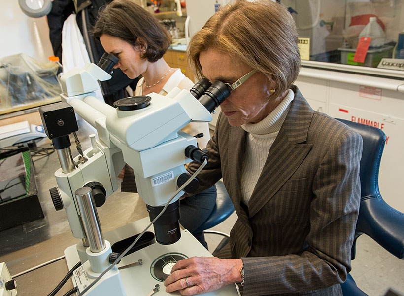
McGovern Investigator Ann Graybiel (right) with former Research Scientist Jill Crittenden. Photo: Justin Knight

Surprise discovery

To understand the essential roles of CDGI, Crittenden first engineered “knockout” mice that lack the gene encoding CDGI. Then the Graybiel team began looking for abnormalities in the CDGI knockout mice that could be tied to the loss of CDGI’s function.

Initially, they noticed that the rodent ear-tag IDs often fell off in the knockout mice, an observation that ultimately led to the surprise discovery by the Graybiel team and others that CDGI is expressed in blood platelets and is responsible for a bleeding disorder in humans, dogs, and other animals. The CDGI knockout mice were otherwise healthy and seemed just like their “wildtype” brothers and sisters, which did not carry the gene mutation. To figure out the role of CDGI in the brain, the Graybiel team would have to scrutinize the mice more closely.

Challenging the striatum

Both the CDGI knockout and wildtype mice were given an extensive set of behavioral and neurological tests and the CDGI mice showed deficits in two tests designed to challenge the striatum.

In one test, mice must find their way through a maze by relying on egocentric (i.e. self-referential) cues, such as their turning right or turning left, and not competing allocentric (i.e. external) cues, such as going toward a bright poster on the wall. Egocentric cues are thought to be processed by the striatum whereas allocentric cues are thought to rely on the hippocampus.

In a second test of striatal function, mice learned various gait patterns to match different patterns of rungs on their running wheel, a task designed to test the mouse’s ability to learn and remember a motor sequence.

The CDGI mice learned both of these striatal tasks more slowly than their wildtype siblings, suggesting that the CDGI mice might perform normally in general tests of behavior because they are able to compensate for striatal deficits by using other brain regions such as the hippocampus to solve standard tasks.

The team then decided to give the mice a completely different type of test that relies on the striatum. Because the striatum is strongly activated by drugs of abuse, which elevate dopamine and drive motor habits, Crittenden and collaborator Morgane Thomsen (now at the University of Copenhagen) looked to see whether the CDGI knockout mice respond normally to amphetamine and cocaine.

Psychomotor stimulants like cocaine and amphetamine normally induce a mixture of hyperactive behaviors such as pacing and focused repetitive behaviors like skin-picking (also called stereotypy or punding in humans). The researchers found however, that the drug-induced behaviors in the CDGI knockout mice were less varied than the normal mice and consisted of abnormally prolonged stereotypy, as though the mice were unable to switch between behaviors. The researchers were able to map the abnormal behavior to CDGI function in the striatum by showing that the same vulnerability to drug-induced stereotypy was observed in mice that were engineered to delete CDGI in the striatum after birth (“conditional knockouts”), but to otherwise have normal CDGI throughout the body.

Controlling cravings

In addition to exhibiting prolonged, repetitive behaviors, the CDGI knockout mice had a vulnerability to self-administer drugs. Although previous research had shown that treatments that activate the M1 acetylcholine receptor can block cocaine self-administration, the team found that this therapy was ineffective in CDGI knockout mice. Knockouts continued to self-administer cocaine (suggesting increased craving for the drug) at the same rate before and after M1 receptor activation treatment, even though the treatment succeeded with their sibling control mice. The researchers concluded that CDGI is critically important for controlling repetitive behaviors and the ability to stop self-administration of addictive stimulants.

mouse brain images
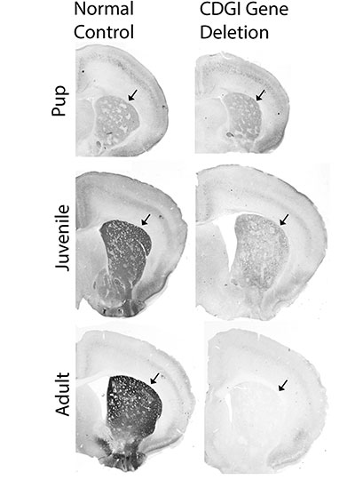
Brain sections from control mice (left) and mice engineered for deletion of the CDGI gene after birth. The expression of CDGI in the striatum (arrows) grows stronger as mice grow from pups to adulthood in control mice, but is gradually lost in the CDGI engineered mice (“conditional knockouts”). Image courtesy of the researchers

To better understand how CDGI is linked to the M1 receptor at the cellular level, the team turned to slice physiologists, scientists who record the electrical activity of neurons in brain slices. Their recordings showed that striatal neurons from CDGI knockouts fail to undergo the normal, expected electrophysiological changes after receiving treatments that target the M1 receptor. In particular, the neurons of the striatum that function broadly to stop ongoing behaviors, did not integrate cellular signals properly and failed to undergo “long-term potentiation,” a type of neuronal plasticity thought to underlie learning.

The new findings suggest that excessive repetitive movements are controlled by M1 receptor signaling through CDGI in indirect pathway neurons of the striatum, a neuronal subtype that degenerates in Huntington’s disease and is affected by dopamine loss and l-DOPA replacement therapy in Parkinson’s disease.

“The M1 acetylcholine receptor is a target for therapeutic drug development in treating cognitive and behavioral problems in multiple disorders, but progress has been severely hampered by off-target side-effects related to the wide-spread expression of the M1 receptor,” Graybiel explains. “Our findings suggest that CDGI offers the possibility for forebrain-specific targeting of M1 receptor signaling cascades that are of interest for blocking pathologically repetitive and unwanted behaviors that are common to numerous brain disorders including Huntington’s disease, drug addiction, autism, and schizophrenia as well as drug-induced dyskinesias. We hope that this work can help therapeutic development for these major health problems.”

This work was funded by the James W. (1963) and Patricia T. Poitras Fund, the William N. & Bernice E. Bumpus Foundation, the Saks Kavanaugh Foundation, the Simons Foundation, and the National Institute of Health.

Gene changes linked to severe repetitive behaviors

Extreme repetitive behaviors such as hand-flapping, body-rocking, skin-picking and sniffing are common to a number of brain disorders including autism, schizophrenia, Huntington’s disease, and drug addiction. These behaviors, termed stereotypies, are also apparent in animal models of drug addiction and autism.

In a new study published in the European Journal of Neuroscience, researchers at the McGovern Institute have identified genes that are activated in the brain prior to the initiation of these severe repetitive behaviors.

“Our lab has found a small set of genes that are regulated in relation to the development of stereotypic behaviors in an animal model of drug addiction,” says MIT Institute Professor Ann Graybiel, who is the senior author of the paper. “We were surprised and interested to see that one of these genes is a susceptibility gene for schizophrenia. This finding might help to understand the biological basis of repetitive, stereotypic behaviors as seen in a range of neurologic and neuropsychiatric disorders, and in otherwise ‘typical’ people under stress.”

A shared molecular pathway

In work led by research scientist Jill Crittenden, researchers in the Graybiel lab exposed mice to amphetamine, a psychomotor stimulant that drives hyperactivity and confined stereotypies in humans and in laboratory animals and that is used to model symptoms of schizophrenia.

They found that stimulant exposure that drives the most prolonged repetitive behaviors lead to activation of genes regulated by Neuregulin 1, a signaling molecule that is important for a variety of cellular functions including neuronal development and plasticity. Neuregulin 1 gene mutations are risk factors for schizophrenia.

The new findings highlight a shared molecular and circuit pathway for stereotypies that are caused by drugs of abuse and in brain disorders, and have implications for why stimulant intoxication is a risk factor for the onset of schizophrenia.

“Experimental treatment with amphetamine has long been used in studies on rodents and other animals in tests to find better treatments for schizophrenia in humans, because there are some behavioral similarities across the two otherwise very different contexts,” explains Graybiel, who is also an investigator at the McGovern Institute and a professor of brain and cognitive sciences at MIT. “It was striking to find Neuregulin 1 — potentially one hint to shared mechanisms underlying some of these similarities.”

Drug exposure linked to repetitive behaviors

Although many studies have measured gene expression changes in animal models of drug addiction, this study is the first to evaluate genome-wide changes specifically associated with restricted repetitive behaviors.

Stereotypies are difficult to measure without labor-intensive, direct observation, because they consist of fine movements and idiosyncratic behaviors. In this study, the authors administered amphetamine (or saline control) to mice and then measured with photobeam-breaks how much they ran around. The researchers identified prolonged periods when the mice were not running around (e.g. were potentially engaged in confined stereotypies), and then they videotaped the mice during these periods to observationally score the severity of restricted repetitive behaviors (e.g. sniffing or licking stereotypies).

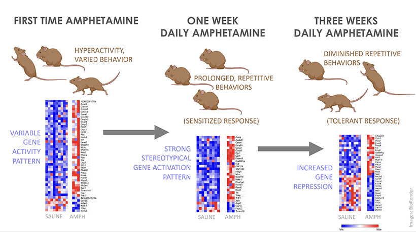
They gave amphetamine to each mouse once a day for 21 days and found that, on average, mice showed very little stereotypy on the first day of drug exposure but that, by the seventh day of exposure, all of the mice showed a prolonged period of stereotypy that gradually became shorter and shorter over the subsequent two weeks.

Graphical abstract
The authors compared gene expression changes in the brains of mice treated with amphetamine for one day, seven days or 21 days. By the twenty-first day of treatment, the stereotypy behaviors were less intense as was the gene upregulation – fewer genes were strongly activated, and more were repressed, relative to the other treatments.

“We were surprised to see the stereotypy diminishing after one week of treatment. We had actually planned a study based on our expectation that the repetitive behaviors would become more intense, but then we realized that this was an opportunity to look at what gene changes were unique to that day of high stereotypy,” says first author Jill Crittenden.

The authors compared gene expression changes in the brains of mice treated with amphetamine for one day, seven days or 21 days. They hypothesized that the gene changes associated specifically with high-stereotypy-associated seven days of drug treatment were the most likely to underlie extreme repetitive behaviors and could identify risk-factor genes for such symptoms in disease.

A shared anatomical pathway

Previous work from the Graybiel lab has shown that stereotypy is directly correlated to circumscribed gene activation in the striatum, a forebrain region that is key for habit formation. In animals with the most intense stereotypy, most of the striatum does not show gene activation, but immediate early gene induction remains high in clusters of cells called striosomes. Striosomes have recently been shown to have powerful control over cells that release dopamine, a neuromodulator that is severely disrupted in drug addiction and in schizophrenia. Strikingly, striosomes contain high levels of Neuregulin 1.

“Our new data suggest that the upregulation of Neuregulin-responsive genes in animals with severely repetitive behaviors reflects gene changes in the striosomal neurons that control the release of dopamine,” Crittenden explains. “Dopamine can directly impact whether an animal repeats an action or explores new actions, so our study highlights a potential role for a striosomal circuit in controlling action-selection in health and in neuropsychiatric disease.”

Patterns of behavior and gene expression

Striatal gene expression levels were measured by sequencing messenger RNAs (mRNAs) in dissected brain tissue. mRNAs are read out from “active” genes to instruct protein-synthesis machinery in how to make the protein that corresponds to the gene’s sequence. Proteins are the main constituents of a cell, thereby controlling each cell’s function. The number of times a particular mRNA sequence is found reflects the frequency at which the gene was being read out at the time that the cellular material was collected.

To identify genes that were read out into mRNA before the period of prolonged stereotypy, the researchers collected brain tissue 20 minutes after amphetamine injection, which is about 30 minutes before peak stereotypy. They then identified which genes had significantly different levels of corresponding mRNAs in drug-treated mice than in mice treated with saline.

A wide variety of genes showed modest mRNA increases after the first amphetamine exposure, which induced mild hyperactivity and a range of behaviors such as walking, sniffing and rearing in the mice.

By the seventh day of treatment, all of the mice were engaged for prolonged periods in one specific repetitive behavior, such as sniffing the wall. Likewise, there were fewer genes that were activated by the seventh day relative to the first treatment day, but they were strongly activated in all mice that received the stereotypy-inducing amphetamine treatment.

By the twenty-first day of treatment, the stereotypy behaviors were less intense as was the gene upregulation – fewer genes were strongly activated, and more were repressed, relative to the other treatments. “It seemed that the mice had developed tolerance to the drug, both in terms of their behavioral response and in terms of their gene activation response,” says Crittenden.

“Trying to seek patterns of gene regulation starting with behavior is correlative work, and we did not prove ‘causality’ in this first small study,” explains Graybiel. “But we hope that the striking parallels between the scope and selectivity of the mRNA and behavioral changes that we detected will help in further work on the tremendously challenging goal of treating addiction.”

This work was funded by the National Institute of Child Health and Human Development, the Saks-Kavanaugh Foundation, the Broderick Fund for Phytocannabinoid Research at MIT, the James and Pat Poitras Research Fund, The Simons Foundation and The Stanley Center for Psychiatric Research at the Broad Institute.

The pursuit of reward

View the interactive version of this story in our Spring 2021 issue of BrainScan.

The brain circuits that influence our decisions, cognitive functions, and ultimately, our actions are intimately connected with the circuits that give rise to our motivations. By exploring these relationships, scientists at McGovern are seeking knowledge that might suggest new strategies for changing our habits or treating motivation-disrupting conditions such as depression and addiction.

Risky decisions

MIT Institute Professor Ann Graybiel. Photo: Justin Knight

In Ann Graybiel’s lab, researchers have been examining how the brain makes choices that carry both positive and negative consequences — deciding to take on a higher-paying but more demanding job, for example. Psychologists call these dilemmas approach-avoidance conflicts, and resolving them not only requires weighing the good versus the bad, but also motivation to engage with the decision.

Emily Hueske, a research scientist in the Graybiel lab, explains that everyone has their own risk tolerance when it comes to such decisions, and certain psychiatric conditions, including depression and anxiety disorders, can shift the tipping point at which a person chooses to “approach” or “avoid.”

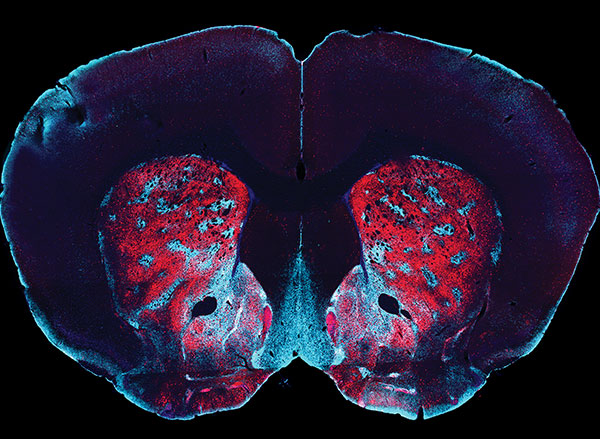
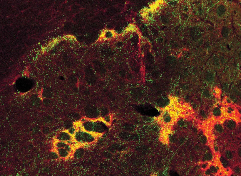
Studies have shown that neurons in the striatum (see image below), a region deep in the brain involved in both motivation and movement, activate as we grapple with these decisions. Graybiel traced this activity even further, to tiny compartments within the striatum called striosomes.

(She discovered striosomes many years ago and has been studying their function for decades.)

A motivational switch

In 2015, Graybiel’s team manipulated striosome signaling within genetically engineered mice and changed the way animals behave in approach-avoidance conflict situations. Taking cues from an assessment used to evaluate approach-avoidance behavior in patients, they presented mice with opportunities to obtain chocolate while experiencing unwelcome exposure in a brightly lit area.

Experimentally activating neurons in striosomes had a dramatic effect, causing mice to venture into brightly lit areas that they would normally avoid. With striosomal circuits switched on, “this animal all of a sudden is like a different creature,” Graybiel says.

Two years later, they found that chronic stress and other factors can also disrupt this signaling and change the choices animals make.

An image of the mouse striatum showing clusters of striosomes (red and yellow). Image: Graybiel lab

Age of ennui

This November, Alexander Friedman, who worked as a research scientist in the Graybiel lab, and Hueske reported in Cell that they found an age-related decline in motivation-modulated learning in mice and rats. Neurons within striosomes became more active than the cells that surround them as animals learned to assign positive and negative values to potential choices. And older mice were less engaged than their younger counterparts in the type of learning required to make these cost-benefit analyses. A similar lack of motivation was observed in a mouse model of Huntington’s disease, a neurodegenerative disorder that is often associated with mood
disturbances in patients.

“This coincides with our previous findings that striosomes are critically important for decisions that involve a conflict.”

“This coincides with our previous findings that striosomes are critically important for decisions that involve a conflict,” says Friedman, who is now an assistant professor at the University of Texas at El Paso.

Graybiel’s team is continuing to investigate these uniquely positioned compartments in the brain, expecting to shed light on the mechanisms that underlie both learning and motivation.

“There’s no learning without motivation, and in fact, motivation can be influenced by learning,” Hueske says. “The more you learn, the more excited you might be to engage in the task. So the two are intertwined.”

The aging brain

Researchers in John Gabrieli’s lab are also seeking to understand the circuits that link motivation to learning, and recently, his team reported that they, too, had found an age-related decline in motivation-modulated learning.

Studies in young adults have shown that memory improves when the brain circuits that process motivation and memory interact. Gabrieli and neurologist Maiya Geddes, who worked in Gabrieli’s lab as a postdoctoral fellow, wondered whether this holds true in older adults, particularly as memory declines.

To find out, the team recruited 40 people to participate in a brain imaging study. About half of the participants were between the ages of 18 and 30, while the others were between the ages of 49 and 84. While inside an fMRI scanner, each participant was asked to commit certain words to memory and told their success would determine how much money they received for participating in the experiment.

Diminished drive

MRI scan
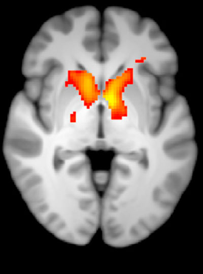
Younger adults show greater activation in the reward-related regions of the brain during incentivized memory tasks compared to older adults. Image: Maiya Geddes

Not surprisingly, when participants were asked 24 hours later to recall the words, the younger group performed better overall than the older group. In young people, incentivized memory tasks triggered activity in parts of the brain involved in both memory and motivation. But in older adults, while these two parts of the brain could be activated independently, they did not seem to be communicating with one another.

“It seemed that the older adults, at least in terms of their brain response, did care about the kind of incentives that we were offering,” says Geddes, who is now an assistant professor at McGill University. “But for whatever reason, that wasn’t allowing them to benefit in terms of improved memory performance.”

Since the study indicates the brain still can anticipate potential rewards, Geddes is now exploring whether other sources of motivation, such as social rewards, might more effectively increase healthful decisions and behaviors in older adults.

Circuit control

Understanding how the brain generates and responds to motivation is not only important for improving learning strategies. Lifestyle choices such as exercise and social engagement can help people preserve cognitive function and improve their quality of life as they age, and Gabrieli says activating the right motivational circuits could help encourage people to implement healthy changes.

By pinpointing these motivational circuits in mice, Graybiel hopes that her research will lead to better treatment strategies for people struggling with motivational challenges, including Parkinson’s disease. Her team is now exploring whether striosomes serve as part of a value-sensitive switch, linking our intentions to dopamine-containing neurons in the midbrain that can modulate our actions.

“Perhaps this motivation is critical for the conflict resolution, and striosomes combine two worlds, dopaminergic motivation and cortical knowledge, resulting in motivation to learn,” Friedman says.

“Now we know that these challenges have a biological basis, and that there are neural circuits that can promote or reduce our feeling of motivational energy,” explains Graybiel. “This realization in itself is a major step toward learning how we can control these circuits both behaviorally and by highly selective therapeutic targeting.”

Two MIT Brain and Cognitive Sciences faculty members earn funding from the G. Harold and Leila Y. Mathers Foundation

Two MIT neuroscientists have received grants from the G. Harold and Leila Y. Mathers Foundation to screen for genes that could help brain cells withstand Parkinson’s disease and to map how gene expression changes in the brain in response to drugs of abuse.

Myriam Heiman, an associate professor in MIT’s Department of Brain and Cognitive Sciences and a core member of the Picower Institute for Learning and Memory and the Broad Institute of MIT and Harvard, and Alan Jasanoff, who is also a professor in biological engineering, brain and cognitive sciences, nuclear science and engineering and an associate investigator at the McGovern Institute for Brain Research, each received three-year awards that formally begin January 1, 2021.

Jasanoff, who also directs MIT’s Center for Neurobiological Engineering, is known for developing sensors that monitor molecular hallmarks of neural activity in the living brain, in real time, via noninvasive MRI brain scanning. One of the MRI-detectable sensors that he has developed is for dopamine, a neuromodulator that is key to learning what behaviors and contexts lead to reward. Addictive drugs artificially drive dopamine release, thereby hijacking the brain’s reward prediction system. Studies have shown that dopamine and drugs of abuse activate gene transcription in specific brain regions, and that this gene expression changes as animals are repeatedly exposed to drugs. Despite the important implications of these neuroplastic changes for the process of addiction, in which drug-seeking behaviors become compulsive, there are no effective tools available to measure gene expression across the brain in real time.

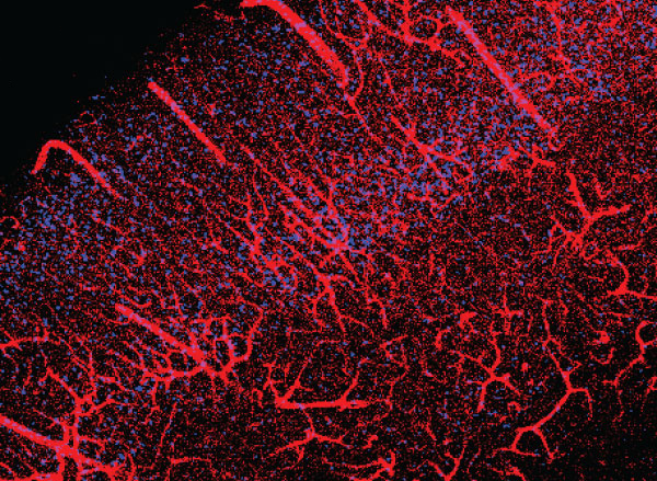
Cerebral vasculature in mouse brain. The Jasanoff lab hopes to develop a method for mapping gene expression the brain with related labeling characteristics .
Image: Alan Jasanoff

With the new Mathers funding, Jasanoff is developing new MRI-detectable sensors for gene expression. With these cutting-edge tools, Jasanoff proposes to make an activity atlas of how the brain responds to drugs of abuse, both upon initial exposure and over repeated doses that simulate the experiences of drug addicted individuals.

“Our studies will relate drug-induced brain activity to longer term changes that reshape the brain in addiction,” says Jasanoff. “We hope these studies will suggest new biomarkers or treatments.”

Dopamine-producing neurons in a brain region called the substantia nigra are known to be especially vulnerable to dying in Parkinson’s disease, leading to the severe motor difficulties experienced during the progression of the incurable, chronic neurodegenerative disorder. The field knows little about what puts specific cells at such dire risk, or what molecular mechanisms might help them resist the disease. In her research on Huntington’s disease, another incurable neurodegenerative disorder in which a specific neuron population in the striatum is especially vulnerable, Heiman has been able to use an innovative method her lab pioneered to discover genes whose expression promotes neuron survival, yielding potential new drug targets. The technique involves conducting an unbiased screen in which her lab knocks out each of the 22,000 genes expressed in the mouse brain one by one in neurons in disease model mice and healthy controls. The technique allows her to determine which genes, when missing, contribute to neuron death amid disease and therefore which genes are particularly needed for survival. The products of those genes can then be evaluated as drug targets. With the new Mathers award, Heiman plans to apply the method to study Parkinson’s disease.

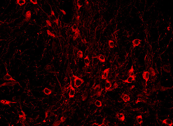
An immunofluorescence image taken in a brain region called the substantia nigra (SN) highlights tyrosine hydroxylase, a protein expressed by dopamine neurons. This type of neuron in the SN is especially vulnerable to neurodegeneration in Parkinson’s disease. Image: Preston Ge/Heiman Lab

“There is currently no molecular explanation for the brain cell loss seen in Parkinson’s disease or a cure for this devastating disease,” Heiman said. “This award will allow us to perform unbiased, genome-wide genetic screens in the brains of mouse models of Parkinson’s disease, probing for genes that allow brain cells to survive the effects of cellular perturbations associated with Parkinson’s disease. I’m extremely grateful for this generous support and recognition of our work from the Mathers Foundation, and hope that our study will elucidate new therapeutic targets for the treatment and even prevention of Parkinson’s disease.”

Using machine learning to track the pandemic’s impact on mental health

Dealing with a global pandemic has taken a toll on the mental health of millions of people. A team of MIT and Harvard University researchers has shown that they can measure those effects by analyzing the language that people use to express their anxiety online.

Using machine learning to analyze the text of more than 800,000 Reddit posts, the researchers were able to identify changes in the tone and content of language that people used as the first wave of the Covid-19 pandemic progressed, from January to April of 2020. Their analysis revealed several key changes in conversations about mental health, including an overall increase in discussion about anxiety and suicide.

“We found that there were these natural clusters that emerged related to suicidality and loneliness, and the amount of posts in these clusters more than doubled during the pandemic as compared to the same months of the preceding year, which is a grave concern,” says Daniel Low, a graduate student in the Program in Speech and Hearing Bioscience and Technology at Harvard and MIT and the lead author of the study.

The analysis also revealed varying impacts on people who already suffer from different types of mental illness. The findings could help psychiatrists, or potentially moderators of the Reddit forums that were studied, to better identify and help people whose mental health is suffering, the researchers say.

“When the mental health needs of so many in our society are inadequately met, even at baseline, we wanted to bring attention to the ways that many people are suffering during this time, in order to amplify and inform the allocation of resources to support them,” says Laurie Rumker, a graduate student in the Bioinformatics and Integrative Genomics PhD Program at Harvard and one of the authors of the study.

Satrajit Ghosh, a principal research scientist at MIT’s McGovern Institute for Brain Research, is the senior author of the study, which appears in the Journal of Internet Medical Research. Other authors of the paper include Tanya Talkar, a graduate student in the Program in Speech and Hearing Bioscience and Technology at Harvard and MIT; John Torous, director of the digital psychiatry division at Beth Israel Deaconess Medical Center; and Guillermo Cecchi, a principal research staff member at the IBM Thomas J. Watson Research Center.

A wave of anxiety

The new study grew out of the MIT class 6.897/HST.956 (Machine Learning for Healthcare), in MIT’s Department of Electrical Engineering and Computer Science. Low, Rumker, and Talkar, who were all taking the course last spring, had done some previous research on using machine learning to detect mental health disorders based on how people speak and what they say. After the Covid-19 pandemic began, they decided to focus their class project on analyzing Reddit forums devoted to different types of mental illness.

“When Covid hit, we were all curious whether it was affecting certain communities more than others,” Low says. “Reddit gives us the opportunity to look at all these subreddits that are specialized support groups. It’s a really unique opportunity to see how these different communities were affected differently as the wave was happening, in real-time.”

The researchers analyzed posts from 15 subreddit groups devoted to a variety of mental illnesses, including schizophrenia, depression, and bipolar disorder. They also included a handful of groups devoted to topics not specifically related to mental health, such as personal finance, fitness, and parenting.

Using several types of natural language processing algorithms, the researchers measured the frequency of words associated with topics such as anxiety, death, isolation, and substance abuse, and grouped posts together based on similarities in the language used. These approaches allowed the researchers to identify similarities between each group’s posts after the onset of the pandemic, as well as distinctive differences between groups.

The researchers found that while people in most of the support groups began posting about Covid-19 in March, the group devoted to health anxiety started much earlier, in January. However, as the pandemic progressed, the other mental health groups began to closely resemble the health anxiety group, in terms of the language that was most often used. At the same time, the group devoted to personal finance showed the most negative semantic change from January to April 2020, and significantly increased the use of words related to economic stress and negative sentiment.

They also discovered that the mental health groups affected the most negatively early in the pandemic were those related to ADHD and eating disorders. The researchers hypothesize that without their usual social support systems in place, due to lockdowns, people suffering from those disorders found it much more difficult to manage their conditions. In those groups, the researchers found posts about hyperfocusing on the news and relapsing back into anorexia-type behaviors since meals were not being monitored by others due to quarantine.

Using another algorithm, the researchers grouped posts into clusters such as loneliness or substance use, and then tracked how those groups changed as the pandemic progressed. Posts related to suicide more than doubled from pre-pandemic levels, and the groups that became significantly associated with the suicidality cluster during the pandemic were the support groups for borderline personality disorder and post-traumatic stress disorder.

The researchers also found the introduction of new topics specifically seeking mental health help or social interaction. “The topics within these subreddit support groups were shifting a bit, as people were trying to adapt to a new life and focus on how they can go about getting more help if needed,” Talkar says.

While the authors emphasize that they cannot implicate the pandemic as the sole cause of the observed linguistic changes, they note that there was much more significant change during the period from January to April in 2020 than in the same months in 2019 and 2018, indicating the changes cannot be explained by normal annual trends.

Mental health resources

This type of analysis could help mental health care providers identify segments of the population that are most vulnerable to declines in mental health caused by not only the Covid-19 pandemic but other mental health stressors such as controversial elections or natural disasters, the researchers say.

Additionally, if applied to Reddit or other social media posts in real-time, this analysis could be used to offer users additional resources, such as guidance to a different support group, information on how to find mental health treatment, or the number for a suicide hotline.

“Reddit is a very valuable source of support for a lot of people who are suffering from mental health challenges, many of whom may not have formal access to other kinds of mental health support, so there are implications of this work for ways that support within Reddit could be provided,” Rumker says.

The researchers now plan to apply this approach to study whether posts on Reddit and other social media sites can be used to detect mental health disorders. One current project involves screening posts in a social media site for veterans for suicide risk and post-traumatic stress disorder.

The research was funded by the National Institutes of Health and the McGovern Institute.