Study reveals a universal pattern of brain wave frequencies

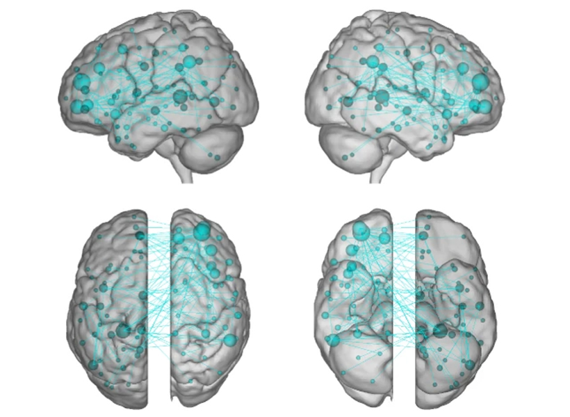
Throughout the brain’s cortex, neurons are arranged in six distinctive layers, which can be readily seen with a microscope. A team of MIT and Vanderbilt University neuroscientists has now found that these layers also show distinct patterns of electrical activity, which are consistent over many brain regions and across several animal species, including humans.

The researchers found that in the topmost layers, neuron activity is dominated by rapid oscillations known as gamma waves. In the deeper layers, slower oscillations called alpha and beta waves predominate. The universality of these patterns suggests that these oscillations are likely playing an important role across the brain, the researchers say.

“When you see something that consistent and ubiquitous across cortex, it’s playing a very fundamental role in what the cortex does,” says Earl Miller, the Picower Professor of Neuroscience, a member of MIT’s Picower Institute for Learning and Memory, and one of the senior authors of the new study.

Imbalances in how these oscillations interact with each other may be involved in brain disorders such as attention deficit hyperactivity disorder, the researchers say.

“Overly synchronous neural activity is known to play a role in epilepsy, and now we suspect that different pathologies of synchrony may contribute to many brain disorders, including disorders of perception, attention, memory, and motor control. In an orchestra, one instrument played out of synchrony with the rest can disrupt the coherence of the entire piece of music,” says Robert Desimone, director of MIT’s McGovern Institute for Brain Research and one of the senior authors of the study.

André Bastos, an assistant professor of psychology at Vanderbilt University, is also a senior author of the open-access paper, which appears today in Nature Neuroscience. The lead authors of the paper are MIT research scientist Diego Mendoza-Halliday and MIT postdoc Alex Major.

Layers of activity

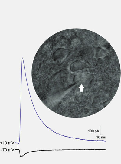
The human brain contains billions of neurons, each of which has its own electrical firing patterns. Together, groups of neurons with similar patterns generate oscillations of electrical activity, or brain waves, which can have different frequencies. Miller’s lab has previously shown that high-frequency gamma rhythms are associated with encoding and retrieving sensory information, while low-frequency beta rhythms act as a control mechanism that determines which information is read out from working memory.

His lab has also found that in certain parts of the prefrontal cortex, different brain layers show distinctive patterns of oscillation: faster oscillation at the surface and slower oscillation in the deep layers. One study, led by Bastos when he was a postdoc in Miller’s lab, showed that as animals performed working memory tasks, lower-frequency rhythms generated in deeper layers regulated the higher-frequency gamma rhythms generated in the superficial layers.

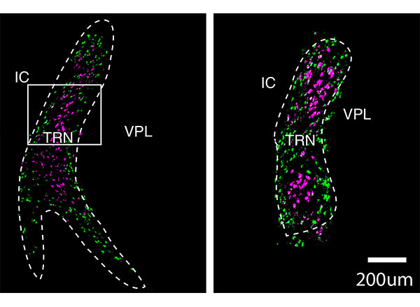
In addition to working memory, the brain’s cortex also is the seat of thought, planning, and high-level processing of emotion and sensory information. Throughout the regions involved in these functions, neurons are arranged in six layers, and each layer has its own distinctive combination of cell types and connections with other brain areas.

“The cortex is organized anatomically into six layers, no matter whether you look at mice or humans or any mammalian species, and this pattern is present in all cortical areas within each species,” Mendoza-Halliday says. “Unfortunately, a lot of studies of brain activity have been ignoring those layers because when you record the activity of neurons, it’s been difficult to understand where they are in the context of those layers.”

In the new paper, the researchers wanted to explore whether the layered oscillation pattern they had seen in the prefrontal cortex is more widespread, occurring across different parts of the cortex and across species.

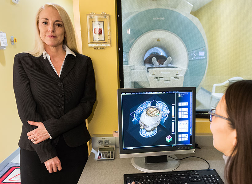
Using a combination of data acquired in Miller’s lab, Desimone’s lab, and labs from collaborators at Vanderbilt, the Netherlands Institute for Neuroscience, and the University of Western Ontario, the researchers were able to analyze 14 different areas of the cortex, from four mammalian species. This data included recordings of electrical activity from three human patients who had electrodes inserted in the brain as part of a surgical procedure they were undergoing.

Recording from individual cortical layers has been difficult in the past, because each layer is less than a millimeter thick, so it’s hard to know which layer an electrode is recording from. For this study, electrical activity was recorded using special electrodes that record from all of the layers at once, then feed the data into a new computational algorithm the authors designed, termed FLIP (frequency-based layer identification procedure). This algorithm can determine which layer each signal came from.

“More recent technology allows recording of all layers of cortex simultaneously. This paints a broader perspective of microcircuitry and allowed us to observe this layered pattern,” Major says. “This work is exciting because it is both informative of a fundamental microcircuit pattern and provides a robust new technique for studying the brain. It doesn’t matter if the brain is performing a task or at rest and can be observed in as little as five to 10 seconds.”

Across all species, in each region studied, the researchers found the same layered activity pattern.

“We did a mass analysis of all the data to see if we could find the same pattern in all areas of the cortex, and voilà, it was everywhere. That was a real indication that what had previously been seen in a couple of areas was representing a fundamental mechanism across the cortex,” Mendoza-Halliday says.

Maintaining balance

The findings support a model that Miller’s lab has previously put forth, which proposes that the brain’s spatial organization helps it to incorporate new information, which carried by high-frequency oscillations, into existing memories and brain processes, which are maintained by low-frequency oscillations. As information passes from layer to layer, input can be incorporated as needed to help the brain perform particular tasks such as baking a new cookie recipe or remembering a phone number.

“The consequence of a laminar separation of these frequencies, as we observed, may be to allow superficial layers to represent external sensory information with faster frequencies, and for deep layers to represent internal cognitive states with slower frequencies,” Bastos says. “The high-level implication is that the cortex has multiple mechanisms involving both anatomy and oscillations to separate ‘external’ from ‘internal’ information.”

Under this theory, imbalances between high- and low-frequency oscillations can lead to either attention deficits such as ADHD, when the higher frequencies dominate and too much sensory information gets in, or delusional disorders such as schizophrenia, when the low frequency oscillations are too strong and not enough sensory information gets in.

“The proper balance between the top-down control signals and the bottom-up sensory signals is important for everything the cortex does,” Miller says. “When the balance goes awry, you get a wide variety of neuropsychiatric disorders.”

The researchers are now exploring whether measuring these oscillations could help to diagnose these types of disorders. They are also investigating whether rebalancing the oscillations could alter behavior — an approach that could one day be used to treat attention deficits or other neurological disorders, the researchers say.

The researchers also hope to work with other labs to characterize the layered oscillation patterns in more detail across different brain regions.

“Our hope is that with enough of that standardized reporting, we will start to see common patterns of activity across different areas or functions that might reveal a common mechanism for computation that can be used for motor outputs, for vision, for memory and attention, et cetera,” Mendoza-Halliday says.

The research was funded by the U.S. Office of Naval Research, the U.S. National Institutes of Health, the U.S. National Eye Institute, the U.S. National Institute of Mental Health, the Picower Institute, a Simons Center for the Social Brain Postdoctoral Fellowship, and a Canadian Institutes of Health Postdoctoral Fellowship.

Refining mental health diagnoses

Maedbh King came to MIT to make a difference in mental health. As a postdoctoral fellow in the K. Lisa Yang Integrative Computational Neuroscience (ICoN) Center, she is building computer models aimed at helping clinicians improve diagnosis and treatment, especially for young people with neurodevelopmental and psychiatric disorders.

Tapping two large patient-data sources, King is working to analyze critical biological and behavioral information to better categorize patients’ mental health conditions, including autism spectrum disorder, attention-deficit hyperactivity disorder (ADHD), anxiety, and suicidal thoughts — and to provide more predictive approaches to addressing them. Her strategy reflects the center’s commitment to a holistic understanding of human brain function using theoretical and computa-tional neuroscience.

“Today, treatment decisions for psychiatric disorders are derived entirely from symptoms, which leaves clinicians and patients trying one treatment and, if it doesn’t work, trying another,” says King. “I hope to help change that.”

King grew up in Dublin, Ireland, and studied psychology in college; gained neuroimaging and programming skills while earning a master’s degree from Western University in Canada; and received her doctorate from the University of California, Berkeley, where she built maps and models of the human brain. In fall 2022, King joined the lab of Satrajit Ghosh, a McGovern Institute principal research scientist whose team uses neuroimaging, speech communication, and machine learning to improve assessments and treatments for mental health and neurological disorders.

Big-data insights

King is pursuing several projects using the Healthy Brain Network, a landmark mental health study of children and adolescents in New York City. She and lab colleagues are extracting data from cognitive and other assessments — such as language patterns, favorite school subjects, and family mental illness history — from roughly 4,000 participants to provide more-nuanced understanding of their neurodevelopmental disorders, such as autism or ADHD.

“Computational models are powerful. They can identify patterns that can’t be obtained with the human eye through electronic records,” says King.

With this database, one can develop “very rich clinical profiles of these young people,” including their challenges and adaptive strengths, King explains. “We’re interested in placing these participants within a spectrum of symptoms, rather than just providing a binary label of, ‘has this disorder’ or ‘doesn’t have it.’ It’s an effort to subtype based on these phenotypic assessments.”

In other research, King is developing tools to detect risk factors for suicide among adolescents. Working with psychiatrists at Children’s Hospital of Philadelphia, she is using detailed questionnaires from some 20,000 youths who visited the hospital’s emergency department over several years; about one-tenth had tried to take their own lives. The questionnaires collect information about demographics, lifestyle, relationships, and other aspects of patients’ lives.

“One of the big questions the physicians want to answer is, Are there any risk predictors we can identify that can ultimately prevent, or at least mitigate, future suicide attempts?” King says. “Computational models are powerful. They can identify patterns that can’t be obtained with the human eye through electronic records.”

King is passionate about producing findings to help practitioners, whether they’re clinicians, teachers, parents, or policy makers, and the populations they’re studying. “This applied work,” she says, “should be communicated in a way that can be useful.

Personal pursuits

This story originally appeared in the Fall 2022 issue of BrainScan.

***

Many neuroscientists were drawn to their careers out of curiosity and wonder. Their deep desire to understand how the brain works drew them into the lab and keeps them coming back, digging deeper and exploring more each day. But for some, the work is more personal.

Several McGovern faculty say they entered their field because someone in their lives was dealing with a brain disorder that they wanted to better understand. They are committed to unraveling the basic biology of those conditions, knowing that knowledge is essential to guide the development of better treatments.

The distance from basic research to clinical progress is shortening, and many young neuroscientists hope not just to deepen scientific understanding of the brain, but to have direct impact on the lives of patients. Some want to know why people they love are suffering from neurological disorders or mental illness; others seek to understand the ways in which their own brains work differently than others. But above all, they want better treatments for people affected by such disorders.

Seeking answers

That’s true for Kian Caplan, a graduate student in MIT’s Department of Brain and Cognitive Sciences who was diagnosed with Tourette syndrome around age 13. At the time, learning that the repetitive, uncontrollable movements and vocal tics he had been making for most of his life were caused by a neurological disorder was something of a relief. But it didn’t take long for Caplan to realize his diagnosis came with few answers.

Graduate student Kian Caplan studies the brain circuits associated with Tourette syndrome and obsessive-compulsive disorder in Guoping Feng and Fan Wang’s labs at the McGovern Institute. Photo: Steph Stevens

Tourette syndrome has been estimated to occur in about six of every 1,000 children, but its neurobiology remains poorly understood.

“The doctors couldn’t really explain why I can’t control the movements and sounds I make,” he says. “They couldn’t really explain why my symptoms wax and wane, or why the tics I have aren’t always the same.”

That lack of understanding is not just frustrating for curious kids like Caplan. It means that researchers have been unable to develop treatments that target the root cause of Tourette syndrome. Drugs that dampen signaling in parts of the brain that control movement can help suppress tics, but not without significant side effects. Caplan has tried those drugs. For him, he says, “they’re not worth the suppression.”

Advised by Fan Wang and McGovern Associate Director Guoping Feng, Caplan is looking for answers. A mouse model of obsessive-compulsive disorder developed in Feng’s lab was recently found to exhibit repetitive movements similar to those of people with Tourette syndrome, and Caplan is working to characterize those tic-like movements. He will use the mouse model to examine the brain circuits underlying the two conditions, which often co-occur in people. Broadly, researchers think Tourette syndrome arises due to dysregulation of cortico-striatal-thalamo-cortical circuits, which connect distant parts of the brain to control movement. Caplan and Wang suspect that the brainstem — a structure found where the brain connects to the spinal cord, known for organizing motor movement into different modules — is probably involved, too.

Wang’s research group studies the brainstem’s role in movement, but she says that like most researchers, she hadn’t considered its role in Tourette syndrome until Caplan joined her lab. That’s one reason Caplan, who has long been a mentor and advocate for students with neurodevelopmental disorders, thinks neuroscience needs more neurodiversity.

“I think we need more representation in basic science research by the people who actually live with those conditions,” he says. Their experiences can lead to insights that may be inaccessible to others, he says, but significant barriers in academia often prevent this kind of representation. Caplan wants to see institutions make systemic changes to ensure that neurodiverse and otherwise minority individuals are able to thrive in academia. “I’m not an exception,” he says, “there should be more people like me here, but the present system makes that incredibly difficult.”

Overcoming adversity

Like Caplan, Lace Riggs faced significant challenges in her pursuit to study the brain. She grew up in Southern California’s Inland Empire, where issues of social disparity, chronic stress, drug addiction, and mental illness were a part of everyday life.

Postdoctoral fellow Lace Riggs studies the origins of neurodevelopmental conditions in Guoping Feng’s lab at the McGovern Institute. Photo: Lace Riggs

“Living in severe poverty and relying on government assistance without access to adequate education and resources led everyone I know and love to suffer tremendously, myself included,” says Riggs, a postdoctoral fellow in the Feng lab.

“There are not a lot of people like me who make it to this stage,” says Riggs, who has lost friends and family members to addiction, mental illness, and suicide. “There’s a reason for that,” she adds. “It’s really, really difficult to get through the educational system and to overcome socioeconomic barriers.”

Today, Riggs is investigating the origins of neurodevelopmental conditions, hoping to pave the way to better treatments for brain disorders by uncovering the molecular changes that alter the structure and function of neural circuits.

Riggs says that the adversities she faced early in life offered valuable insights in the pursuit of these goals. She first became interested in the brain because she wanted to understand how our experiences have a lasting impact on who we are — including in ways that leave people vulnerable to psychiatric problems.

“While the need for more effective treatments led me to become interested in psychiatry, my fascination with the brain’s unique ability to adapt is what led me to neuroscience,” says Riggs.

After finishing high school, Riggs attended California State University in San Bernardino and became the only member of her family to attend university or attempt a four-year degree. Today, she spends her days working with mice that carry mutations linked to autism or ADHD in humans, studying the animals’ behavior and monitoring their neural activity. She expects that aberrant neural circuit activity in these conditions may also contribute to mood disorders, whose origins are harder to tease apart because they often arise when genetic and environmental factors intersect. Ultimately, Riggs says, she wants to understand how our genes dictate whether an experience will alter neural signaling and impact mental health in a long-lasting way.

Riggs uses patch clamp electrophysiology to record the strength of inhibitory and excitatory synaptic input onto individual neurons (white arrow) in an animal model of autism. Image: Lace Riggs

“If we understand how these long-lasting synaptic changes come about, then we might be able to leverage these mechanisms to develop new and more effective treatments.”

While the turmoil of her childhood is in the past, Riggs says it is not forgotten — in part, because of its lasting effects on her own mental health.  She talks openly about her ongoing struggle with social anxiety and complex post-traumatic stress disorder because she is passionate about dismantling the stigma surrounding these conditions. “It’s something I have to deal with every day,” Riggs says. That means coping with symptoms like difficulty concentrating, hypervigilance, and heightened sensitivity to stress. “It’s like a constant hum in the background of my life, it never stops,” she says.

“I urge all of us to strive, not only to make scientific discoveries to move the field forward,” says Riggs, “but to improve the accessibility of this career to those whose lived experiences are required to truly accomplish that goal.”

Circuit that focuses attention brings in wide array of inputs

In a new brain-wide circuit tracing study, scientists at MIT’s Picower Institute for Learning and Memory focused selective attention on a circuit that governs, fittingly enough, selective attention. The comprehensive maps they produced illustrate how broadly the mammalian brain incorporates and integrates information to focus its sensory resources on its goals.

Working in mice, the team traced thousands of inputs into the circuit, a communication loop between the anterior cingulate cortex (ACC) and the lateral posterior (LP) thalamus. In primates the LP is called the pulvinar. Studies in humans and nonhuman primates have indicated that the byplay of these two regions is critical for brain functions like being able to focus on an object of interest in a crowded scene, says study co-lead author Yi Ning Leow, a graduate student in the lab of senior author Mriganka Sur, the Newton Professor in MIT’s Department of Brain and Cognitive Sciences. Research has implicated dysfunction in the circuit in attention-affecting disorders such as autism and attention deficit/hyperactivity disorder.

The new study in the Journal of Comparative Neurology extends what’s known about the circuit by detailing it in mice, Leow says, importantly showing that the mouse circuit is closely analogous to the primate version even if the LP is proportionately smaller and less evolved than the pulvinar.

“In these rodent models we were able to find very similar circuits,” Leow says. “So we can possibly study these higher-order functions in mice as well. We have a lot more genetic tools in mice so we are better able to look at this circuit.”

The study, also co-led by former MIT undergraduate Blake Zhou, therefore provides a detailed roadmap in the experimentally accessible mouse model for understanding how the ACC and LP cooperate to produce selective attention. For instance, now that Leow and Zhou have located all the inputs that are wired into the circuit, Leow is tapping into those feeds to eavesdrop on the information they are carrying. Meanwhile, she is correlating that information flow with behavior.

“This study lays the groundwork for understanding one of the most important, yet most elusive, components of brain function, namely our ability to selectively attend to one thing out of several, as well as switch attention,” Sur says.

Using virally mediated circuit-tracing techniques pioneered by co-author Ian Wickersham, principal research scientist in brain and cognitive sciences and the McGovern Institute for Brain Research at MIT, the team found distinct sources of input for the ACC and the LP. Generally speaking, the detailed study finds that the majority of inputs to the ACC were from frontal cortex areas that typically govern goal-directed planning, and from higher visual areas. The bulk of inputs to the LP, meanwhile, were from deeper regions capable of providing context such as the mouse’s needs, location and spatial cues, information about movement, and general information from a mix of senses.

So even though focusing attention might seem like a matter of controlling the senses, Leow says, the circuit pulls in a lot of other information as well.

“We’re seeing that it’s not just sensory — there are so many inputs that are coming from non-sensory areas as well, both sub-cortically and cortically,” she says. “It seems to be integrating a lot of different aspects that might relate to the behavioral state of the animal at a given time. It provides a way to provide a lot of internal and special context for that sensory information.”

Given the distinct sets of inputs to each region, the ACC may be tasked with focusing attention on a desired object, while the LP is modulating how the ACC goes about making those computations, accounting for what’s going on both inside and outside the animal. Decoding just what that incoming contextual information is, and what the LP tells the ACC, are the key next steps, Leow says. Another clear set of questions the study raises are what are the circuit’s outputs. In other words, after it integrates all this information, what does it do with it?

The paper’s other authors are Heather Sullivan and Alexandria Barlowe.

A National Science Scholarship, the National Institutes of Health, and the JPB Foundation provided support for the study.

What’s happening in your brain when you’re spacing out?

This story is adapted from a News@Northeastern post.

We all do it. One second you’re fully focused on the task in front of you, a conversation with a friend, or a professor’s lecture, and the next second your mind is wandering to your dinner plans.

But how does that happen?

“We spend so much of our daily lives engaged in things that are completely unrelated to what’s in front of us,” says Aaron Kucyi, neuroscientist and principal research scientist in the department of psychology at Northeastern. “And we know very little about how it works in the brain.”

So Kucyi and colleagues at Massachusetts General Hospital, Boston University, and the McGovern Institute at MIT started scanning people’s brains using functional magnetic resonance imaging (fMRI) to get an inside look. Their results, published Friday in the journal Nature Communications, add complexity to our understanding of the wandering mind.

It turns out that spacing out might not deserve the bad reputation that it receives. Many more parts of the brain seem to be engaged in mind-wandering than previously thought, supporting the idea that it’s actually a quite dynamic and fundamental function of our psychology.

“Many of those things that we do when we’re spacing out are very adaptive and important to our lives,” says Kucyi, the paper’s first author. We might be drafting an email in our heads while in the shower, or trying to remember the host’s spouse’s name while getting dressed for a party. Moments when our minds wander can allow space for creativity and planning for the future, he says, so it makes sense that many parts of the brain would be engaged in that kind of thinking.

But mind wandering may also be detrimental, especially for those suffering from mental illness, explains the study’s senior author, Susan Whitfield-Gabrieli. “For many of us, mind wandering may be a healthy, positive and constructive experience, like reminiscing about the past, planning for the future, or engaging in creative thinking,” says Whitfield-Gabrieli, a professor of psychology at Northeastern University and a McGovern Institute research affiliate. “But for those suffering from mental illness such as depression, anxiety or psychosis, reminiscing about the past may transform into ruminating about the past, planning for the future may become obsessively worrying about the future and creative thinking may evolve into delusional thinking.”

Identifying the brain circuits associated with mind wandering, she says, may reveal new targets and better treatment options for people suffering from these disorders.

McGovern research affiliate Susan Whitfield-Gabrieli in the Martinos Imaging Center.

Inside the wandering mind

To study wandering minds, the researchers first had to set up a situation in which people were likely to lose focus. They recruited test subjects at the McGovern Institute’s Martinos Imaging Center to complete a simple, repetitive, and rather boring task. With an fMRI scanner mapping their brain activity, participants were instructed to press a button whenever an image of a city scene appeared on a screen in front of them and withhold a response when a mountain image appeared.

Throughout the experiment, the subjects were asked whether they were focused on the task at hand. If a subject said their mind was wandering, the researchers took a close look at their brain scans from right before they reported loss of focus. The data was then fed into a machine-learning algorithm to identify patterns in the neurological connections involved in mind-wandering (called “stimulus-independent, task-unrelated thought” by the scientists).

Scientists previously identified a specialized system in the brain considered to be responsible for mind-wandering. Called the “default mode network,” these parts of the brain activated when someone’s thoughts were drifting away from their immediate surroundings and deactivated when they were focused. The other parts of the brain, that theory went, were quiet when the mind was wandering, says Kucyi.

The researchers used a technique called “connectome-based predictive modeling” to identify patterns in the brain connections involved in mind-wandering. Image courtesy of the researchers.

The “default mode network” did light up in Kucyi’s data. But parts of the brain associated with other functions also appeared to activate when his subjects reported that their minds had wandered.

For example, the “default mode network” and networks in the brain related to controlling or maintaining a train of thought also seemed to be communicating with one another, perhaps helping explain the ability to go down a rabbit hole in your mind when you’re distracted from a task. There was also a noticeable lack of communication between the “default mode network” and the systems associated with sensory input, which makes sense, as the mind is wandering away from the person’s immediate environment.

“It makes sense that virtually the whole brain is involved,” Kucyi says. “Mind-wandering is a very complex operation in the brain and involves drawing from our memory, making predictions about the future, dynamically switching between topics that we’re thinking about, fluctuations in our mood, and engaging in vivid visual imagery while ignoring immediate visual input,” just to name a few functions.

The “default mode network” still seems to be key, Kucyi says. Virtual computer analysis suggests that if you took the regions of the brain in that network out of the equation, the other brain regions would not be able to pick up the slack in mind-wandering.

Kucyi, however, didn’t just want to identify regions of the brain that lit up when someone said their mind was wandering. He also wanted to be able to use that generalized pattern of brain activity to be able to predict whether or not a subject would say that their focus had drifted away from the task in front of them.

That’s where the machine-learning analysis of the data came in. The idea, Kucyi says, is that “you could bring a new person into the scanner and not even ask them whether they were mind-wandering or not, and have a good estimate from their brain data whether they were.”

The ADHD brain

To test the patterns identified through machine learning, the researchers brought in a new set of test subjects – people diagnosed with ADHD. When the fMRI scans lit up the parts of the brain Kucyi and his colleagues had identified as engaged in mind-wandering in the first part of the study, the new test subjects reported that their thoughts had drifted from the images of cities and mountains in front of them. It worked.

Kucyi doesn’t expect fMRI scans to become a new way to diagnose ADHD, however. That wasn’t the goal. Perhaps down the road it could be used to help develop treatments, he suggests. But this study was focused on “informing the biological mechanisms behind it.”

John Gabrieli, a co-author on the study and director of the imaging center at MIT’s McGovern Institute, adds that “there is recent evidence that ADHD patients with more mind-wandering have many more everyday practical and clinical difficulties than ADHD patients with less mind-wandering. This is the first evidence about the brain basis for that important difference, and points to what neural systems ought to be the targets of intervention to help ADHD patients who struggle the most.”

For Kucyi, the study of “mind-wandering” goes beyond ADHD. And the contents of those straying thoughts may be telling, he says.

“We just asked people whether they were focused on the task or away from the task, but we have no idea what they were thinking about,” he says. “What are people thinking about? For example, are those more positive thoughts or negative thoughts?” Such answers, which he hopes to explore in future research, could help scientists better understand other pathologies such as depression and anxiety, which often involve rumination on upsetting or worrisome thoughts.

Whitfield-Gabrieli and her team are already exploring whether behavioral interventions, such as mindfulness based real-time fMRI neurofeedback, can be used to help train people suffering from mental illness to modulate their own brain networks and reduce hallucinations, ruminations, and other troubling symptoms.

“We hope that our research will have clinical implications that extend far beyond the potential for identifying treatment targets for ADHD,” she says.

Using machine learning to track the pandemic’s impact on mental health

Dealing with a global pandemic has taken a toll on the mental health of millions of people. A team of MIT and Harvard University researchers has shown that they can measure those effects by analyzing the language that people use to express their anxiety online.

Using machine learning to analyze the text of more than 800,000 Reddit posts, the researchers were able to identify changes in the tone and content of language that people used as the first wave of the Covid-19 pandemic progressed, from January to April of 2020. Their analysis revealed several key changes in conversations about mental health, including an overall increase in discussion about anxiety and suicide.

“We found that there were these natural clusters that emerged related to suicidality and loneliness, and the amount of posts in these clusters more than doubled during the pandemic as compared to the same months of the preceding year, which is a grave concern,” says Daniel Low, a graduate student in the Program in Speech and Hearing Bioscience and Technology at Harvard and MIT and the lead author of the study.

The analysis also revealed varying impacts on people who already suffer from different types of mental illness. The findings could help psychiatrists, or potentially moderators of the Reddit forums that were studied, to better identify and help people whose mental health is suffering, the researchers say.

“When the mental health needs of so many in our society are inadequately met, even at baseline, we wanted to bring attention to the ways that many people are suffering during this time, in order to amplify and inform the allocation of resources to support them,” says Laurie Rumker, a graduate student in the Bioinformatics and Integrative Genomics PhD Program at Harvard and one of the authors of the study.

Satrajit Ghosh, a principal research scientist at MIT’s McGovern Institute for Brain Research, is the senior author of the study, which appears in the Journal of Internet Medical Research. Other authors of the paper include Tanya Talkar, a graduate student in the Program in Speech and Hearing Bioscience and Technology at Harvard and MIT; John Torous, director of the digital psychiatry division at Beth Israel Deaconess Medical Center; and Guillermo Cecchi, a principal research staff member at the IBM Thomas J. Watson Research Center.

A wave of anxiety

The new study grew out of the MIT class 6.897/HST.956 (Machine Learning for Healthcare), in MIT’s Department of Electrical Engineering and Computer Science. Low, Rumker, and Talkar, who were all taking the course last spring, had done some previous research on using machine learning to detect mental health disorders based on how people speak and what they say. After the Covid-19 pandemic began, they decided to focus their class project on analyzing Reddit forums devoted to different types of mental illness.

“When Covid hit, we were all curious whether it was affecting certain communities more than others,” Low says. “Reddit gives us the opportunity to look at all these subreddits that are specialized support groups. It’s a really unique opportunity to see how these different communities were affected differently as the wave was happening, in real-time.”

The researchers analyzed posts from 15 subreddit groups devoted to a variety of mental illnesses, including schizophrenia, depression, and bipolar disorder. They also included a handful of groups devoted to topics not specifically related to mental health, such as personal finance, fitness, and parenting.

Using several types of natural language processing algorithms, the researchers measured the frequency of words associated with topics such as anxiety, death, isolation, and substance abuse, and grouped posts together based on similarities in the language used. These approaches allowed the researchers to identify similarities between each group’s posts after the onset of the pandemic, as well as distinctive differences between groups.

The researchers found that while people in most of the support groups began posting about Covid-19 in March, the group devoted to health anxiety started much earlier, in January. However, as the pandemic progressed, the other mental health groups began to closely resemble the health anxiety group, in terms of the language that was most often used. At the same time, the group devoted to personal finance showed the most negative semantic change from January to April 2020, and significantly increased the use of words related to economic stress and negative sentiment.

They also discovered that the mental health groups affected the most negatively early in the pandemic were those related to ADHD and eating disorders. The researchers hypothesize that without their usual social support systems in place, due to lockdowns, people suffering from those disorders found it much more difficult to manage their conditions. In those groups, the researchers found posts about hyperfocusing on the news and relapsing back into anorexia-type behaviors since meals were not being monitored by others due to quarantine.

Using another algorithm, the researchers grouped posts into clusters such as loneliness or substance use, and then tracked how those groups changed as the pandemic progressed. Posts related to suicide more than doubled from pre-pandemic levels, and the groups that became significantly associated with the suicidality cluster during the pandemic were the support groups for borderline personality disorder and post-traumatic stress disorder.

The researchers also found the introduction of new topics specifically seeking mental health help or social interaction. “The topics within these subreddit support groups were shifting a bit, as people were trying to adapt to a new life and focus on how they can go about getting more help if needed,” Talkar says.

While the authors emphasize that they cannot implicate the pandemic as the sole cause of the observed linguistic changes, they note that there was much more significant change during the period from January to April in 2020 than in the same months in 2019 and 2018, indicating the changes cannot be explained by normal annual trends.

Mental health resources

This type of analysis could help mental health care providers identify segments of the population that are most vulnerable to declines in mental health caused by not only the Covid-19 pandemic but other mental health stressors such as controversial elections or natural disasters, the researchers say.

Additionally, if applied to Reddit or other social media posts in real-time, this analysis could be used to offer users additional resources, such as guidance to a different support group, information on how to find mental health treatment, or the number for a suicide hotline.

“Reddit is a very valuable source of support for a lot of people who are suffering from mental health challenges, many of whom may not have formal access to other kinds of mental health support, so there are implications of this work for ways that support within Reddit could be provided,” Rumker says.

The researchers now plan to apply this approach to study whether posts on Reddit and other social media sites can be used to detect mental health disorders. One current project involves screening posts in a social media site for veterans for suicide risk and post-traumatic stress disorder.

The research was funded by the National Institutes of Health and the McGovern Institute.

Mapping the brain’s sensory gatekeeper

Many people with autism experience sensory hypersensitivity, attention deficits, and sleep disruption. One brain region that has been implicated in these symptoms is the thalamic reticular nucleus (TRN), which is believed to act as a gatekeeper for sensory information flowing to the cortex.

A team of researchers from MIT and the Broad Institute of MIT and Harvard has now mapped the TRN in unprecedented detail, revealing that the region contains two distinct subnetworks of neurons with different functions. The findings could offer researchers more specific targets for designing drugs that could alleviate some of the sensory, sleep, and attention symptoms of autism, says Guoping Feng, one of the leaders of the research team.

These cross-sections of the thalamic reticular nucleus (TRN) show two distinct populations of neurons, labeled in purple and green. A team of researchers from MIT and the Broad Institute of MIT and Harvard has now mapped the TRN in unprecedented detail.
Image: courtesy of the researchers

“The idea is that you could very specifically target one group of neurons, without affecting the whole brain and other cognitive functions,” says Feng, the James W. and Patricia Poitras Professor of Neuroscience at MIT and a member of MIT’s McGovern Institute for Brain Research.

Feng; Zhanyan Fu, associate director of neurobiology at the Broad Institute’s Stanley Center for Psychiatric Research; and Joshua Levin, a senior group leader at the Broad Institute, are the senior authors of the study, which appears today in Nature. The paper’s lead authors are former MIT postdoc Yinqing Li, former Broad Institute postdoc Violeta Lopez-Huerta, and Broad Institute research scientist Xian Adiconis.

Distinct populations

When sensory input from the eyes, ears, or other sensory organs arrives in our brains, it goes first to the thalamus, which then relays it to the cortex for higher-level processing. Impairments of these thalamo-cortical circuits can lead to attention deficits, hypersensitivity to noise and other stimuli, and sleep problems.

One of the major pathways that controls information flow between the thalamus and the cortex is the TRN, which is responsible for blocking out distracting sensory input. In 2016, Feng and MIT Assistant Professor Michael Halassa, who is also an author of the new Nature paper, discovered that loss of a gene called Ptchd1 significantly affects TRN function. In boys, loss of this gene, which is carried on the X chromosome, can lead to attention deficits, hyperactivity, aggression, intellectual disability, and autism spectrum disorders.

In that study, the researchers found that when the Ptchd1 gene was knocked out in mice, the animals showed many of the same behavioral defects seen in human patients. When it was knocked out only in the TRN, the mice showed only hyperactivity, attention deficits, and sleep disruption, suggesting that the TRN is responsible for those symptoms.

In the new study, the researchers wanted to try to learn more about the specific types of neurons found in the TRN, in hopes of finding new ways to treat hyperactivity and attention deficits. Currently, those symptoms are most often treated with stimulant drugs such as Ritalin, which have widespread effects throughout the brain.

“Our goal was to find some specific ways to modulate the function of thalamo-cortical output and relate it to neurodevelopmental disorders,” Feng says. “We decided to try using single-cell technology to dissect out what cell types are there, and what genes are expressed. Are there specific genes that are druggable as a target?”

To explore that possibility, the researchers sequenced the messenger RNA molecules found in neurons of the TRN, which reveals genes that are being expressed in those cells. This allowed them to identify hundreds of genes that could be used to differentiate the cells into two subpopulations, based on how strongly they express those particular genes.

They found that one of these cell populations is located in the core of the TRN, while the other forms a very thin layer surrounding the core. These two populations also form connections to different parts of the thalamus, the researchers found. Based on those connections, the researchers hypothesize that cells in the core are involved in relaying sensory information to the brain’s cortex, while cells in the outer layer appear to help coordinate information that comes in through different senses, such as vision and hearing.

“Druggable targets”

The researchers now plan to study the varying roles that these two populations of neurons may have in a variety of neurological symptoms, including attention deficits, hypersensitivity, and sleep disruption. Using genetic and optogenetic techniques, they hope to determine the effects of activating or inhibiting different TRN cell types, or genes expressed in those cells.

“That can help us in the future really develop specific druggable targets that can potentially modulate different functions,” Feng says. “Thalamo-cortical circuits control many different things, such as sensory perception, sleep, attention, and cognition, and it may be that these can be targeted more specifically.”

This approach could also be useful for treating attention or hypersensitivity disorders even when they aren’t caused by defects in TRN function, the researchers say.

“TRN is a target where if you enhance its function, you might be able to correct problems caused by impairments of the thalamo-cortical circuits,” Feng says. “Of course we are far away from the development of any kind of treatment, but the potential is that we can use single-cell technology to not only understand how the brain organizes itself, but also how brain functions can be segregated, allowing you to identify much more specific targets that modulate specific functions.”

The research was funded by the Simons Center for the Social Brain at MIT, the Hock E. Tan and K. Lisa Yang Center for Autism Research at MIT, the James and Patricia Poitras Center for Psychiatric Disorders Research at MIT, the Stanley Center for Psychiatric Research at the Broad Institute, the National Institutes of Health/National Institute for Mental Health, the Klarman Cell Observatory at the Broad Institute, the Pew Foundation, and the Human Frontiers Science Program.

Brain biomarkers predict mood and attention symptoms

Mood and attentional disorders amongst teens are an increasing concern, for parents, society, and for peers. A recent Pew research center survey found conditions such as depression and anxiety to be the number one concern that young students had about their friends, ranking above drugs or bullying.

“We’re seeing an epidemic in teen anxiety and depression,” explains McGovern Research Affiliate Susan Whitfield-Gabrieli.

“Scientists are finding a huge increase in suicide ideation and attempts, something that hit home for me as a mother of teens. Emergency rooms in hospitals now have guards posted outside doors of these teenagers that attempted suicide—this is a pressing issue,” explains Whitfield-Gabrieli who is also director of the Northeastern University Biomedical Imaging Center and a member of the Poitras Center for Psychiatric Disorders Research.

Finding new methods for discovering early biomarkers for risk of psychiatric disorders would allow early interventions and avoid reaching points of crisis such as suicide ideation or attempts. In research published recently in JAMA Psychiatry, Whitfield-Gabrieli and colleagues found that signatures predicting future development of depression and attentional symptoms can be detected in children as young as seven years old.

Long-term view

While previous work had suggested that there may be biomarkers that predict development of mood and attentional disorders, identifying early biomarkers prior to an onset of illness requires following a cohort of pre-teens from a young age, and monitoring them across years. This effort to have a proactive, rather than reactive, approach to the development of symptoms associated with mental disorders is exactly the route Whitfield-Gabrieli and colleagues took.

“One of the exciting aspects of this study is that the cohort is not pre-selected for already having symptoms of psychiatric disorders themselves or even in their family,” explained Whitfield-Gabrieli. “It’s an unbiased cohort that we followed over time.”

McGovern research affiliate Susan Whitfield-Gabrieli has discovered early brain biomarkers linked to psychiatric disorders.

In some past studies, children were pre-selected, for example a major depressive disorder diagnosis in the parents, but Whitfield-Gabrieli and colleagues, Silvia Bunge from Berkeley and Laurie Cutting from Vanderbilt, recruited a range of children without preconditions, and examined them at age 7, then again 4 years later. The researchers examined resting state functional connectivity, and compared this to scores on the child behavioral checklist (CBCL), allowing them to relate differences in the brain to a standardized analysis of behavior that can be linked to psychiatric disorders. The CBCL is used both in research and in the clinic and his highly predictive of disorders including ADHD, so that changes in the brain could be related to changes in a widely used clinical scoring system.

“Over the four years, some people got worse, some got better, and some stayed the same according the CBCL. We could relate this directly to differences in brain networks, and could identify at age 7 who would get worse,” explained Whitfield-Gabrieli.

Brain network changes

The authors analyzed differences in resting state network connectivity, regions across the brain that rise and fall in activity level together, as visualized using fMRI. Reduced connectivity between these regions may allow us to get a handle on reduced “top-down” control of neural circuits. The dorsolateral prefrontal region is linked to executive function, external attention, and emotional control. Increased connection with the medial prefrontal cortex is known to be present in attention deficit hyperactivity disorder (ADHD), while a reduced connection to a different brain region, the sgACC, is seen in major depressive disorder. The question remained as to whether these changes can be seen prior to the onset of diagnosable attentional or mood disorders.

Whitfield-Gabrieli and colleagues found that these resting state networks varied in the brains of children that would later develop anxiety/depression and ADHD symptoms. Weaker scores in connectivity between the dorsolateral and medial prefrontal cortical regions tended to be seen in children whose attention scores went on to improve. Analysis of the resting state networks above could differentiate those who would have typical attentional behavior by age 11 versus those that went on to develop ADHD.

Whitfield-Gabrieli has replicated this finding in an independent sample of children and she is continuing to expand the analysis and check the results, as well as follow this cohort into the future. Should changes in resting state networks be a consistent biomarker, the next step is to initiate interventions prior to the point of crisis.

“We’ve recently been able to use mindfulness interventions, and show these reduce self-perceived stress and amygdala activation in response to fear, and we are also testing the effect of exercise interventions,” explained Whitfield-Gabrieli. “The hope is that by using predictive biomarkers we can augment children’s lifestyles with healthy interventions that can prevent risk converting to a psychiatric disorder.”

Controlling attention with brain waves

Having trouble paying attention? MIT neuroscientists may have a solution for you: Turn down your alpha brain waves. In a new study, the researchers found that people can enhance their attention by controlling their own alpha brain waves based on neurofeedback they receive as they perform a particular task.

The study found that when subjects learned to suppress alpha waves in one hemisphere of their parietal cortex, they were able to pay better attention to objects that appeared on the opposite side of their visual field. This is the first time that this cause-and-effect relationship has been seen, and it suggests that it may be possible for people to learn to improve their attention through neurofeedback.

Desimone lab study shows that people can boost attention by manipulating their own alpha brain waves with neurofeedback training.

“There’s a lot of interest in using neurofeedback to try to help people with various brain disorders and behavioral problems,” says Robert Desimone, director of MIT’s McGovern Institute for Brain Research. “It’s a completely noninvasive way of controlling and testing the role of different types of brain activity.”

It’s unknown how long these effects might last and whether this kind of control could be achieved with other types of brain waves, such as beta waves, which are linked to Parkinson’s disease. The researchers are now planning additional studies of whether this type of neurofeedback training might help people suffering from attentional or other neurological disorders.

Desimone is the senior author of the paper, which appears in Neuron on Dec. 4. McGovern Institute postdoc Yasaman Bagherzadeh is the lead author of the study. Daniel Baldauf, a former McGovern Institute research scientist, and Dimitrios Pantazis, a McGovern Institute principal research scientist, are also authors of the paper.

Alpha and attention

There are billions of neurons in the brain, and their combined electrical signals generate oscillations known as brain waves. Alpha waves, which oscillate in the frequency of 8 to 12 hertz, are believed to play a role in filtering out distracting sensory information.

Previous studies have shown a strong correlation between attention and alpha brain waves, particularly in the parietal cortex. In humans and in animal studies, a decrease in alpha waves has been linked to enhanced attention. However, it was unclear if alpha waves control attention or are just a byproduct of some other process that governs attention, Desimone says.

To test whether alpha waves actually regulate attention, the researchers designed an experiment in which people were given real-time feedback on their alpha waves as they performed a task. Subjects were asked to look at a grating pattern in the center of a screen, and told to use mental effort to increase the contrast of the pattern as they looked at it, making it more visible.

During the task, subjects were scanned using magnetoencephalography (MEG), which reveals brain activity with millisecond precision. The researchers measured alpha levels in both the left and right hemispheres of the parietal cortex and calculated the degree of asymmetry between the two levels. As the asymmetry between the two hemispheres grew, the grating pattern became more visible, offering the participants real-time feedback.

McGovern postdoc Yasaman sits in a magnetoencephalography (MEG) scanner. Photo: Justin Knight

Although subjects were not told anything about what was happening, after about 20 trials (which took about 10 minutes), they were able to increase the contrast of the pattern. The MEG results indicated they had done so by controlling the asymmetry of their alpha waves.

“After the experiment, the subjects said they knew that they were controlling the contrast, but they didn’t know how they did it,” Bagherzadeh says. “We think the basis is conditional learning — whenever you do a behavior and you receive a reward, you’re reinforcing that behavior. People usually don’t have any feedback on their brain activity, but when we provide it to them and reward them, they learn by practicing.”

Although the subjects were not consciously aware of how they were manipulating their brain waves, they were able to do it, and this success translated into enhanced attention on the opposite side of the visual field. As the subjects looked at the pattern in the center of the screen, the researchers flashed dots of light on either side of the screen. The participants had been told to ignore these flashes, but the researchers measured how their visual cortex responded to them.

One group of participants was trained to suppress alpha waves in the left side of the brain, while the other was trained to suppress the right side. In those who had reduced alpha on the left side, their visual cortex showed a larger response to flashes of light on the right side of the screen, while those with reduced alpha on the right side responded more to flashes seen on the left side.

“Alpha manipulation really was controlling people’s attention, even though they didn’t have any clear understanding of how they were doing it,” Desimone says.

Persistent effect

After the neurofeedback training session ended, the researchers asked subjects to perform two additional tasks that involve attention, and found that the enhanced attention persisted. In one experiment, subjects were asked to watch for a grating pattern, similar to what they had seen during the neurofeedback task, to appear. In some of the trials, they were told in advance to pay attention to one side of the visual field, but in others, they were not given any direction.

When the subjects were told to pay attention to one side, that instruction was the dominant factor in where they looked. But if they were not given any cue in advance, they tended to pay more attention to the side that had been favored during their neurofeedback training.

In another task, participants were asked to look at an image such as a natural outdoor scene, urban scene, or computer-generated fractal shape. By tracking subjects’ eye movements, the researchers found that people spent more time looking at the side that their alpha waves had trained them to pay attention to.

“It is promising that the effects did seem to persist afterwards,” says Desimone, though more study is needed to determine how long these effects might last.

The research was funded by the McGovern Institute.

Guoping Feng

Listening to Synapses

Guoping Feng studies the development and function of synapses – the interconnections between neurons – and their disruption in brain disorders. He uses molecular genetics combined with behavioral and electrophysiological methods to study the molecular components of the synapse and to understand how disruptions in these components can lead to neurodevelopmental and psychiatric disease. By understanding the molecular, cellular, and circuit changes underlying brain disorders, the Feng lab hopes one day to help develop new and effective treatments for brain disorders.

Virtual Tour of Feng Lab