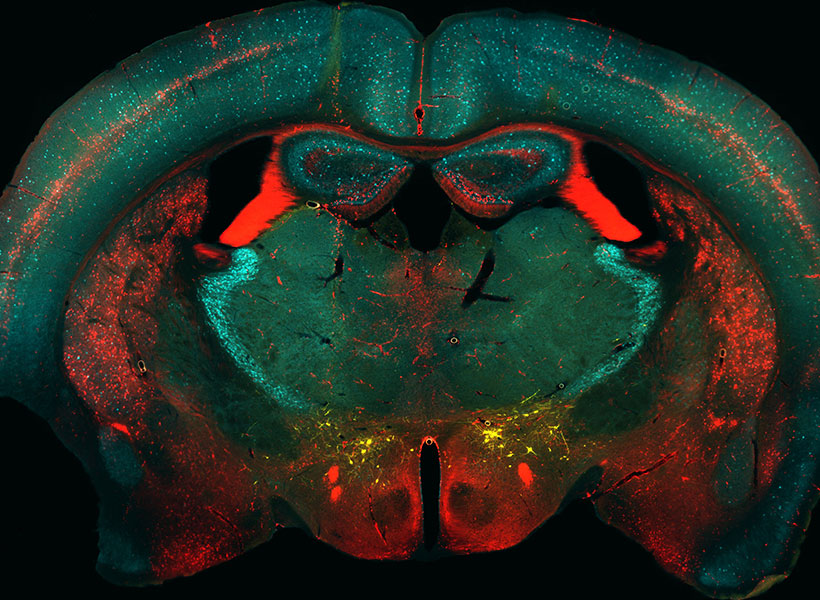
New MRI probe can reveal more of the brain’s inner workings

Using a novel probe for functional magnetic resonance imaging (fMRI), MIT biological engineers have devised a way to monitor individual populations of neurons and reveal how they interact with each other.

Similar to how the gears of a clock interact in specific ways to turn the clock’s hands, different parts of the brain interact to perform a variety of tasks, such as generating behavior or interpreting the world around us. The new MRI probe could potentially allow scientists to map those networks of interactions.

“With regular fMRI, we see the action of all the gears at once. But with our new technique, we can pick up individual gears that are defined by their relationship to the other gears, and that’s critical for building up a picture of the mechanism of the brain,” says Alan Jasanoff, an MIT professor of biological engineering, brain and cognitive sciences, and nuclear science and engineering.

Using this technique, which involves genetically targeting the MRI probe to specific populations of cells in animal models, the researchers were able to identify neural populations involved in a circuit that responds to rewarding stimuli. The new MRI probe could also enable studies of many other brain circuits, the researchers say.

Jasanoff, who is also an associate investigator at the McGovern Institute, is the senior author of the study, which appears today in Nature Neuroscience. The lead authors of the paper are recent MIT PhD recipient Souparno Ghosh and former MIT research scientist Nan Li.

Tracing connections

Traditional fMRI imaging measures changes to blood flow in the brain, as a proxy for neural activity. When neurons receive signals from other neurons, it triggers an influx of calcium, which causes a diffusible gas called nitric oxide to be released. Nitric oxide acts in part as a vasodilator that increases blood flow to the area.

Imaging calcium directly can offer a more precise picture of brain activity, but that type of imaging usually requires fluorescent chemicals and invasive procedures. The MIT team wanted to develop a method that could work across the brain without that type of invasiveness.

“If we want to figure out how brain-wide networks of cells and brain-wide mechanisms function, we need something that can be detected deep in tissue and preferably across the entire brain at once,” Jasanoff says. “The way that we chose to do that in this study was to essentially hijack the molecular basis of fMRI itself.”

The researchers created a genetic probe, delivered by viruses, that codes for a protein that sends out a signal whenever the neuron is active. This protein, which the researchers called NOSTIC (nitric oxide synthase for targeting image contrast), is an engineered form of an enzyme called nitric oxide synthase. The NOSTIC protein can detect elevated calcium levels that arise during neural activity; it then generates nitric oxide, leading to an artificial fMRI signal that arises only from cells that contain NOSTIC.

The probe is delivered by a virus that is injected into a particular site, after which it travels along axons of neurons that connect to that site. That way, the researchers can label every neural population that feeds into a particular location.

“When we use this virus to deliver our probe in this way, it causes the probe to be expressed in the cells that provide input to the location where we put the virus,” Jasanoff says. “Then, by performing functional imaging of those cells, we can start to measure what makes input to that region take place, or what types of input arrive at that region.”

Turning the gears

In the new study, the researchers used their probe to label populations of neurons that project to the striatum, a region that is involved in planning movement and responding to reward. In rats, they were able to determine which neural populations send input to the striatum during or immediately following a rewarding stimulus — in this case, deep brain stimulation of the lateral hypothalamus, a brain center that is involved in appetite and motivation, among other functions.

One question that researchers have had about deep brain stimulation of the lateral hypothalamus is how wide-ranging the effects are. In this study, the MIT team showed that several neural populations, located in regions including the motor cortex and the entorhinal cortex, which is involved in memory, send input into the striatum following deep brain stimulation.

“It’s not simply input from the site of the deep brain stimulation or from the cells that carry dopamine. There are these other components, both distally and locally, that shape the response, and we can put our finger on them because of the use of this probe,” Jasanoff says.

During these experiments, neurons also generate regular fMRI signals, so in order to distinguish the signals that are coming specifically from the genetically altered neurons, the researchers perform each experiment twice: once with the probe on, and once following treatment with a drug that inhibits the probe. By measuring the difference in fMRI activity between these two conditions, they can determine how much activity is present in probe-containing cells specifically.

The researchers now hope to use this approach, which they call hemogenetics, to study other networks in the brain, beginning with an effort to identify some of the regions that receive input from the striatum following deep brain stimulation.

“One of the things that’s exciting about the approach that we’re introducing is that you can imagine applying the same tool at many sites in the brain and piecing together a network of interlocking gears, which consist of these input and output relationships,” Jasanoff says. “This can lead to a broad perspective on how the brain works as an integrated whole, at the level of neural populations.”

The research was funded by the National Institutes of Health and the MIT Simons Center for the Social Brain.

School of Engineering welcomes new faculty

The School of Engineering is welcoming 17 new faculty members to its departments, institutes, labs, and centers. With research and teaching activities ranging from the development of robotics and machine learning technologies to modeling the impact of elevated carbon dioxide levels on vegetation, they are poised to make significant contributions in new directions across the school and to a wide range of research efforts around the Institute.

“I am delighted to welcome our wonderful new faculty,” says Anantha Chandrakasan, dean of the MIT School of Engineering and Vannevar Bush Professor of Electrical Engineering and Computer Science. “Their impact as talented educators, researchers, collaborators, and mentors will be felt across the School of Engineering and beyond as they strengthen our engineering community.”

Among the new faculty members are four from the Department of Electrical Engineering and Computer Science (EECS), which jointly reports into the School of Engineering and the MIT Stephen A. Schwarzman College of Computing.

Iwnetim “Tim” Abate will join the Department of Materials Science and Engineering in July 2023. He is currently both a Miller and Presidential Postdoctoral Fellow at the University of California at Berkeley. He received his MS and PhD in materials science and engineering from Stanford University and BS in physics from Minnesota State University at Moorhead. He also has research experience in industry (IBM) and at national labs (Los Alamos and SLAC National Accelerator Laboratories). Utilizing computational and experimental approaches in tandem, his research program at MIT will focus on the intersection of material chemistry, electrochemistry, and condensed matter physics to develop solutions for climate change and smart agriculture, including next-generation battery and sensor devices. Abate is also a co-founder and president of a nonprofit organization, SciFro Inc., working on empowering the African youth and underrepresented minorities in the United States to solve local problems through scientific research and innovation. He will continue working on expanding the vision and impact of SciFro with the MIT community. Abate received the Dan Cubicciotti Award of the Electrochemical Society, the EDGE and DARE graduate fellowships, the United Technologies Research Center fellowship, the John Stevens Jr. Memorial Award and the Justice, Equity, Diversity and Inclusion Graduation Award from Stanford University. He will hold the Toyota Career Development Professorship at MIT.

Kaitlyn Becker will join the Department of Mechanical Engineering as an assistant professor in August 2022. Becker received her PhD in materials science and mechanical engineering from Harvard University in 2021 and previously worked in industry as a manufacturing engineer at Cameron Health and a senior engineer for Nano Terra, Inc. She is a postdoc at the Harvard University School of Engineering and Applied Sciences and is also currently a senior glassblowing instructor in the Department of Materials Science and Engineering at MIT. Becker works on adaptive soft robots for grasping and manipulation of delicate structures from the desktop to the deep sea. Her research focuses on novel soft robotic platforms, adding functionality through innovations at the intersection of design and fabrication. She has developed novel fabrication methodologies and mechanical programming methods for large integrated arrays of soft actuators capable of collective manipulation and locomotion, and demonstrated integration of microfluidic circuits to control arrays of multichannel, two-degrees-of-freedom soft actuators. Becker received the National Science Foundation Graduate Research Fellowship in 2015, the Microsoft Graduate Women’s Scholarship in 2015, the Winston Chen Graduate Fellowship in 2015, and the Courtlandt S. Gross Memorial Scholarship in 2014.

Brandon J. DeKosky joined the Department of Chemical Engineering as an assistant professor in a newly introduced joint faculty position between the department and the Ragon Institute of MGH, MIT, and Harvard in September 2021. He received his BS in chemical engineering from University of Kansas and his PhD in chemical engineering from the University of Texas at Austin. He then did postdoctoral research at the Vaccine Research Center of the National Institute of Infectious Diseases. In 2017, Brandon launched his independent academic career as an assistant professor at the University of Kansas in a joint position with the Department of Chemical Engineering and the Department of Pharmaceutical Chemistry. He was also a member of the bioengineering graduate program. His research program focuses on developing and applying a suite of new high-throughput experimental and computational platforms for molecular analysis of adaptive immune responses, to accelerate precision drug discovery. He has received several notable recognitions, which include receipt of the NIH K99 Path to Independence and NIH DP5 Early Independence awards, the Cellular and Molecular Bioengineering Rising Star Award from the Biomedical Engineering Society, and the Career Development Award from the Congressionally Directed Medical Research Program’s Peer Reviewed Cancer Research Program.

Mohsen Ghaffari will join the Department of Electrical Engineering and Computer Science in April 2022. He received his BS from the Sharif University of Technology, and his MS and PhD in EECS from MIT. His research focuses on distributed and parallel algorithms for large graphs. Ghaffari received the ACM Doctoral Dissertation Honorable Mention Award, the ACM-EATCS Principles of Distributed Computing Doctoral Dissertation Award, and the George M. Sprowls Award for Best Computer Science PhD thesis at MIT. Before coming to MIT, he was on the faculty at ETH Zurich, where he received a prestigious European Research Council Starting Grant.

Aristide Gumyusenge joined the Department of Materials Science and Engineering in January. He is currently a postdoc at Stanford University working with Professor Zhenan Bao and Professor Alberto Salleo. He received a BS in chemistry from Wofford College in 2015 and a PhD in chemistry from Purdue University in 2019. His research background and interests are in semiconducting polymers, their processing and characterization, and their unique role in the future of electronics. Particularly, he has tackled longstanding challenges in operation stability of semiconducting polymers under extreme heat and has pioneered high-temperature plastic electronics. He has been selected as a PMSE Future Faculty Scholar (2021), the GLAM Postdoctoral Fellow (2020-22), and the MRS Arthur Nowick and Graduate Student Gold Awardee (2019), among other recognitions. At MIT, he will lead the Laboratory of Organic Materials for Smart Electronics (OMSE Lab). Through polymer design, novel processing strategies, and large-area manufacturing of electronic devices, he is interested in relating molecular design to device performance, especially transistor devices able to mimic and interface with biological systems. He will hold the Merton C. Flemings Career Development Professorship.

Mina Konakovic Lukovic will join the Department of Electrical Engineering and Computer Science as an assistant professor in July 2022. She received her BS and MS from the University of Belgrade, Faculty of Mathematics. She earned her PhD in 2019 in the School of Computer and Communication Sciences at the Swiss Federal Institute of Technology Lausanne, advised by Professor Mark Pauly. Currently a Schmidt Science Postdoctoral Fellow in MIT’s Computer Science and Artificial Intelligence Laboratory (CSAIL), she has been mentored by Professor Wojciech Matusik. Her research focuses on computer graphics, computational fabrication, 3D geometry processing, and machine learning, including architectural geometry and the design of programmable materials. She received the ACM SIGGRAPH Outstanding Doctoral Dissertation Honorable Mention, the Eurographics PhD Award, and was recently awarded the 2021 SIAM Activity Group on Geometric Design Early Career Prize.

Darcy McRose will join the Department of Civil and Environmental Engineering as an assistant professor in August 2022. She completed a BS in Earth systems at Stanford and a PhD in geosciences at Princeton University. Darcy is currently conducting postdoctoral work at Caltech, where she is mentored by Professor Dianne Newman in the divisions of Biology and Biological Engineering and Geological and Planetary Sciences. Her research program focuses on microbe-environment interactions and their effects on biogeochemical cycles, and incorporates techniques ranging from microbial physiology and genetics to geochemistry. A particular emphasis for this work is the production and use of secondary metabolites and small molecules in soils and sediments. McRose received the Caltech BBE Division postdoctoral fellowship in 2019 and is currently a Simons Foundation Marine Microbial Ecology postdoctoral fellow as well as a L’Oréal USA for Women in Science fellow.

Qin (Maggie) Qi joined the Department of Chemical Engineering as an assistant professor in January 2022. She received two BS degrees in chemical engineering and in operations research from Cornell University, before moving on to Stanford for her PhD. She then took on a postdoc position at Harvard University School of Engineering and Applied Sciences and the Wyss Institute. Maggie’s proposed research includes combining extensive theoretical and computational work on predictive models that guide experimental design. She seeks to investigate particle-cell biomechanics and function for better targeted cell-based therapies. She also plans to design microphysiological systems that elucidate hydrodynamics in complex organs, including delivery of drugs to the eye, and to examine ionic liquids as complex fluids for biomaterial design. She aims to push the boundaries of fluid mechanics, transport phenomena, and soft matter for human health and to innovate precision health care solutions. Maggie received the T.S. Lo Graduate Fellowship and the Stanford Graduate Fellowship in Science and Engineering. Among her accomplishments, Maggie was a participant in the inaugural class of the MIT Rising Stars in ChemE Program in 2018.

Manish Raghavan will join the MIT Sloan School of Management and the Department of Electrical Engineering and Computer Science as an assistant professor in September 2022. He shares a joint appointment with the MIT Schwarzman College of Computing. He received a bachelor’s degree in electrical engineering and computer science from the University of California at Berkeley, and PhD from the Computer Science department at Cornell University. Prior to joining MIT, he was a postdoc at the Harvard Center for Research on Computation and Society. His research interests lie in the application of computational techniques to domains of social concern, including algorithmic fairness and behavioral economics, with a particular focus on the use of algorithmic tools in the hiring pipeline. He is also a member of Cornell’s Artificial Intelligence, Policy, and Practice initiative and Mechanism Design for Social Good.

Ritu Raman joined the Department of Mechanical Engineering as an assistant professor and Brit and Alex d’Arbeloff Career Development Chair in August 2021. Raman received her PhD in mechanical engineering from the University of Illinois at Urbana-Champaign as an NSF Graduate Research Fellow in 2016 and completed a postdoctoral fellowship with Professor Robert Langer at MIT, funded by a NASEM Ford Foundation Fellowship and a L’Oréal USA For Women in Science Fellowship. Raman’s lab designs adaptive living materials powered by assemblies of living cells for applications ranging from medicine to machines. Currently, she is focused on using biological materials and engineering tools to build living neuromuscular tissues. Her goal is to help restore mobility to those who have lost it after disease or trauma and to deploy biological actuators as functional components in machines. Raman published the book Biofrabrication with MIT Press in September 2021. She was in the MIT Technology Review “35 Innovators Under 35” 2019 class, the Forbes “30 Under 30” 2018 class, and has received numerous awards including being named a National Academy of Sciences Kavli Frontiers of Science Fellow in 2020 and receiving the Science and Sartorius Prize for Regenerative Medicine and Cell Therapy in 2019. Ritu has championed many initiatives to empower women in science, including being named an AAAS IF/THEN ambassador and founding the Women in Innovation and Stem Database at MIT (WISDM).

Nidhi Seethapathi joined the Department of Brain and Cognitive Sciences and the Department of Electrical Engineering and Computer Science in January 2022. She shares a joint appointment with the MIT Schwarzman College of Computing. She received a bachelor’s degree in mechanical engineering from Veermata Jijabai Technological Institute and a PhD from the Movement Lab at Ohio State University. Her research interests include building computational predictive models of human movement with applications to autonomous and robot-aided neuromotor rehabilitation. In her work, she uses a combination of tools and approaches from dynamics, control theory, and machine learning. During her PhD, she was a Schlumberger Foundation Faculty for the Future Fellow. She then worked as a postdoc in the Kording Lab at University of Pennsylvania, developing data-driven tools for autonomous neuromotor rehabilitation, in collaboration with the Rehabilitation Robotics Lab.

Vincent Sitzmann will join the Department of Electrical Engineering and Computer Science as an assistant professor in July 2022. He earned his BS from the Technical University of Munich in 2015, his MS from Stanford in 2017, and his PhD from Stanford in 2020. At MIT, he will be the principal investigator of the Scene Representation Group, where he will lead research at the intersection of machine learning, graphics, neural rendering, and computer vision to build algorithms that learn to reconstruct, understand, and interact with 3D environments from incomplete observations the way humans can. Currently, Vincent is a postdoc at the MIT Computer Science and Artificial Intelligence Laboratory with Josh Tenenbaum, Bill Freeman, and Fredo Durand. Along with multiple scholarships and fellowships, he has been recognized with the NeurIPS Honorable Mention: Outstanding New Directions in 2019.

Tess Smidt joined the Department of Electrical Engineering and Computer Science as an assistant professor in September 2021. She earned her SB in physics from MIT in 2012 and her PhD in physics from the University of California at Berkeley in 2018. She is the principal investigator of the Atomic Architects group at the Research Laboratory of Electronics, where she works at the intersection of physics, geometry, and machine learning to design algorithms that aid in the understanding and design of physical systems. Her research focuses on machine learning that incorporates physical and geometric constraints, with applications to materials design. Prior to joining the MIT EECS faculty, she was the 2018 Alvarez Postdoctoral Fellow in Computing Sciences at Lawrence Berkeley National Laboratory and a software engineering intern on the Google Accelerated Sciences team, where she developed Euclidean symmetry equivariant neural networks which naturally handle 3D geometry and geometric tensor data.

Loza Tadesse will join the Department of Mechanical Engineering as an assistant professor in July 2023. She received her PhD in bioengineering from Stanford University in 2021 and previously was a medical student at St. Paul Hospital Millennium Medical College in Ethiopia. She is currently a postdoc at the University of California at Berkeley. Tadesse’s past research combines Raman spectroscopy and machine learning to develop a rapid, all-optical, and label-free bacterial diagnostic and antibiotic susceptibility testing system that aims to circumvent the time-consuming culturing step in “gold standard” methods. She aims to establish a research program that develops next-generation point-of-care diagnostic devices using spectroscopy, optical, and machine learning tools for application in resource limited clinical settings such as developing nations, military sites, and space exploration. Tadesse has been listed as a 2022 Forbes “30 Under 30” in health care, received many awards including the Biomedical Engineering Society (BMES) Career Development Award, the Stanford DARE Fellowship and the Gates Foundation “Call to Action” $200,000 grant for SciFro Inc., an educational nonprofit in Ethiopia, which she co-founded.

César Terrer joined the Department of Civil and Environmental Engineering as an assistant professor in July 2021. He obtained his PhD in ecosystem ecology and climate change from Imperial College London, where he started working at the interface between experiments and models to better understand the effects of elevated carbon dioxide on vegetation. His research has advanced the understanding on the effects of carbon dioxide in terrestrial ecosystems, the role of soil nutrients in a climate change context, and plant-soil interactions. Synthesizing observational data from carbon dioxide experiments and satellites through meta-analysis and machine learning, César has found that microbial interactions between plants and soils play a major role in the carbon cycle at a global scale, affecting the speed of global warming.

Haruko Wainwright joined the Department of Nuclear Science and Engineering as an assistant professor in January 2021. She received her BEng in engineering physics from Kyoto University, Japan in 2003, her MS in nuclear engineering in 2006, her MA in statistics in 2010, and her PhD in nuclear engineering in 2010 from University of California at Berkeley. Before joining MIT, she was a staff scientist in the Earth and Environmental Sciences Area at Lawrence Berkeley National Laboratory and an adjunct professor in nuclear engineering at UC Berkeley. Her research focuses on environmental modeling and monitoring technologies, with a particular emphasis on nuclear waste and nuclear-related contamination. She has been developing Bayesian methods for multi-type multiscale data integration and model-data integration. She leads and co-leads multiple interdisciplinary projects, including the U.S. Department of Energy’s Advanced Long-term Environmental Monitoring Systems (ALTEMIS) project, and the Artificial Intelligence for Earth System Predictability (AI4ESP) initiative.

Martin Wainwright will join the Department of Electrical Engineering and Computer Science in July 2022. He received a bachelor’s degree in mathematics from University of Waterloo, Canada, and PhD in EECS from MIT. Prior to joining MIT, he was the Chancellor’s Professor at the University of California at Berkeley, with a joint appointment between the Department of Statistics and the Department of EECS. His research interests include high-dimensional statistics, statistical machine learning, information theory, and optimization theory. Among other awards, he has received the COPSS Presidents’ Award (2014) from the Joint Statistical Societies, the David Blackwell Lectureship (2017), and Medallion Lectureship (2013) from the Institute of Mathematical Statistics, and Best Paper awards from the IEEE Signal Processing Society and IEEE Information Theory Society. He was a Section Lecturer at the International Congress of Mathematicians in 2014.


Augmented: The journey of Hugh Herr

Augmented is a Nova PBS documentary that premiered in February 2022, featuring Hugh Herr, the co-director of the K. Lisa Yang Center for Bionics at MIT.

Follow the dramatic personal journey of Hugh Herr, a biophysicist working to create brain-controlled robotic limbs. At age 17, Herr’s legs were amputated after a climbing accident. Frustrated by the crude prosthetic limbs he was given, Herr set out to remedy their design, leading him to a career as an inventor of innovative prosthetic devices. Now, Herr is teaming up with an injured climber and a surgeon at a leading Boston hospital to test a new approach to surgical amputation that allows prosthetic limbs to move and feel like the real thing. Herr’s journey is a powerful tale of innovation and the inspiring story of a personal tragedy transformed into a life-long quest to help others.

Read more at PBS.org.

David Ginty named winner of 2022 Scolnick Prize

Harvard neurobiologist David Ginty, winner of the 2022 Scolnick Prize.

The McGovern Institute for Brain Research announced today that Harvard neurobiologist David D. Ginty has been selected for the 2022 Edward M. Scolnick Prize in Neuroscience. Ginty, who is the Edward R. and Anne G. Lefler Professor of Neurobiology at Harvard Medical School, is being recognized for his fundamental discoveries into the neural mechanisms underlying the sense of touch. The Scolnick Prize is awarded annually by the McGovern Institute for outstanding advances in neuroscience.

“David Ginty has made seminal contributions in basic research that also have important translational implications,” says Robert Desimone, McGovern Institute Director and chair of the selection committee. “His rigorous research has led us to understand how the peripheral nervous system encodes the overall perception of touch, and how molecular mechanisms underlying this can fail in disease states.”

Ginty obtained his PhD in 1989 with Edward Seidel where he studied cell proliferation factors. He went on to a postdoctoral fellowship researching nerve growth factor with John Wagner at the Dana-Farber Cancer Institute and, upon Wagner’s departure to Cornell, transferred to Michael Greenberg’s lab at Harvard Medical School. There, he dissected intracellular signaling pathways for neuronal growth factors and neurotransmitters and developed key antibody reagents to detect activated forms of transcription factors. These antibody tools are now used by labs around the world in the research of neuronal plasticity and brain disorders, including Alzheimer’s disease and schizophrenia.

In 1995, Ginty started his own laboratory at Johns Hopkins University with a focus on the development and functional organization of the peripheral nervous system. Ginty’s group created and applied the latest genetic engineering techniques in mice to uncover how the peripheral nervous system develops and is organized at the molecular, cellular and circuit levels to perceive touch. Most notably, using gene targeting combined with electrophysiological, behavioral and anatomical analyses, the Ginty lab untangled properties and functions of the different types of touch neurons, termed low- and high-threshold mechanoreceptors, that convey distinct aspects of stimulus information from the skin to the central nervous system. Ginty and colleagues also discovered organizational principles of spinal cord and brainstem circuits dedicated to touch information processing, and that integration of signals from the different mechanoreceptor types begins within spinal cord networks before signal transmission to the brain.

In 2013, Ginty joined the faculty of Harvard Medical School where his team applied their genetic tools and techniques to probe the neural basis of touch sensitivity disorders. They discovered properties and functions of peripheral sensory neurons, spinal cord circuits, and ascending pathways that transmit noxious, painful stimuli from the periphery to the brain. They also asked whether abnormalities in peripheral nervous system function lead to touch over-reactivity in cases of autism or in neuropathic pain caused by nerve injury, chemotherapy, or diabetes, where even a soft touch can be aversive or painful. His team found that sensory abnormalities observed in several mouse models of autism spectrum disorder could be traced to peripheral mechanosensory neurons. They also found that reducing the activity of peripheral sensory neurons prevented tactile over-reactivity in these models and even, in some cases, lessened anxiety and abnormal social behaviors. These findings provided a plausible explanation for how sensory dysfunction may contribute to physiological and cognitive impairments in autism. Importantly, this laid the groundwork for a new approach and initiative to identify new potential therapies for disorders of touch and pain.

Ginty was named a Howard Hughes Medical Institute Investigator in 2000 and was elected to the American Academy of Arts and Sciences in 2015 and the National Academy of Sciences in 2017. He shared Columbia University’s Alden W. Spencer Prize with Ardem Patapoutian in 2017 and was awarded the Society for Neuroscience Julius Axelrod Prize in 2021. Ginty is also known for exceptional mentorship. He directed the neuroscience graduate program at Johns Hopkins from 2006 to 2013 and now serves as the associate director of Harvard’s neurobiology graduate program.

The McGovern Institute will award the Scolnick Prize to Ginty on Wednesday, June 1, 2022. At 4:00 pm he will deliver a lecture entitled “The sensory neurons of touch: beauty is skin deep,” to be followed by a reception at the McGovern Institute, 43 Vassar Street (building 46, room 3002) in Cambridge. The event is free and open to the public; registration is required.

Singing in the brain

Press Mentions

For the first time, MIT neuroscientists have identified a population of neurons in the human brain that lights up when we hear singing, but not other types of music.

These neurons, found in the auditory cortex, appear to respond to the specific combination of voice and music, but not to either regular speech or instrumental music. Exactly what they are doing is unknown and will require more work to uncover, the researchers say.

“The work provides evidence for relatively fine-grained segregation of function within the auditory cortex, in a way that aligns with an intuitive distinction within music,” says Sam Norman-Haignere, a former MIT postdoc who is now an assistant professor of neuroscience at the University of Rochester Medical Center.

The work builds on a 2015 study in which the same research team used functional magnetic resonance imaging (fMRI) to identify a population of neurons in the brain’s auditory cortex that responds specifically to music. In the new work, the researchers used recordings of electrical activity taken at the surface of the brain, which gave them much more precise information than fMRI.

“There’s one population of neurons that responds to singing, and then very nearby is another population of neurons that responds broadly to lots of music. At the scale of fMRI, they’re so close that you can’t disentangle them, but with intracranial recordings, we get additional resolution, and that’s what we believe allowed us to pick them apart,” says Norman-Haignere.

Norman-Haignere is the lead author of the study, which appears today in the journal Current Biology. Josh McDermott, an associate professor of brain and cognitive sciences, and Nancy Kanwisher, the Walter A. Rosenblith Professor of Cognitive Neuroscience, both members of MIT’s McGovern Institute for Brain Research and Center for Brains, Minds and Machines (CBMM), are the senior authors of the study.

Neural recordings

In their 2015 study, the researchers used fMRI to scan the brains of participants as they listened to a collection of 165 sounds, including different types of speech and music, as well as everyday sounds such as finger tapping or a dog barking. For that study, the researchers devised a novel method of analyzing the fMRI data, which allowed them to identify six neural populations with different response patterns, including the music-selective population and another population that responds selectively to speech.

In the new study, the researchers hoped to obtain higher-resolution data using a technique known as electrocorticography (ECoG), which allows electrical activity to be recorded by electrodes placed inside the skull. This offers a much more precise picture of electrical activity in the brain compared to fMRI, which measures blood flow in the brain as a proxy of neuron activity.

“With most of the methods in human cognitive neuroscience, you can’t see the neural representations,” Kanwisher says. “Most of the kind of data we can collect can tell us that here’s a piece of brain that does something, but that’s pretty limited. We want to know what’s represented in there.”

Electrocorticography cannot be typically be performed in humans because it is an invasive procedure, but it is often used to monitor patients with epilepsy who are about to undergo surgery to treat their seizures. Patients are monitored over several days so that doctors can determine where their seizures are originating before operating. During that time, if patients agree, they can participate in studies that involve measuring their brain activity while performing certain tasks. For this study, the MIT team was able to gather data from 15 participants over several years.

For those participants, the researchers played the same set of 165 sounds that they used in the earlier fMRI study. The location of each patient’s electrodes was determined by their surgeons, so some did not pick up any responses to auditory input, but many did. Using a novel statistical analysis that they developed, the researchers were able to infer the types of neural populations that produced the data that were recorded by each electrode.

“When we applied this method to this data set, this neural response pattern popped out that only responded to singing,” Norman-Haignere says. “This was a finding we really didn’t expect, so it very much justifies the whole point of the approach, which is to reveal potentially novel things you might not think to look for.”

That song-specific population of neurons had very weak responses to either speech or instrumental music, and therefore is distinct from the music- and speech-selective populations identified in their 2015 study.

Music in the brain

In the second part of their study, the researchers devised a mathematical method to combine the data from the intracranial recordings with the fMRI data from their 2015 study. Because fMRI can cover a much larger portion of the brain, this allowed them to determine more precisely the locations of the neural populations that respond to singing.

“This way of combining ECoG and fMRI is a significant methodological advance,” McDermott says. “A lot of people have been doing ECoG over the past 10 or 15 years, but it’s always been limited by this issue of the sparsity of the recordings. Sam is really the first person who figured out how to combine the improved resolution of the electrode recordings with fMRI data to get better localization of the overall responses.”

The song-specific hotspot that they found is located at the top of the temporal lobe, near regions that are selective for language and music. That location suggests that the song-specific population may be responding to features such as the perceived pitch, or the interaction between words and perceived pitch, before sending information to other parts of the brain for further processing, the researchers say.

The researchers now hope to learn more about what aspects of singing drive the responses of these neurons. They are also working with MIT Professor Rebecca Saxe’s lab to study whether infants have music-selective areas, in hopes of learning more about when and how these brain regions develop.

The research was funded by the National Institutes of Health, the U.S. Army Research Office, the National Science Foundation, the NSF Science and Technology Center for Brains, Minds, and Machines, the Fondazione Neurone, the Howard Hughes Medical Institute, and the Kristin R. Pressman and Jessica J. Pourian ’13 Fund at MIT.

On a mission to alleviate chronic pain

About 50 million Americans suffer from chronic pain, which interferes with their daily life, social interactions, and ability to work. MIT Professor Fan Wang wants to develop new ways to help relieve that pain, by studying and potentially modifying the brain’s own pain control mechanisms.

Her recent work has identified an “off switch” for pain, located in the brain’s amygdala. She hopes that finding ways to control this switch could lead to new treatments for chronic pain.

“Chronic pain is a major societal issue,” Wang says. “By studying pain-suppression neurons in the brain’s central amygdala, I hope to create a new therapeutic approach for alleviating pain.”

Wang, who joined the MIT faculty in January 2021, is also the leader of a new initiative at the McGovern Institute for Brain Research that is studying drug addiction, with the goal of developing more effective treatments for addiction.

“Opioid prescription for chronic pain is a major contributor to the opioid epidemic. With the Covid pandemic, I think addiction and overdose are becoming worse. People are more anxious, and they seek drugs to alleviate such mental pain,” Wang says. “As scientists, it’s our duty to tackle this problem.”

Sensory circuits

Wang, who grew up in Beijing, describes herself as “a nerdy child” who loved books and math. In high school, she took part in science competitions, then went on to study biology at Tsinghua University. She arrived in the United States in 1993 to begin her PhD at Columbia University. There, she worked on tracing the connection patterns of olfactory receptor neurons in the lab of Richard Axel, who later won the Nobel Prize for his discoveries of odorant receptors and how the olfactory system is organized.

After finishing her PhD, Wang decided to switch gears. As a postdoc at the University of California at San Francisco and then Stanford University, she began studying how the brain perceives touch.

In 2003, Wang joined the faculty at Duke University School of Medicine. There, she began developing techniques to study the brain circuits that underlie the sense of touch, tracing circuits that carry sensory information from the whiskers of mice to the brain. She also studied how the brain integrates movements of touch organs with signals of sensory stimuli to generate perception (such as using stretching movements to sense elasticity).

As she pursued her sensory perception studies, Wang became interested in studying pain perception, but she felt she needed to develop new techniques to tackle it. While at Duke, she invented a technique called CANE (capturing activated neural ensembles), which can identify networks of neurons that are activated by a particular stimulus.

Using this approach in mice, she identified neurons that become active in response to pain, but so many neurons across the brain were activated that it didn’t offer much useful information. As a way to indirectly get at how the brain controls pain, she decided to use CANE to explore the effects of drugs used for general anesthesia. During general anesthesia, drugs render a patient unconscious, but Wang hypothesized that the drugs might also shut off pain perception.

“At that time, it was just a wild idea,” Wang recalls. “I thought there may be other mechanisms — that instead of just a loss of consciousness, anesthetics may do something to the brain that actually turns pain off.”

Support for the existence of an “off switch” for pain came from the observation that wounded soldiers on a battlefield can continue to fight, essentially blocking out pain despite their injuries.

In a study of mice treated with anesthesia drugs, Wang discovered that the brain does have this kind of switch, in an unexpected location: the amygdala, which is involved in regulating emotion. She showed that this cluster of neurons can turn off pain when activated, and when it is suppressed, mice become highly sensitive to ordinary gentle touch.

“There’s a baseline level of activity that makes the animals feel normal, and when you activate these neurons, they’ll feel less pain. When you silence them, they’ll feel more pain,” Wang says.

Turning off pain

That finding, which Wang reported in 2020, raised the possibility of somehow modulating that switch in humans to try to treat chronic pain. This is a long-term goal of Wang’s, but more work is required to achieve it, she says. Currently her lab is working on analyzing the RNA expression patterns of the neurons in the cluster she identified. They also are measuring the neurons’ electrical activity and how they interact with other neurons in the brain, in hopes of identifying circuits that could be targeted to tamp down the perception of pain.

One way of modulating these circuits could be to use deep brain stimulation, which involves implanting electrodes in certain areas of the brain. Focused ultrasound, which is still in early stages of development and does not require surgery, could be a less invasive alternative.

Another approach Wang is interested in exploring is pairing brain stimulation with a context such as looking at a smartphone app. This kind of pairing could help train the brain to shut off pain using the app, without the need for the original stimulation (deep brain stimulation or ultrasound).

“Maybe you don’t need to constantly stimulate the brain. You may just need to reactivate it with a context,” Wang says. “After a while you would probably need to be restimulated, or reconditioned, but at least you have a longer window where you don’t need to go to the hospital for stimulation, and you just need to use a context.”

Wang, who was drawn to MIT in part by its focus on fostering interdisciplinary collaborations, is now working with several other McGovern Institute members who are taking different angles to try to figure out how the brain generates the state of craving that occurs in drug addiction, including opioid addiction.

“We’re going to focus on trying to understand this craving state: how it’s created in the brain and how can we sort of erase that trace in the brain, or at least control it. And then you can neuromodulate it in real time, for example, and give people a chance to get back their control,” she says.

Dendrites may help neurons perform complicated calculations

Within the human brain, neurons perform complex calculations on information they receive. Researchers at MIT have now demonstrated how dendrites — branch-like extensions that protrude from neurons — help to perform those computations.

The researchers found that within a single neuron, different types of dendrites receive input from distinct parts of the brain, and process it in different ways. These differences may help neurons to integrate a variety of inputs and generate an appropriate response, the researchers say.

In the neurons that the researchers examined in this study, it appears that this dendritic processing helps cells to take in visual information and combine it with motor feedback, in a circuit that is involved in navigation and planning movement.

“Our hypothesis is that these neurons have the ability to pick out specific features and landmarks in the visual environment, and combine them with information about running speed, where I’m going, and when I’m going to start, to move toward a goal position,” says Mark Harnett, an associate professor of brain and cognitive sciences, a member of MIT’s McGovern Institute for Brain Research, and the senior author of the study.

Mathieu Lafourcade, a former MIT postdoc, is the lead author of the paper, which appears today in Neuron.

Complex calculations

Any given neuron can have dozens of dendrites, which receive synaptic input from other neurons. Neuroscientists have hypothesized that these dendrites can act as compartments that perform their own computations on incoming information before sending the results to the body of the neuron, which integrates all these signals to generate an output.

Previous research has shown that dendrites can amplify incoming signals using specialized proteins called NMDA receptors. These are voltage-sensitive neurotransmitter receptors that are dependent on the activity of other receptors called AMPA receptors. When a dendrite receives many incoming signals through AMPA receptors at the same time, the threshold to activate nearby NMDA receptors is reached, creating an extra burst of current.

This phenomenon, known as supralinearity, is believed to help neurons distinguish between inputs that arrive close together or farther apart in time or space, Harnett says.

In the new study, the MIT researchers wanted to determine whether different types of inputs are targeted specifically to different types of dendrites, and if so, how that would affect the computations performed by those neurons. They focused on a population of neurons called pyramidal cells, the principal output neurons of the cortex, which have several different types of dendrites. Basal dendrites extend below the body of the neuron, apical oblique dendrites extend from a trunk that travels up from the body, and tuft dendrites are located at the top of the trunk.

Harnett and his colleagues chose a part of the brain called the retrosplenial cortex (RSC) for their studies because it is a good model for association cortex — the type of brain cortex used for complex functions such as planning, communication, and social cognition. The RSC integrates information from many parts of the brain to guide navigation, and pyramidal neurons play a key role in that function.

In a study of mice, the researchers first showed that three different types of input come into pyramidal neurons of the RSC: from the visual cortex into basal dendrites, from the motor cortex into apical oblique dendrites, and from the lateral nuclei of the thalamus, a visual processing area, into tuft dendrites.

“Until now, there hasn’t been much mapping of what inputs are going to those dendrites,” Harnett says. “We found that there are some sophisticated wiring rules here, with different inputs going to different dendrites.”

A range of responses

The researchers then measured electrical activity in each of those compartments. They expected that NMDA receptors would show supralinear activity, because this behavior has been demonstrated before in dendrites of pyramidal neurons in both the primary sensory cortex and the hippocampus.

In the basal dendrites, the researchers saw just what they expected: Input coming from the visual cortex provoked supralinear electrical spikes, generated by NMDA receptors. However, just 50 microns away, in the apical oblique dendrites of the same cells, the researchers found no signs of supralinear activity. Instead, input to those dendrites drives a steady linear response. Those dendrites also have a much lower density of NMDA receptors.

“That was shocking, because no one’s ever reported that before,” Harnett says. “What that means is the apical obliques don’t care about the pattern of input. Inputs can be separated in time, or together in time, and it doesn’t matter. It’s just a linear integrator that’s telling the cell how much input it’s getting, without doing any computation on it.”

Those linear inputs likely represent information such as running speed or destination, Harnett says, while the visual information coming into the basal dendrites represents landmarks or other features of the environment. The supralinearity of the basal dendrites allows them to perform more sophisticated types of computation on that visual input, which the researchers hypothesize allows the RSC to flexibly adapt to changes in the visual environment.

In the tuft dendrites, which receive input from the thalamus, it appears that NMDA spikes can be generated, but not very easily. Like the apical oblique dendrites, the tuft dendrites have a low density of NMDA receptors. Harnett’s lab is now studying what happens in all of these different types of dendrites as mice perform navigation tasks.

The research was funded by a Boehringer Ingelheim Fonds PhD Fellowship, the National Institutes of Health, the James W. and Patricia T. Poitras Fund, the Klingenstein-Simons Fellowship Program, a Vallee Scholar Award, and a McKnight Scholar Award.

Seven new faculty join the MIT School of Science

This winter, seven new faculty members join the MIT School of Science in the departments of Biology and Brain and Cognitive Sciences.

Siniša Hrvatin studies how animals initiate, regulate, and survive states of stasis, such as torpor and hibernation. To survive extreme environments, many animals have evolved the ability to decrease metabolic rate and body temperature and enter dormant states. His long-term goal is to harness the potential of these biological adaptations to advance medicine. Previously, he identified the neurons that regulate mouse torpor and established a platform for the development of cell-type-specific viral drivers.
Hrvatin earned his bachelor’s degree in biochemical sciences in 2007 and his PhD in stem cell and regenerative medicine in 2013, both from Harvard University. He was then a postdoc in bioengineering at MIT and a postdoc in neurobiology at Harvard Medical School. Hrvatin returns to MIT as an assistant professor of biology and a member of the Whitehead Institute for Biomedical Research.

Sara Prescott investigates how sensory inputs from within the body control mammalian physiology and behavior. Specifically, she uses mammalian airways as a model system to explore how the cells that line the surface of the body communicate with parts of the nervous system. For example, what mechanisms elicit a reflexive cough? Prescott’s research considers the critical questions of how airway insults are detected, encoded, and adapted to mammalian airways with the ultimate goal of providing new ways to treat autonomic dysfunction.

Prescott earned her bachelor’s degree in molecular biology from Princeton University in 2008 followed by her PhD in developmental biology from Stanford University in 2016. Prior to joining MIT, she was a postdoc at Harvard Medical School and Howard Hughes Medical Institute. The Department of Biology welcomes Prescott as an assistant professor.
Alison Ringel is a T-cell immunologist with a background in biochemistry, biophysics, and structural biology. She investigates how environmental factors such as aging, metabolism, and diet impact tumor progress and the immune responses that cause tumor control. By mapping the environment around a tumor on a cellular level, she seeks to gain a molecular understanding of cancer risk factors.

Ringel received a bachelor’s degree in molecular biology, biochemistry, and physics from Wesleyan University, then a PhD in molecular biophysics from John Hopkins University School of Medicine. Previously, Ringel was a postdoc in the Department of Cell Biology at Harvard Medical School. She joins MIT as an assistant professor in the Department of Biology and a core member of the Ragon Institute of MGH, MIT and Harvard.

Francisco J. Sánchez-Rivera PhD ’16 investigates genetic variation with a focus on cancer. He integrates genome engineering technologies, genetically-engineered mouse models (GEMMs), and single cell lineage tracing and omics approaches in order to understand the mechanics of cancer development and evolution. With state-of-the-art technologies — including a CRISPR-based genome editing system he developed as a graduate student at MIT — he hopes to make discoveries in cancer genetics that will shed light on disease progression and pave the way for better therapeutic treatments.

Sánchez-Rivera received his bachelor’s degree in microbiology from the University of Puerto Rico at Mayagüez followed by a PhD in biology from MIT. He then pursued postdoctoral studies at Memorial Sloan Kettering Cancer Center supported by a HHMI Hanna Gray Fellowship. Sánchez-Rivera returns to MIT as an assistant professor in the Department of Biology and a member of the Koch Institute for Integrative Cancer Research at MIT.

Nidhi Seethapathi builds predictive models to help understand human movement with a combination of theory, computational modeling, and experiments. Her research focuses on understanding the objectives that govern movement decisions, the strategies used to execute movement, and how new movements are learned. By studying movement in real-world contexts using creative approaches, Seethapathi aims to make discoveries and develop tools that could improve neuromotor rehabilitation.

Seethapathi earned her bachelor’s degree in mechanical engineering from the Veermata Jijabai Technological Institute followed by her PhD in mechanical engineering from Ohio State University. In 2018, she continued to the University of Pennsylvania where she was a postdoc. She joins MIT as an assistant professor in the Department of Brain and Cognitive Sciences with a shared appointment in the Department of Electrical Engineering and Computer Science at the MIT Schwarzman College of Computing.

Hernandez Moura Silva researches how the immune system supports tissue physiology. Silva focuses on macrophages, a type of immune cell involved in tissue homeostasis. He plans to establish new strategies to explore the effects and mechanisms of such immune-related pathways, his research ultimately leading to the development of therapeutic approaches to treat human diseases.

Silva earned a bachelor’s degree in biological sciences and a master’s degree in molecular biology from the University of Brasilia. He continued to complete a PhD in immunology at the University of São Paulo School of Medicine: Heart Institute. Most recently, he acted as the Bernard Levine Postdoctoral Fellow in immunology and immuno-metabolism at the New York University School of Medicine: Skirball Institute of Biomolecular Medicine. Silva joins MIT as an assistant professor in the Department of Biology and a core member of the Ragon Institute.

Yadira Soto-Feliciano PhD ’16 studies chromatin — the complex of DNA and proteins that make up chromosomes. She combines cancer biology and epigenetics to understand how certain proteins affect gene expression and, in turn, how they impact the development of cancer and other diseases. In decoding the chemical language of chromatin, Soto-Feliciano pursues a basic understanding of gene regulation that could improve the clinical management of diseases associated with their dysfunction.

Soto-Feliciano received her bachelor’s degree in chemistry from the University of Puerto Rico at Mayagüez followed by a PhD in biology from MIT, where she was also a research fellow with the Koch Institute. Most recently, she was the Damon Runyon-Sohn Pediatric Cancer Postdoctoral Fellow at The Rockefeller University. Soto-Feliciano returns to MIT as an assistant professor in the Department of Biology and a member of the Koch Institute.

A new approach to curbing cocaine use

Cocaine, opioids, and other drugs of abuse disrupt the brain’s reward system, often shifting users’ priorities to obtaining more drug above all else. For people battling addiction, this persistent craving is notoriously difficult to overcome—but new research from scientists at MIT’s McGovern Institute and collaborators points toward a therapeutic strategy that could help.

Researchers in MIT Institute Professor Ann Graybiel’s lab and collaborators at the University of Copenhagen and Vanderbilt University report in a January 25, 2022 online publication in the journal Addiction Biology that activating a signaling molecule in the brain known as muscarinic receptor 4 (M4) causes rodents to reduce cocaine self-administration and simultaneously choose a food treat over cocaine.

M4 receptors are found on the surface of neurons in the brain, where they alter signaling in response to the neurotransmitter acetylcholine. They are plentiful in the striatum, a brain region that Graybiel’s lab has shown is deeply involved in habit formation. They are of interest to addiction researchers because, along with a related receptor called M1, which is also abundant in the striatum, they often seem to act in opposition to the neurotransmitter dopamine.

Drugs of abuse stimulate the brain’s habit circuits by allowing dopamine to build up in the brain. With chronic use, that circuitry can become less sensitive to dopamine, so experiences that were once rewarding become less pleasurable and users are driven to seek higher doses of their drug. Attempts to directly block the dopamine system have not been found to be an effective way of treating addiction and can have unpleasant or dangerous side-effects, so researchers are seeking an alternative strategy to restore balance within the brain’s reward circuitry. “Another way to tweak that system is to activate these muscarinic receptors,” explains Jill Crittenden, a research scientist in the Graybiel lab.

New pathways to treatment

At the University of Copenhagen, neuroscientist Morgane Thomsen has found that activating the M1 receptor causes rodents to choose a food treat over cocaine. In the new work, she showed that a drug that selectively activates the M4 receptor has a similar effect.

When rats that have been trained to self-administer cocaine are given an M4-activating compound, they immediately reduce their drug use, actively choosing food instead. Thomsen found that this effect grew stronger over a seven-day course of treatment, with cocaine use declining day by day. When the M4-activating treatment was stopped, rats quickly resumed their prior cocaine-seeking behavior.

While Thomsen’s experiments have now shown that animals’ cocaine use can be reduced by activating either M1 or M4, it’s clear that the two muscarinic receptors don’t modulate cocaine use in the same way. M1 activation works on a different time scale, taking some time to kick in, but leaving some lasting effects even after the treatment has been discontinued.

Experiments with genetically modified mice developed in Graybiel’s lab confirm that the two receptors influence drug-seeking behavior via different molecular pathways. Previously, the team discovered that activating M1 has no effect on cocaine-seeking in mice that lack a signaling molecule called CalDAG-GEFI. M4 activation, however, reduces cocaine consumption regardless of whether CalDAG-GEFI is present. “The CalDAG-GEFI is completely essential for the M1 effect to happen, but doesn’t appear to play any role in the M4 effect,” Thomsen says. “So that really separates the pathways. In both the behavior and the neurobiology, it’s two different ways that we can modulate the cocaine effects.” The findings suggest that activating M4 could help people with substance abuse disorders overcome their addiction, and that such a strategy might be even more effective if combined with activation of the M1 receptor.

Graybiel’s lab first became interested in CalDAG-GEFI in the late 1990s, when they discovered that it was unusually abundant in the main compartment of the brain’s striatum. Their research revealed the protein to be important for controlling movement and even uncovered an essential role in blood clotting—but CalDAG-GEFI’s impacts on behavior remained elusive for a long time. Graybiel says it’s gratifying that this long-standing interest has now shed light on a potential therapeutic strategy for substance abuse disorder. Her lab will continue investigating the molecular pathways that underlie addiction as part of the McGovern Institute’s new addiction initiative.

Assessing connections in the brain’s reading network

When we read, information zips between language processing centers in different parts of the brain, traveling along neural highways in the white matter. This coordinated activity allows us to decipher words and comprehend their meaning. Many neuroscientists suspect that variations in white matter may underlie differences in reading ability, and hope that by determining which white matter tracts are involved, they will be able to guide the development of more effective interventions for children who struggle with reading skills.

In a January 14, 2022, online publication in the journal NeuroImage, scientists at MIT’s McGovern Institute report on the largest brain imaging study to date to evaluate the relationship between white matter structure and reading ability. Their findings suggest that if white matter deficiencies are a significant cause of reading disability, new strategies will be needed to pin them down.

White matter is composed of bundles of insulated nerve fibers. It can be thought of as the internet of the brain, says senior author John Gabrieli, the Grover Hermann Professor of Health Sciences and Technology at MIT. “It’s the connectivity: the way that the brain communicates at some distance to orchestrate higher-level thoughts, and abilities like reading,” explains Gabrieli, who is also a professor of brain and cognitive sciences and an investigator at the McGovern Institute.

The left inferior cerebellar peduncle, a white matter tract that connects the cerebellum to the brainstem and spinal cord. Image: Steven Meisler

Long-distance connections

To visualize white matter and study its structure, neuroscientists use an imaging technique called diffusion-weighted imaging (DWI). Images are collected in an MRI scanner by tracking the movements of water molecules in the brain. A key measure used to interpret these images is fractional anisotropy (FA), which varies with many physical features of nerve fibers, such as their density, diameter, and degree of insulation. Although FA does not measure any of these properties directly, it is considered an indicator of structural integrity within white matter tracts.

Several studies have found the FA of one or more white matter tracts to be lower in children with low reading scores or dyslexia than in children with stronger reading abilities. But those studies are small—usually involving only a few dozen children—and their findings are inconsistent. So it has been difficult to attribute reading problems to poor connections between specific parts of the brain.

Hoping to glean more conclusive results, Gabrieli and Steven Meisler, a graduate student in the Harvard Program in Speech and Hearing Bioscience and Technology who is completing his doctoral work in the Gabrieli lab, turned to a large collection of high-quality brain images available through the Child Mind Institute’s Healthy Brain Network. Using DWI images collected from 686 children and state-of-the-art methods of analysis, they assessed the FA of 20 white matter tracts that are thought to be important for reading.

The children represented in the dataset had diverse reading abilities, but surprisingly, when they compared children with and without reading disability, Meisler and Gabrieli found no significant differences in the FA of any of the 20 tracts. Nor did they find any correlation between white matter FA and children’s overall reading scores.

More detailed analysis did link reading ability to the FA of two particular white matter tracts. The researchers only detected the correlation when they narrowed their analysis to children older than eight, who are usually reading to learn, rather than learning to read. Within this group, they found two white matter tracts whose FA was lower in children who struggled with a specific reading skill: reading “pseudowords.” The ability to read nonsense words is used to assess knowledge of the relationship between letters and sounds, since real words can be recognized instead through experience and memory.

The right superior longitudinal fasciculus, a white matter tract that connects frontal brain regions to parietal areas. The research team found that fractional anisotropy (FA) of the right superior longitudinal fasciculus and the left inferior cerebellar peduncles (shown above) correlated positively with pseudoword reading ability among children ages 9 and older. Image: Steven Meisler

The first of these tracts connects language processing centers in the frontal and parietal brain regions. The other contains fibers that connect that the brainstem with the cerebellum, and may help control the eye movements needed to see and track words. The FA differences that Meisler and Gabrieli linked to reading scores were small, and it’s not yet clear what they mean. Since less cohesive structure in these two tracts was linked to lower pseudoword-reading scores only in older children, it may be a consequence of living with a reading disability rather than a cause, Meisler says.

The findings don’t rule out a role for white matter structure in reading disability, but they do suggest that researchers will need a different approach to find relevant features. “Our results suggest that FA does not relate to reading abilities as much as previously thought,” Meisler says. In future studies, he says, researchers will likely need to take advantage of more advanced methods of image analysis to assess features that more directly reflect white matter’s ability to serve as a conduit of information.