Twelve with MIT ties elected to the National Academy of Medicine for 2023

The National Academy of Medicine announced the election of 100 new members to join their esteemed ranks in 2023, among them five MIT faculty members and seven additional affiliates.

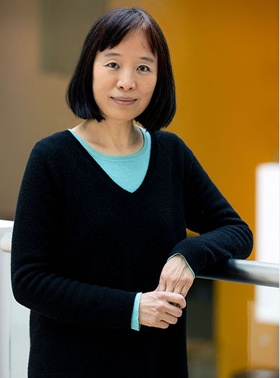
MIT professors Daniel Anderson, Regina Barzilay, Guoping Feng, Darrell Irvine, and Morgen Shen were among the new members. Justin Hanes PhD ’96, Said Ibrahim MBA ’16, and Jennifer West ’92, along with three former students in the Harvard-MIT Program in Health Sciences and Technology (HST) — Michael Chiang, Siddhartha Mukherjee, and Robert Vonderheide — were also elected, as was Yi Zhang, an associate member of The Broad Institute of MIT and Harvard.

Election to the academy is considered one of the highest honors in the fields of health and medicine and recognizes individuals who have demonstrated outstanding professional achievement and commitment to service, the academy noted in announcing the election of its new members.

MIT faculty

Daniel G. Anderson, professor in the Department of Chemical Engineering and the Institute for Medical Engineering and Science, was elected “for pioneering the area of non-viral gene therapy and cellular delivery. His work has resulted in fundamental scientific advances; over 500 papers, patents, and patent applications; and the creation of companies, products, and technologies that are now in the clinic.” Anderson is an affiliate of the Broad Institute of MIT and Harvard and of the Ragon Institute at MGH, MIT and Harvard.

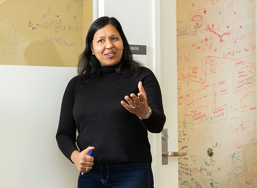
Regina Barzilay, the School of Engineering Distinguished Professor for AI and Health within the Department of Electrical Engineering and Computer Science at MIT, was elected “for the development of machine learning tools that have been transformational for breast cancer screening and risk assessment, and for the development of molecular design tools broadly utilized for drug discovery.” Barzilay is the AI faculty lead within the MIT Abdul Latif Jameel Clinic for Machine Learning in Health and an affiliate of the Computer Science and Artificial Intelligence Laboratory and Institute for Medical Engineering and Science.

Guoping Feng, the associate director of the McGovern Institute for Brain Research, James W. (1963) and Patricia T. Professor of Neuroscience in MIT’s Department of Brain and Cognitive Sciences, and an affiliate of the Broad Institute of MIT and Harvard, was elected “for his breakthrough discoveries regarding the pathological mechanisms of neurodevelopmental and psychiatric disorders, providing foundational knowledges and molecular targets for developing effective therapeutics for mental illness such as OCD, ASD, and ADHD.”

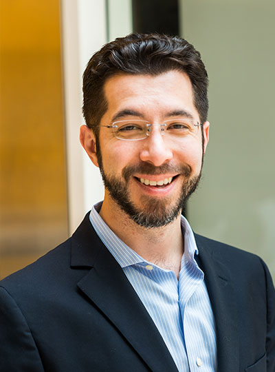
Darrell J. Irvine ’00, the Underwood-Prescott Professor of Biological Engineering and Materials Science at MIT and a member of the Koch Institute for Integrative Cancer Research, was elected “for the development of novel methods for delivery of immunotherapies and vaccines for cancer and infectious diseases.”

Morgan Sheng, professor of neuroscience in the Department of Brain and Cognitive Sciences, with affiliations in the McGovern Institute and The Picower Institute for Learning and Memory at MIT, as well as the Broad Institute of MIT and Harvard, was elected “for transforming the understanding of excitatory synapses. He revealed the postsynaptic density as a protein network controlling synaptic signaling and morphology; established the paradigm of signaling complexes organized by PDZ scaffolds; and pioneered the concept of localized regulation of mitochondria, apoptosis, and complement for targeted synapse elimination.”

Additional MIT affiliates

Michael F. Chiang, a former student in the Harvard-MIT Program in Health Sciences and Technology (HST) who is now director of the National Eye Institute of the National Institutes of Health, was honored “for pioneering applications of biomedical informatics to ophthalmology in artificial intelligence, telehealth, pediatric retinal disease, electronic health records, and data science, including methodological and diagnostic advances in AI for pediatric retinopathy of prematurity, and for contributions to developing and implementing the largest ambulatory care registry in the United States.”

Justin Hanes PhD ’96, who earned his PhD from the MIT Department of Chemical Engineering and is now a professor at Johns Hopkins University, was honored “for pioneering discoveries and inventions of innovative drug delivery technologies, especially mucosal, ocular, and central nervous system drug delivery systems; and for international leadership in research and education at the interface of engineering, medicine, and entrepreneurship, leading to clinical translation of drug delivery technologies.”

Said Ibrahim MBA ’16, a graduate of the MIT Sloan School of Management who is now a senior vice president and chair, department of medicine at the Zucker School of Medicine at Hofstra/Northwell, was honored for influential “health services research on racial disparities in elective joint replacement that has provided a national model for advancing health equity research beyond the identification of inequities and toward their remediation, and for his research that has been leveraged to engage diverse and innovative emerging scholars.”

Siddhartha Mukherjee, a former student in HST who is now an associate professor of medicine at Columbia University School of Medicine, was honored “for contributing important research in the immunotherapy of myeloid malignancies, such as acute myeloid leukemia, for establishing international centers for immunotherapy for childhood cancers, and for the discovery of tissue-resident stem cells.”

Robert H. Vonderheide, a former student in HST who is now a professor and vice dean at the Perelman School of Medicine and vice president of cancer programs at the University of Pennsylvania Health System, was honored “for developing immune combination therapies for patients with pancreatic cancer by driving proof-of-concept from lab to clinic, then leading national, randomized clinical trials for therapy, maintenance, and interception; and for improving access of minority individuals to clinical trials while directing an NCI comprehensive cancer center.”

Jennifer West ’92, a graduate of the MIT Department of Chemical Engineering who is now a professor of biomedical engineering and dean of the School of Engineering and Applied Science at the University of Virginia at Charlottesville, was honored “for the invention, development, and translation of novel biomaterials including bioactive, photopolymerizable hydrogels and theranostic nanoparticles.”

Yi Zhang, associate member of the Broad Institute, was honored “for making fundamental contributions to the epigenetics field through systematic identification and characterization of chromatin modifying enzymes, including EZH2, JmjC, and Tet. His proof-of-principle work on EZH2 inhibitors led to the founding of Epizyme and eventual making of tazemetostat, a drug approved for epithelioid sarcoma and follicular lymphoma.”

“It is my honor to welcome this truly exceptional class of new members to the National Academy of Medicine,” said NAM President Victor J. Dzau. “Their contributions to health and medicine are unparalleled, and their leadership and expertise will be essential to helping the NAM tackle today’s urgent health challenges, inform the future of health care, and ensure health equity for the benefit of all around the globe.”

Four McGovern Investigators receive NIH BRAIN Initiative grants

In the human brain, 86 billion neurons form more than 100 trillion connections with other neurons at junctions called synapses. Scientists at the McGovern Institute are working with their collaborators to develop technologies to map these connections across the brain, from mice to humans.

Today, the National Institutes of Health (NIH) announced a new program to support research projects that have the potential to reveal an unprecedented and dynamic picture of the connected networks in the brain. Four of these NIH-funded research projects will take place in McGovern labs.

BRAIN Initiative

In 2013, the Obama administration announced the Brain Research Through Advancing Innovative Neurotechnologies® (BRAIN) Initiative, a public-private research effort to support the development and application of new technologies to understand brain function.

Today, the NIH announced its third project supported by the BRAIN Initiative, called BRAIN Initiative Connectivity Across Scales (BRAIN CONNECTS). The new project complements two previous large-scale projects, which together aim to transform neuroscience research by generating wiring diagrams that can span entire brains across multiple species. These detailed wiring diagrams can help uncover the logic of the brain’s neural code, leading to a better understanding of how this circuitry makes us who we are and how it could be rewired to treat brain diseases.

BRAIN CONNECTS at McGovern

The initial round of BRAIN CONNECTS awards will support researchers at more than 40 university and research institutions across the globe with 11 grants totaling $150 million over five years. Four of these grants have been awarded to McGovern researchers Guoping Feng, Ila Fiete, Satra Ghosh, and Ian Wickersham, whose projects are outlined below:

BRAIN CONNECTS: Comprehensive regional projection map of marmoset with single axon and cell type resolution
Team: Guoping Feng (McGovern Institute, MIT), Partha Mitra (Cold Spring Harbor Laboratory), Xiao Wang (Broad Institute), Ian Wickersham (McGovern Institute, MIT)

Summary: This project will establish an integrated experimental-computational platform to create the first comprehensive brain-wide mesoscale connectivity map in a non-human primate (NHP), the common marmoset (Callithrix jacchus). It will do so by tracing axonal projections of RNA barcode-identified neurons brain-wide in the marmoset, utilizing a sequencing-based imaging method that also permits simultaneous transcriptomic cell typing of the identified neurons. This work will help bridge the gap between brain-wide mesoscale connectivity data available for the mouse from a decade of mapping efforts using modern techniques and the absence of comparable data in humans and NHPs.

BRAIN CONNECTS: A center for high-throughput integrative mouse connectomics
Team: Jeff Lichtman (Harvard University), Ila Fiete (McGovern Institute, MIT), Sebastian Seung (Princeton University), David Tank (Princeton University), Hongkui Zeng (Allen Institute), Viren Jain (Google), Greg Jeffries (Oxford University)

Summary: This project aims to produce a large-scale synapse-level brain map (connectome) that includes all the main areas of the mouse hippocampus. This region is of clinical interest because it is an essential part of the circuit underlying spatial navigation and memory and the earliest impairments and degeneration related to Alzheimer’s disease.

BRAIN CONNECTS: The center for Large-scale Imaging of Neural Circuits (LINC)
Team: Anastasia Yendiki (MGH), Satra Ghosh (McGovern, MIT), Suzanne Haber (University of Rochester), Elizabeth Hillman (Columbia University)

Summary: This project will generate connectional diagrams of the monkey and human brain at unprecedented resolutions. These diagrams will be linked both to the neuroanatomic literature and to in vivo neuroimaging techniques, bridging between the rigor of the former and the clinical relevance of the latter. The data to be generated by this project will advance our understanding of brain circuits that are implicated in motor and psychiatric disorders, and that are targeted by deep-brain stimulation to treat these disorders.

BRAIN CONNECTS: Mapping brain-wide connectivity of neuronal types using barcoded connectomics
Team: Xiaoyin Chen (Allen Institute), Ian Wickersham (McGovern Institute, MIT), and Justus Kebschull of JHU

Summary: This project aims to optimize and develop barcode sequencing-based neuroanatomical techniques to achieve brain-wide, high-throughput, highly multiplexed mapping of axonal projections and synaptic connectivity of neuronal types at cellular resolution in primate brains. The team will work together to apply these techniques to generate an unprecedented multi-resolution map of brain-wide projections and synaptic inputs of neurons in the macaque visual cortex at cellular resolution.


Study decodes surprising approach mice take in learning

Neuroscience discoveries ranging from the nature of memory to treatments for disease have depended on reading the minds of mice, so researchers need to truly understand what the rodents’ behavior is telling them during experiments. In a new study that examines learning from reward, MIT researchers deciphered some initially mystifying mouse behavior, yielding new ideas about how mice think and a mathematical tool to aid future research.

The task the mice were supposed to master is simple: Turn a wheel left or right to get a reward and then recognize when the reward direction switches. When neurotypical people play such “reversal learning” games they quickly infer the optimal approach: stick with the direction that works until it doesn’t and then switch right away. Notably, people with schizophrenia struggle with the task. In the new study in PLOS Computational Biology, mice surprised scientists by showing that while they were capable of learning the “win-stay, lose-shift” strategy, they nonetheless refused to fully adopt it.

“It is not that mice cannot form an inference-based model of this environment—they can,” said corresponding author Mriganka Sur, Newton Professor in The Picower Institute for Learning and Memory and MIT’s Department of Brain and Cognitive Sciences (BCS). “The surprising thing is that they don’t persist with it. Even in a single block of the game where you know the reward is 100 percent on one side, every so often they will try the other side.”

While the mouse motif of departing from the optimal strategy could be due to a failure to hold it in memory, said lead author and Sur Lab graduate student Nhat Le, another possibility is that mice don’t commit to the “win-stay, lose-shift” approach because they don’t trust that their circumstances will remain stable or predictable. Instead, they might deviate from the optimal regime to test whether the rules have changed. Natural settings, after all, are rarely stable or predictable.

“I’d like to think mice are smarter than we give them credit for,” Le said.

But regardless of which reason may cause the mice to mix strategies, added co-senior author Mehrdad Jazayeri, Associate Professor in BCS and the McGovern Institute for Brain Research, it is important for researchers to recognize that they do and to be able to tell when and how they are choosing one strategy or another.

“This study highlights the fact that, unlike the accepted wisdom, mice doing lab tasks do not necessarily adopt a stationary strategy and it offers a computationally rigorous approach to detect and quantify such non-stationarities,” he said. “This ability is important because when researchers record the neural activity, their interpretation of the underlying algorithms and mechanisms may be invalid when they do not take the animals’ shifting strategies into account.”

Tracking thinking

The research team, which also includes co-author Murat Yildirim, a former Sur lab postdoc who is now an assistant professor at the Cleveland Clinic Lerner Research Institute, initially expected that the mice might adopt one strategy or the other. They simulated the results they’d expect to see if the mice either adopted the optimal strategy of inferring a rule about the task, or more randomly surveying whether left or right turns were being rewarded. Mouse behavior on the task, even after days, varied widely but it never resembled the results simulated by just one strategy.

To differing, individual extents, mouse performance on the task reflected variance along three parameters: how quickly they switched directions after the rule switched, how long it took them to transition to the new direction, and how loyal they remained to the new direction. Across 21 mice, the raw data represented a surprising diversity of outcomes on a task that neurotypical humans uniformly optimize. But the mice clearly weren’t helpless. Their average performance significantly improved over time, even though it plateaued below the optimal level.

In the task, the rewarded side switched every 15-25 turns. The team realized the mice were using more than one strategy in each such “block” of the game, rather than just inferring the simple rule and optimizing based on that inference. To disentangle when the mice were employing that strategy or another, the team harnessed an analytical framework called a Hidden Markov Model (HMM), which can computationally tease out when one unseen state is producing a result vs. another unseen state. Le likens it to what a judge on a cooking show might do: inferring which chef contestant made which version of a dish based on patterns in each plate of food before them.

Before the team could use an HMM to decipher their mouse performance results, however, they had to adapt it. A typical HMM might apply to individual mouse choices, but here the team modified it to explain choice transitions over the course of whole blocks. They dubbed their modified model the blockHMM. Computational simulations of task performance using the blockHMM showed that the algorithm is able to infer the true hidden states of an artificial agent. The authors then used this technique to show the mice were persistently blending multiple strategies, achieving varied levels of performance.

“We verified that each animal executes a mixture of behavior from multiple regimes instead of a behavior in a single domain,” Le and his co-authors wrote. “Indeed 17/21 mice used a combination of low, medium and high-performance behavior modes.”

Further analysis revealed that the strategies afoot were indeed the “correct” rule inference strategy and a more exploratory strategy consistent with randomly testing options to get turn-by-turn feedback.

Now that the researchers have decoded the peculiar approach mice take to reversal learning, they are planning to look more deeply into the brain to understand which brain regions and circuits are involved. By watching brain cell activity during the task, they hope to discern what underlies the decisions the mice make to switch strategies.

By examining reversal learning circuits in detail, Sur said, it’s possible the team will gain insights that could help explain why people with schizophrenia show diminished performance on reversal learning tasks. Sur added that some people with autism spectrum disorders also persist with newly unrewarded behaviors longer than neurotypical people, so his lab will also have that phenomenon in mind as they investigate.

Yildirim, too, is interested in examining potential clinical connections.

“This reversal learning paradigm fascinates me since I want to use it in my lab with various preclinical models of neurological disorders,” he said. “The next step for us is to determine the brain mechanisms underlying these differences in behavioral strategies and whether we can manipulate these strategies.”

Funding for the study came from The National Institutes of Health, the Army Research Office, a Paul and Lilah Newton Brain Science Research Award, the Massachusetts Life Sciences Initiative, The Picower Institute for Learning and Memory and The JPB Foundation.

One scientist’s journey from the Middle East to MIT

Smiling man holidng paper in a room.
Ubadah Sabbagh, soon after receiving his US citizenship papers, in April 2023. Photo: Ubadah Sabbagh

“I recently exhaled a breath I’ve been holding in for nearly half my life. After applying over a decade ago, I’m finally an American. This means so many things to me. Foremost, it means I can go back to the the Middle East, and see my mama and the family, for the first time in 14 years.” — McGovern Institute Postdoctoral Associate Ubadah Sabbagh, X (formerly Twitter) post, April 27, 2023

The words sit atop a photo of Ubadah Sabbagh, who joined the lab of Guoping Feng, James W. (1963) and Patricia T. Poitras Professor at MIT, as a postdoctoral associate in 2021. Sabbagh, a Syrian national, is dressed in a charcoal grey jacket, a keffiyeh loose around his neck, and holding his US citizenship papers, which he began applying for when he was 19 and an undergraduate at the University of Missouri-Kansas City (UMKC) studying biology and bioinformatics.

In the photo he is 29.

A clarity of vision

Sabbagh’s journey from the Middle East to his research position at MIT has been marked by determination and courage, a multifaceted curiosity, and a role as a scientist-writer/scientist-advocate.  He is particularly committed to the importance of humanity in science.

“For me, a scientist is a person who is not only in the lab but also has a unique perspective to contribute to society,” he says. “The scientific method is an idea, and that can be objective. But the process of doing science is a human endeavor, and like all human endeavors, it is inherently both social and political.”

At just 30 years of age, some of Sabbagh’s ideas have disrupted conventional thinking about how science is done in the United States. He believes nations should do science not primarily to compete, for example, but to be aspirational.

“It is our job to make our work accessible to the public, to educate and inform, and to help ground policy,” he says. “In our technologically advanced society, we need to raise the baseline for public scientific intuition so that people are empowered and better equipped to separate truth from myth.”

Two men sitting at a booth wearing headphones.
Ubadah Sabbagh is interviewed for Max Planck Forida’s Neurotransmissions podcast at the 2023 Society for Neuroscience conference in San Diego. Photo: Max Planck Florida

His research and advocacy work have won him accolades, including the 2023 Young Arab Pioneers Award from the Arab Youth Center and the 2020 Young Investigator Award from the American Society of Neurochemistry. He was also named to the 2021 Forbes “30 under 30” list, the first Syrian to be selected in the Science category.

A path to knowledge

Sabbagh’s path to that knowledge began when, living on his own at age 16, he attended Longview Community College, in Kansas City, often juggling multiple jobs. It continued at UMKC, where he fell in love with biology and had his first research experience with bioinformatician Gerald Wyckoff at the same time the civil war in Syria escalated, with his family still in the Middle East. “That was a rough time for me,” he says. “I had a lot of survivor’s guilt: I am here, I have all of this stability and security compared to what they have, and while they had suffocation, I had opportunity. I need to make this mean something positive, not just for me, but in as broad a way as possible for other people.”

Child smiles in front of scientific poster.
Ubadah Sabbagh, age 9, presents his first scientific poster. Photo: Ubadah Sabbagh

The war also sparked Sabbagh’s interest in human behavior—“where it originates, what motivates people to do things, but in a biological, not a psychological way,” he says. “What circuitry is engaged? What is the infrastructure of the brain that leads to X, Y, Z?”

His passion for neuroscience blossomed as a graduate student at Virginia Tech, where he earned his PhD in translational biology, medicine, and health. There, he received a six-year NIH F99/K00 Award, and under the mentorship of neuroscientist at the Fralin Biomedical Research Institute he researched the connections between the eye and the brain, specifically, mapping the architecture of the principle neurons in a region of the thalamus essential to visual processing.

“The retina, and the entire visual system, struck me as elegant, with beautiful layers of diverse cells found at every node,” says Sabbagh, his own eyes lighting up.

His research earned him a coveted spot on the Forbes “30 under 30” list, generating enormous visibility, including in the Arab world, adding visitors to his already robust X (formerly Twitter) account, which has more than 9,200 followers. “The increased visibility lets me use my voice to advocate for the things I care about,” he says.

“I need to make this mean something positive, not just for me, but in as broad a way as possible for other people.” — Ubadah Sabbagh

Those causes range from promoting equity and inclusion in science to transforming the American system of doing science for the betterment of science and the scientists themselves. He cofounded the nonprofit Black in Neuro to celebrate and empower Black scholars in neuroscience, and he continues to serve on the board. He is the chair of an advisory committee for the Society for Neuroscience (SfN), recommending ways SfN can better address the needs of its young members, and a member of the Advisory Committee to the National Institutes of Health (NIH) Director working group charged with re-envisioning postdoctoral training. He serves on the advisory board of Community for Rigor, a new NIH initiative that aims to teach scientific rigor at national scale and, in his spare time, he writes articles about the relationship of science and policy for publications including Scientific American and the Washington Post.

Still, there have been obstacles. The same year Sabbagh received the NIH F99/K00 Award, he faced major setbacks in his application to become a citizen. He would not try again until 2021, when he had his PhD in hand and had joined the McGovern Institute.

An MIT postdoc and citizenship

Sabbagh dove into his research in Guoping Feng’s lab with the same vigor and outside-the-box thinking that characterized his previous work. He continues to investigate the thalamus, but in a region that is less involved in processing pure sensory signals, such as light and sound, and more focused on cognitive functions of the brain. He aims to understand how thalamic brain areas orchestrate complex functions we carry out every day, including working memory and cognitive flexibility.

“This is important to understand because when this orchestra goes out of tune it can lead to a range of neurological disorders, including autism spectrum disorder and schizophrenia,” he says. He is also developing new tools for studying the brain using genome editing and viral engineering to expand the toolkit available to neuroscientists.

Microscopic image of mouse brain
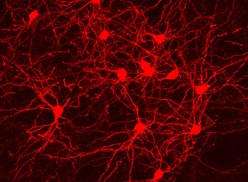
Neurons in a transgenic mouse brain labeled by Sabbagh using genome editing technology in the Feng lab. Image: Ubadah Sabbagh

The environment at the McGovern Institute is also a source of inspiration for Sabbagh’s research. “The scale and scope of work being done at McGovern is remarkable. It’s an exciting place for me to be as a neuroscientist,” said Sabbagh. “Besides being intellectually enriching, I’ve found great community here – something that’s important to me wherever I work.”

Returning to the Middle East

Profile of scientist Ubadah Sabbagh speaking at a table.
McGovern postdoc Ubadah Sabbagh at the 2023 Young Arab Pioneers Award ceremony in Abu Dhabi. Photo: Arab Youth Center

While at an advisory meeting at the NIH, Sabbagh learned he had been selected as a Young Arab Pioneer by the Arab Youth Center and was flown the next day to Abu Dhabi for a ceremony overseen by Her Excellency Shamma Al Mazrui, Cabinet Member and Minister of Community Development in the United Arab Emirates. The ceremony recognized 20 Arab youth from around the world in sectors ranging from scientific research to entrepreneurship and community development. Sabbagh’s research “presented a unique portrayal of creative Arab youth and an admirable representation of the values of youth beyond the Arab world,” said Sadeq Jarrar, executive director of the center.

“There I was, among other young Arab leaders, learning firsthand about their efforts, aspirations, and their outlook for the future,” says Sabbagh, who was deeply inspired by the experience.

Just a month earlier, his passport finally secured, Sabbagh had reunited with his family in the Middle East after more than a decade in the United States. “I had been away for so long,” he said, describing the experience as a “cultural reawakening.”

Woman hands man an award on stage.
Ubadah Sabbagh receives a Young Arab Pioneer Award by Her Excellency Shamma Al Mazrui, Cabinet Member and Minister of Community Development in the United Arab Emirates. Photo: Arab Youth Center

Sabbagh saw a gaping need he had not been aware of when he left 14 years earlier, as a teen. “The Middle East had such a glorious intellectual past,” he says. “But for years people have been leaving to get their advanced scientific training, and there is no adequate infrastructure to support them if they want to go back.” He wondered: What if there were a scientific renaissance in the region? How would we build infrastructure to cultivate local minds and local talent? What if the next chapter of the Middle East included being a new nexus of global scientific advancements?

“I felt so inspired,” he says. “I have a longing, someday, to meaningfully give back.”

Scientists discover how mutations in a language gene produce speech deficits

Mutations of a gene called Foxp2 have been linked to a type of speech disorder called apraxia that makes it difficult to produce sequences of sound. A new study from MIT and National Yang Ming Chiao Tung University sheds light on how this gene controls the ability to produce speech.

In a study of mice, the researchers found that mutations in Foxp2 disrupt the formation of dendrites and neuronal synapses in the brain’s striatum, which plays important roles in the control of movement. Mice with these mutations also showed impairments in their ability to produce the high-frequency sounds that they use to communicate with other mice.

Those malfunctions arise because Foxp2 mutations prevent the proper assembly of motor proteins, which move molecules within cells, the researchers found.

“These mice have abnormal vocalizations, and in the striatum there are many cellular abnormalities,” says Ann Graybiel, an MIT Institute Professor, a member of MIT’s McGovern Institute for Brain Research, and an author of the paper. “This was an exciting finding. Who would have thought that a speech problem might come from little motors inside cells?”

Fu-Chin Liu PhD ’91, a professor at National Yang Ming Chiao Tung University in Taiwan, is the senior author of the study, which appears today in the journal Brain. Liu and Graybiel also worked together on a 2016 study of the potential link between Foxp2 and autism spectrum disorder. The lead authors of the new Brain paper are Hsiao-Ying Kuo and Shih-Yun Chen of National Yang Ming Chiao Tung University.

Speech control

Children with Foxp2-associated apraxia tend to begin speaking later than other children, and their speech is often difficult to understand. The disorder is believed to arise from impairments in brain regions, such as the striatum, that control the movements of the lips, mouth, and tongue. Foxp2 is also expressed in the brains of songbirds such as zebra finches and is critical to those birds’ ability to learn songs.

Foxp2 encodes a transcription factor, meaning that it can control the expression of many other target genes. Many species express Foxp2, but humans have a special form of Foxp2. In a 2014 study, Graybiel and colleagues found evidence that the human form of Foxp2, when expressed in mice, allowed the mice to accelerate the switch from declarative to procedural types of learning.

In that study, the researchers showed that mice engineered to express the human version of Foxp2, which differs from the mouse version by only two DNA base pairs, were much better at learning mazes and performing other tasks that require turning repeated actions into behavioral routines. Mice with human-like Foxp2 also had longer dendrites — the slender extensions that help neurons form synapses — in the striatum, which is involved in habit formation as well as motor control.

In the new study, the researchers wanted to explore how the Foxp2 mutation that has been linked with apraxia affects speech production, using ultrasonic vocalizations in mice as a proxy for speech. Many rodents and other animals such as bats produce these vocalizations to communicate with each other.

While previous studies, including the work by Liu and Graybiel in 2016, had suggested that Foxp2 affects dendrite growth and synapse formation, the mechanism for how that occurs was not known. In the new study, led by Liu, the researchers investigated one proposed mechanism, which is that Foxp2 affects motor proteins.

One of these molecular motors is the dynein protein complex, a large cluster of proteins that is responsible for shuttling molecules along microtubule scaffolds within cells.

“All kinds of molecules get shunted around to different places in our cells, and that’s certainly true of neurons,” Graybiel says. “There’s an army of tiny molecules that move molecules around in the cytoplasm or put them into the membrane. In a neuron, they may send molecules from the cell body all the way down the axons.”

A delicate balance

The dynein complex is made up of several other proteins. The most important of these is a protein called dynactin1, which interacts with microtubules, enabling the dynein motor to move along microtubules. In the new study, the researchers found that dynactin1 is one of the major targets of the Foxp2 transcription factor.

The researchers focused on the striatum, one of the regions where Foxp2 is most often found, and showed that the mutated version of Foxp2 is unable to suppress dynactin1 production. Without that brake in place, cells generate too much dynactin1. This upsets the delicate balance of dynein-dynactin1, which prevents the dynein motor from moving along microtubules.

Those motors are needed to shuttle molecules that are necessary for dendrite growth and synapse formation on dendrites. With those molecules stranded in the cell body, neurons are unable to form synapses to generate the proper electrophysiological signals they need to make speech production possible.

Mice with the mutated version of Foxp2 had abnormal ultrasonic vocalizations, which typically have a frequency of around 22 to 50 kilohertz. The researchers showed that they could reverse these vocalization impairments and the deficits in the molecular motor activity, dendritic growth, and electrophysiological activity by turning down the gene that encodes dynactin1.

Mutations of Foxp2 can also contribute to autism spectrum disorders and Huntington’s disease, through mechanisms that Liu and Graybiel previously studied in their 2016 paper and that many other research groups are now exploring. Liu’s lab is also investigating the potential role of abnormal Foxp2 expression in the subthalamic nucleus of the brain as a possible factor in Parkinson’s disease.

The research was funded by the Ministry of Science and Technology of Taiwan, the Ministry of Education of Taiwan, the U.S. National Institute of Mental Health, the Saks Kavanaugh Foundation, the Kristin R. Pressman and Jessica J. Pourian ’13 Fund, and Stephen and Anne Kott.

Yang Dan named winner of the 2023 Scolnick Prize in Neuroscience

The McGovern Institute announced today that the 2023 Edward M. Scolnick Prize in Neuroscience will be awarded to neurobiologist Yang Dan. Dan holds the Nan Fung Life Sciences Chancellor’s Chair in Neuroscience at the University of California, Berkeley, and has been a Howard Hughes Investigator since 2008. The Scolnick Prize is awarded annually by the McGovern Institute for outstanding achievements in neuroscience.

“Yang Dan’s systems-level experimentation to identify the cell types and circuits that control sleep cycles represents the highest level of neuroscience research,” says Robert Desimone, McGovern Institute director and chair of the selection committee. “Her work has defined precise mechanisms for how motor behaviors are suppressed during sleep and activated during arousal, with potential implications for the design of more targeted sedatives and the treatment of sleep disorders.”

Significance of sleep

Dan received a BS in Physics in 1988 from Peking University in China. She then moved to the US to obtain her PhD in neurobiology from Columbia University, in 1994, under the mentorship of Professor Mu-Ming Poo. Her doctoral research focused on mechanisms of plasticity at the neuromuscular synapse and was published in Science, Nature, and Neuron. During this time, she showed that the quantal release of neurotransmitters is not unique to neuronal cell types and, as one example, that retrograde signaling from muscle cells regulates the synaptic strength of the neuromuscular junction. For her postdoctoral training, Dan joined Clay Reid’s lab at The Rockefeller University and then accompanied Reid’s move to Harvard Medical School a short time later. Within just over two years, Yang had collected and analyzed neuronal recording data to support and develop key computational models of visual information coding – her two papers describing this work have been cited, together, over 900 times.

Yang Dan started her own laboratory in January 1997 when she joined the faculty of UC Berkeley’s Department of Molecular and Cell Biology as an assistant professor; she became a full professor in 2005. Dan’s lab became known for discoveries of how sensory inputs, especially visual inputs, are processed by the brain to influence behavior. Using electrophysiological recordings in model animals and computational analyses, her group worked out rules for how synaptic plasticity and neural connectivity, at the microcircuit and brain-wide level, contribute to learning and goal-directed behaviors.

Sleep recordings in various animal models and humans, shown in a research review by Yang Dan (2019 Annual Review of Neuroscience). (a) In nonmammalian animals such as jellyfish, Caenorhabditis elegans, Drosophila, and zebrafish, locomotor assay is used to measure sleep. (b) Examples of mouse EEG and EMG recordings during wakefulness and NREM and REM sleep. (c) Example polysomnography recordings from a healthy human subject during wakefulness and NREM (stage 3) and phasic REM sleep.

The Dan lab carved out a new research direction upon their discovery of mechanisms controlling rapid eye movement (REM) sleep, a state in which the brain is active and neuroplastic despite minimal sensory input. In their 2015 Nature paper, Dan’s group showed that, in mice, optogenetic activation of inhibitory neurons that project forward from the brainstem to the middle of the brain can instantaneously induce REM sleep. Since then, the Dan lab has published nearly a dozen primary research papers on the sleep-wake cycle that capitalize on the latest neural engineering techniques to record and control specific cell types and circuits in the brain. Most recently, she reported the discovery of neurons in the midbrain that receive wide-ranging inputs to coordinate active suppression of movement during REM and non-REM sleep with the release of movement during arousal. This circuit is key to the ability, known to exist in most animals, to experience sleep and even vivid dreaming without acting out. Dan’s discoveries are paving the way to a holistic understanding, from the molecular to macrocircuit levels, of how our bodies regulate sleep, an evolutionarily conserved behavior that is essential for survival.

Awards and honors

Dan was appointed as a Howard Hughes Medical Institute Investigator in 2008 and elected to the US National Academy of Sciences in 2018. She was awarded the Li Ka Shing Women in Science Award in 2007 and a Research Award for Innovation in Neuroscience from the Society for Neuroscience in 2009. She teaches summer courses at institutes around the world and has mentored 16 graduate students and 27 postdoctoral researchers, 25 of whom now run their own independent laboratories. Currently, Dan serves as an editorial board member on top-ranked science journals including Cell, Neuron, PNAS, and Current Opinion in Neurobiology.

Yang Dan will be awarded the Scolnick Prize on Wednesday, June 7, 2023. At 4:00 pm on that day, she will deliver a lecture titled “The how and why of sleep,” to be followed by a reception at the McGovern Institute, 43 Vassar Street (building 46, room 3002) in Cambridge. The event is free and open to the public.


Partnership with MIT Museum explores relationship between neuroscience and society

What does a healthy relationship between neuroscience and society look like? How do we set the conditions for that relationship to flourish? Researchers and staff at the McGovern Institute and the MIT Museum have been exploring these questions with a five-month planning grant from the Dana Foundation.

Between October 2022 and March 2023, the team tested the potential for an MIT Center for Neuroscience and Society through a series of MIT-sponsored events that were attended by students and faculty of nearby Cambridge Public Schools. The goal of the project was to learn more about what happens when the distinct fields of neuroscience, ethics, and public engagement are brought together to work side-by-side.

Researchers assist volunteer in mock MRI scanner
Gabrieli lab members Sadie Zacharek (left) and Shruti Nishith (right) demonstrate how the MRI mock scanner works with a student volunteer from the Cambridge Public Schools. Photo: Emma Skakel, MIT Museum

Middle schoolers visit McGovern

Over four days in February, more than 90 sixth graders from Rindge Avenue Upper Campus (RAUC) in Cambridge, Massachusetts, visited the McGovern Institute and participated in hands-on experiments and discussions about the ethical, legal, and social implications of neuroscience research. RAUC is one of four middle schools in the city of Cambridge with an economically, racially, and culturally diverse student population. The middle schoolers interacted with an MIT team led by McGovern Scientific Advisor Jill R. Crittenden, including seventeen McGovern neuroscientists, three MIT Museum outreach coordinators, and neuroethicist Stephanie Bird, a member of the Dana Foundation planning grant team.

“It is probably the only time in my life I will see a real human brain.” – RAUC student

The students participated in nine activities each day, including trials of brain-machine interfaces, close-up examinations of preserved human brains, a tour of McGovern’s imaging center in which students watched as their teacher’s brain was scanned, and a visit to the MIT Museum’s interactive Artificial Intelligence Gallery.

Imagine-IT, a brain-machine interface designed by a team of middle school students during a visit to the McGovern Institute.

To close out their visit, students worked in groups alongside experts to invent brain-computer interfaces designed to improve or enhance human abilities. At each step, students were introduced to ethical considerations through consent forms, questions regarding the use of animal and human brains, and the possible impacts of their own designs on individuals and society.

“I admit that prior to these four days, I would’ve been indifferent to the inclusion of children’s voices in a discussion about technically complex ethical questions, simply because they have not yet had any opportunity to really understand how these technologies work,” says one researcher involved in the visit. “But hearing the students’ questions and ideas has changed my perspective. I now believe it is critically important that all age groups be given a voice when discussing socially relevant issues, such as the ethics of brain computer interfaces or artificial intelligence.”


For more information on the proposed MIT Center for Neuroscience and Society, visit the MIT Museum website.

How Huntington’s disease affects different neurons

In patients with Huntington’s disease, neurons in a part of the brain called the striatum are among the hardest-hit. Degeneration of these neurons contributes to patients’ loss of motor control, which is one of the major hallmarks of the disease.

Neuroscientists at MIT have now shown that two distinct cell populations in the striatum are affected differently by Huntington’s disease. They believe that neurodegeneration of one of these populations leads to motor impairments, while damage to the other population, located in structures called striosomes, may account for the mood disorders that are often see in the early stages of the disease.

“As many as 10 years ahead of the motor diagnosis, Huntington’s patients can experience mood disorders, and one possibility is that the striosomes might be involved in these,” says Ann Graybiel, an MIT Institute Professor, a member of MIT’s McGovern Institute for Brain Research, and one of the senior authors of the study.

Using single-cell RNA sequencing to analyze the genes expressed in mouse models of Huntington’s disease and postmortem brain samples from Huntington’s patients, the researchers found that cells of the striosomes and another structure, the matrix, begin to lose their distinguishing features as the disease progresses. The researchers hope that their mapping of the striatum and how it is affected by Huntington’s could help lead to new treatments that target specific cells within the brain.

This kind of analysis could also shed light on other brain disorders that affect the striatum, such as Parkinson’s disease and autism spectrum disorder, the researchers say.

Myriam Heiman, an associate professor in MIT’s Department of Brain and Cognitive Sciences and a member of the Picower Institute for Learning and Memory, and Manolis Kellis, a professor of computer science in MIT’s Computer Science and Artificial Intelligence Laboratory (CSAIL) and a member of the Broad Institute of MIT and Harvard, are also senior authors of the study. Ayano Matsushima, a McGovern Institute research scientist, and Sergio Sebastian Pineda, an MIT graduate student, are the lead authors of the paper, which appears in Nature Communications.

Neuron vulnerability

Huntington’s disease leads to degeneration of brain structures called the basal ganglia, which are responsible for control of movement and also play roles in other behaviors, as well as emotions. For many years, Graybiel has been studying the striatum, a part of the basal ganglia that is involved in making decisions that require evaluating the outcomes of a particular action.

Many years ago, Graybiel discovered that the striatum is divided into striosomes, which are clusters of neurons, and the matrix, which surrounds the striosomes. She has also shown that striosomes are necessary for making decisions that require an anxiety-provoking cost-benefit analysis.

In a 2007 study, Richard Faull of the University of Auckland discovered that in postmortem brain tissue from Huntington’s patients, the striosomes showed a great deal of degeneration. Faull also found that while those patients were alive, many of them had shown signs of mood disorders such as depression before their motor symptoms developed.

To further explore the connections between the striatum and the mood and motor effects of Huntington’s, Graybiel teamed up with Kellis and Heiman to study the gene expression patterns of striosomal and matrix cells. To do that, the researchers used single-cell RNA sequencing to analyze human brain samples and brain tissue from two mouse models of Huntington’s disease.

Within the striatum, neurons can be classified as either D1 or D2 neurons. D1 neurons are involved in the “go” pathway, which initiates an action, and D2 neurons are part of the “no-go” pathway, which suppresses an action. D1 and D2 neurons can both be found within either the striosomes and the matrix.

The analysis of RNA expression in each of these types of cells revealed that striosomal neurons are harder hit by Huntington’s than matrix neurons. Furthermore, within the striosomes, D2 neurons are more vulnerable than D1.

The researchers also found that these four major cell types begin to lose their identifying molecular identities and become more difficult to distinguish from one another in Huntington’s disease. “Overall, the distinction between striosomes and matrix becomes really blurry,” Graybiel says.

Striosomal disorders

The findings suggest that damage to the striosomes, which are known to be involved in regulating mood, may be responsible for the mood disorders that strike Huntington’s patients in the early stages of the disease. Later on, degeneration of the matrix neurons likely contributes to the decline of motor function, the researchers say.

In future work, the researchers hope to explore how degeneration or abnormal gene expression in the striosomes may contribute to other brain disorders.

Previous research has shown that overactivity of striosomes can lead to the development of repetitive behaviors such as those seen in autism, obsessive compulsive disorder, and Tourette’s syndrome. In this study, at least one of the genes that the researchers discovered was overexpressed in the striosomes of Huntington’s brains is also linked to autism.

Additionally, many striosome neurons project to the part of the brain that is most affected by Parkinson’s disease (the substantia nigra, which produces most of the brain’s dopamine).

“There are many, many disorders that probably involve the striatum, and now, partly through transcriptomics, we’re working to understand how all of this could fit together,” Graybiel says.

The research was funded by the Saks Kavanaugh Foundation, the CHDI Foundation, the National Institutes of Health, the Nancy Lurie Marks Family Foundation, the Simons Foundation, the JPB Foundation, the Kristin R. Pressman and Jessica J. Pourian ’13 Fund, and Robert Buxton.

Self-assembling proteins can store cellular “memories”

As cells perform their everyday functions, they turn on a variety of genes and cellular pathways. MIT engineers have now coaxed cells to inscribe the history of these events in a long protein chain that can be imaged using a light microscope.

Cells programmed to produce these chains continuously add building blocks that encode particular cellular events. Later, the ordered protein chains can be labeled with fluorescent molecules and read under a microscope, allowing researchers to reconstruct the timing of the events.

This technique could help shed light on the steps that underlie processes such as memory formation, response to drug treatment, and gene expression.

“There are a lot of changes that happen at organ or body scale, over hours to weeks, which cannot be tracked over time,” says Edward Boyden, the Y. Eva Tan Professor in Neurotechnology, a professor of biological engineering and brain and cognitive sciences at MIT, a Howard Hughes Medical Institute investigator, and a member of MIT’s McGovern Institute for Brain Research and Koch Institute for Integrative Cancer Research.

If the technique could be extended to work over longer time periods, it could also be used to study processes such as aging and disease progression, the researchers say.

Boyden is the senior author of the study, which appears today in Nature Biotechnology. Changyang Linghu, a former J. Douglas Tan Postdoctoral Fellow at the McGovern Institute, who is now an assistant professor at the University of Michigan, is the lead author of the paper.

Cellular history

Biological systems such as organs contain many different kinds of cells, all of which have distinctive functions. One way to study these functions is to image proteins, RNA, or other molecules inside the cells, which provide hints to what the cells are doing. However, most methods for doing this offer only a glimpse of a single moment in time, or don’t work well with very large populations of cells.

“Biological systems are often composed of a large number of different types of cells. For example, the human brain has 86 billion cells,” Linghu says. “To understand those kinds of biological systems, we need to observe physiological events over time in these large cell populations.”

To achieve that, the research team came up with the idea of recording cellular events as a series of protein subunits that are continuously added to a chain. To create their chains, the researchers used engineered protein subunits, not normally found in living cells, that can self-assemble into long filaments.

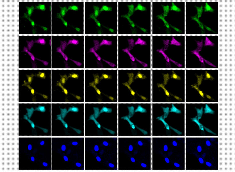
The researchers designed a genetically encoded system in which one of these subunits is continuously produced inside cells, while the other is generated only when a specific event occurs. Each subunit also contains a very short peptide called an epitope tag — in this case, the researchers chose tags called HA and V5. Each of these tags can bind to a different fluorescent antibody, making it easy to visualize the tags later on and determine the sequence of the protein subunits.

For this study, the researchers made production of the V5-containing subunit contingent on the activation of a gene called c-fos, which is involved in encoding new memories. HA-tagged subunits make up most of the chain, but whenever the V5 tag shows up in the chain, that means that c-fos was activated during that time.

“We’re hoping to use this kind of protein self-assembly to record activity in every single cell,” Linghu says. “It’s not only a snapshot in time, but also records past history, just like how tree rings can permanently store information over time as the wood grows.”

Recording events

In this study, the researchers first used their system to record activation of c-fos in neurons growing in a lab dish. The c-fos gene was activated by chemically induced activation of the neurons, which caused the V5 subunit to be added to the protein chain.

To explore whether this approach could work in the brains of animals, the researchers programmed brain cells of mice to generate protein chains that would reveal when the animals were exposed to a particular drug. Later, the researchers were able to detect that exposure by preserving the tissue and analyzing it with a light microscope.

The researchers designed their system to be modular, so that different epitope tags can be swapped in, or different types of cellular events can be detected, including, in principle, cell division or activation of enzymes called protein kinases, which help control many cellular pathways.

The researchers also hope to extend the recording period that they can achieve. In this study, they recorded events for several days before imaging the tissue. There is a tradeoff between the amount of time that can be recorded and the time resolution, or frequency of event recording, because the length of the protein chain is limited by the size of the cell.

“The total amount of information it could store is fixed, but we could in principle slow down or increase the speed of the growth of the chain,” Linghu says. “If we want to record for a longer time, we could slow down the synthesis so that it will reach the size of the cell within, let’s say two weeks. In that way we could record longer, but with less time resolution.”

The researchers are also working on engineering the system so that it can record multiple types of events in the same chain, by increasing the number of different subunits that can be incorporated.

The research was funded by the Hock E. Tan and K. Lisa Yang Center for Autism Research, John Doerr, the National Institutes of Health, the National Science Foundation, the U.S. Army Research Office, and the Howard Hughes Medical Institute.

Season’s Greetings from the McGovern Institute

This year’s holiday video (shown above) was inspired by Ev Fedorenko’s July 2022 Nature Neuroscience paper, which found similar patterns of brain activation and language selectivity across speakers of 45 different languages.

Universal language network

Ev Fedorenko uses the widely translated book “Alice in Wonderland” to test brain responses to different languages. Photo: Caitlin Cunningham

Over several decades, neuroscientists have created a well-defined map of the brain’s “language network,” or the regions of the brain that are specialized for processing language. Found primarily in the left hemisphere, this network includes regions within Broca’s area, as well as in other parts of the frontal and temporal lobes. Although roughly 7,000 languages are currently spoken and signed across the globe, the vast majority of those mapping studies have been done in English speakers as they listened to or read English texts.

To truly understand the cognitive and neural mechanisms that allow us to learn and process such diverse languages, Fedorenko and her team scanned the brains of speakers of 45 different languages while they listened to Alice in Wonderland in their native language. The results show that the speakers’ language networks appear to be essentially the same as those of native English speakers — which suggests that the location and key properties of the language network appear to be universal.

The many languages of McGovern

English may be the primary language used by McGovern researchers, but more than 35 other languages are spoken by scientists and engineers at the McGovern Institute. Our holiday video features 30 of these researchers saying Happy New Year in their native (or learned) language. Below is the complete list of languages included in our video. Expand each accordion to learn more about the speaker of that particular language and the meaning behind their new year’s greeting.