McGovern Fellows recognized with life sciences innovation award

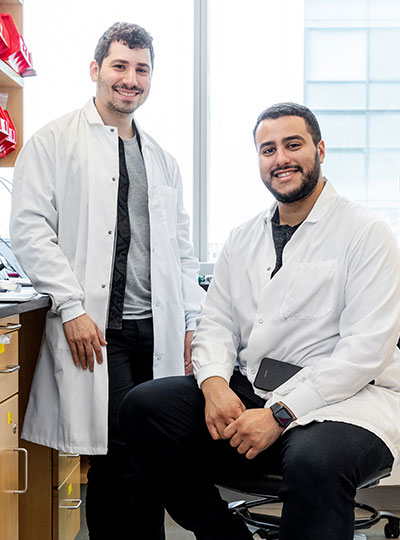
McGovern Institute Fellows Omar Abudayyeh and Jonathan Gootenberg have been named the inaugural recipients of the Termeer Scholars Awards, which recognize “emerging biomedical researchers that represent the future of the biotechnology industry.” The Termeer Foundation is a nonprofit organization focused on connecting life science innovators and catalyzing the creation of new medicines.

“The Termeer Foundation is committed to championing emerging biotechnology leaders and finding people who want to solve the biggest problems in human health,” said Belinda Termeer, president of the Termeer Foundation. “By supporting researchers like Omar and Jonathan, we plant the seeds for future success in individuals who are preparing to make significant contributions in academia and industry.”

The Abudayyeh-Gootenberg lab is developing a suite of new tools to enable next-generation cellular engineering, with uses in basic research, therapeutics and diagnostics. Building off the revolutionary biology of natural biological systems, including mobile genetic elements and CRISPR systems, the team develops new approaches for understanding and manipulating genomes, transcriptomes and cellular fate. The technologies have broad applications, including in oncology, aging and genetic disease.

These tools have been adopted by researchers over the world and formed the basis for four companies that Abudayyeh and Gootenberg have co-founded. They will receive a $50,000 grant to support professional development, knowledge advancement and/or stakeholder engagement and will become part of The Termeer Foundation’s signature Network of Termeer Fellows (first-time CEOs and entrepreneurs) and Mentors (experienced industry leaders).

“The Termeer Foundation is working to improve the long odds of biotechnology by identifying and supporting future biotech leaders; if we help them succeed as leaders, we can help their innovations reach patients,” said Alan Waltws, co-founder of the Termeer Foundation. “While our Termeer Fellows program has supported first time CEOs and entrepreneurs for the past five years, our new Termeer Scholars program will provide much needed support to the researchers whose innovative ideas represent the future of the biotechnology industry – researchers like Omar and Jonathan.”

Abudayyeh and Gootenberg were honored at the Termeer Foundation’s annual dinner in Boston on June 16, 2022.

Convenience-sized RNA editing

Last year, researchers at MIT’s McGovern Institute discovered and characterized Cas7-11, the first CRISPR enzyme capable of making precise, guided cuts to strands of RNA without harming cells in the process. Now, working with collaborators at the University of Tokyo, the same team has revealed that Cas7-11 can be shrunk to a more compact version, making it an even more viable option for editing the RNA inside living cells. The new, compact Cas7-11 was described today in the journal Cell along with a detailed structural analysis of the original enzyme.

“When we looked at the structure, it was clear there were some pieces that weren’t needed which we could actually remove,” says McGovern Fellow Omar Abudayyeh, who led the new work with McGovern Fellow Jonathan Gootenberg and collaborator Hiroshi Nishimasu from the University of Tokyo. “This makes the enzyme small enough that it fits into a single viral vector for therapeutic applications.”

The authors, who also include postdoctoral researcher Nathan Zhou from the McGovern Institute and Kazuki Kato from the University Tokyo, see the new three-dimensional structure of Cas7-11 as a rich resource toanswer questions about the basic biology of the enzymes and reveal other ways to tweak its function in the future.

Targeting RNA

McGovern Fellows Jonathan Gootenberg and Omar Abudayyeh in their lab. Photo: Caitlin Cunningham

Over the past decade, the CRISPR-Cas9 genome editing technology has given researchers the ability to modify the genes inside human cells—a boon for both basic research and the development of therapeutics to reverse disease-causing genetic mutations. But CRISPR-Cas9 only works to alter DNA, and for some research and clinical purposes, editing RNA is more effective or useful.

A cell retains its DNA for life, and passes an identical copy to daughter cells as it duplicates, so any changes to DNA are relatively permanent. However, RNA is a more transient molecule, transcribed from DNA and degraded not long after.

“There are lots of positives about being able to permanently change DNA, especially when it comes to treating an inherited genetic disease,” Gootenberg says. “But for an infection, an injury or some other temporary disease, being able to temporarily modify a gene through RNA targeting makes more sense.”

Until Abudayyeh, Gootenberg and their colleagues discovered and characterized Cas7-11, the only enzyme that could target RNA had a messy side effect; when it recognized a particular gene, the enzyme—Cas13—began cutting up all the RNA around it. This property makes Cas13 effective for diagnostic tests, where it is used to detect the presence of a piece of RNA, but not very useful for therapeutics, where targeted cuts are required.

The discovery of Cas7-11 opened the doors to a more precise form of RNA editing, analogous to the Cas9 enzyme for DNA. However, the massive Cas7-11 protein was too big to fit inside a single viral vector—the empty shell of a virus that researchers typically use to deliver gene editing machinery into patient’s cells.

Structural insight

To determine the overall structure of Cas7-11, Abudayyeh, Gootenberg and Nishimasu used cryo-electron microscopy, which shines beams of electrons on frozen protein samples and measures how the beams are transmitted. The researchers knew that Cas7-11 was like an amalgamation of five separate Cas enzymes, fused into one single gene, but were not sure exactly how those parts folded and fit together.

“The really fascinating thing about Cas7-11, from a fundamental biology perspective, is that it should be all these separate pieces that come together, but instead you have a fusion into one gene,” Gootenberg says. “We really didn’t know what that would look like.”

The structure of Cas7-11, caught in the act of binding both its target tRNA strand and the guide RNA, which directs that binding, revealed how the pieces assembled and which parts of the protein were critical to recognizing and cutting RNA. This kind of structural insight is critical to figuring out how to make Cas7-11 carry out targeted jobs inside human cells.

The structure also illuminated a section of the protein that wasn’t serving any apparent functional role. This finding suggested the researchers could remove it, re-engineering Cas7-11 to make it smaller without taking away its ability to target RNA. Abudayyeh and Gootenberg tested the impact of removing different bits of this section, resulting in a new compact version of the protein, dubbed Cas7-11S. With Cas7-11S in hand, they packaged the system inside a single viral vector, delivered it into mammalian cells and efficiently targeted RNA.

The team is now planning future studies on other proteins that interact with Cas7-11 in the bacteria that it originates from, and also hopes to continue working towards the use of Cas7-11 for therapeutic applications.

“Imagine you could have an RNA gene therapy, and when you take it, it modifies your RNA, but when you stop taking it, that modification stops,” Abudayyeh says. “This is really just the beginning of enabling that tool set.”

This research was funded, in part, by the McGovern Institute Neurotechnology Program, K. Lisa Yang and Hock E. Tan Center for Molecular Therapeutics in Neuroscience, G. Harold & Leila Y. Mathers Charitable Foundation, MIT John W. Jarve (1978) Seed Fund for Science Innovation, FastGrants, Basis for Supporting Innovative Drug Discovery and Life Science Research Program, JSPS KAKENHI, Takeda Medical Research Foundation, and Inamori Research Institute for Science.

New research center focused on brain-body relationship established at MIT

The inextricable link between our brains and our bodies has been gaining increasing recognition among researchers and clinicians over recent years. Studies have shown that the brain-body pathway is bidirectional — meaning that our mental state can influence our physical health and vice versa. But exactly how the two interact is less clear.

A new research center at MIT, funded by a $38 million gift to the McGovern Institute for Brain Research from philanthropist K. Lisa Yang, aims to unlock this mystery by creating and applying novel tools to explore the multidirectional, multilevel interplay between the brain and other body organ systems. This gift expands Yang’s exceptional philanthropic support of human health and basic science research at MIT over the past five years.

“Lisa Yang’s visionary gift enables MIT scientists and engineers to pioneer revolutionary technologies and undertake rigorous investigations into the brain’s complex relationship with other organ systems,” says MIT President L. Rafael Reif.  “Lisa’s tremendous generosity empowers MIT scientists to make pivotal breakthroughs in brain and biomedical research and, collectively, improve human health on a grand scale.”

The K. Lisa Yang Brain-Body Center will be directed by Polina Anikeeva, professor of materials science and engineering and brain and cognitive sciences at MIT and an associate investigator at the McGovern Institute. The center will harness the power of MIT’s collaborative, interdisciplinary life sciences research and engineering community to focus on complex conditions and diseases affecting both the body and brain, with a goal of unearthing knowledge of biological mechanisms that will lead to promising therapeutic options.

“Under Professor Anikeeva’s brilliant leadership, this wellspring of resources will encourage the very best work of MIT faculty, graduate fellows, and research — and ultimately make a real impact on the lives of many,” Reif adds.

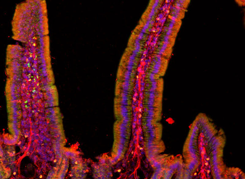
microscope image of gut
Mouse small intestine stained to reveal cell nucleii (blue) and peripheral nerve fibers (red).
Image: Polina Anikeeva, Marie Manthey, Kareena Villalobos

Center goals  

Initial projects in the center will focus on four major lines of research:

  • Gut-Brain: Anikeeva’s group will expand a toolbox of new technologies and apply these tools to examine major neurobiological questions about gut-brain pathways and connections in the context of autism spectrum disorders, Parkinson’s disease, and affective disorders.
  • Aging: CRISPR pioneer Feng Zhang, the James and Patricia Poitras Professor of Neuroscience at MIT and investigator at the McGovern Institute, will lead a group in developing molecular tools for precision epigenomic editing and erasing accumulated “errors” of time, injury, or disease in various types of cells and tissues.
  • Pain: The lab of Fan Wang, investigator at the McGovern Institute and professor of brain and cognitive sciences, will design new tools and imaging methods to study autonomic responses, sympathetic-parasympathetic system balance, and brain-autonomic nervous system interactions, including how pain influences these interactions.
  • Acupuncture: Wang will also collaborate with Hilda (“Scooter”) Holcombe, a veterinarian in MIT’s Division of Comparative Medicine, to advance techniques for documenting changes in brain and peripheral tissues induced by acupuncture in mouse models. If successful, these techniques could lay the groundwork for deeper understandings of the mechanisms of acupuncture, specifically how the treatment stimulates the nervous system and restores function.

A key component of the K. Lisa Yang Brain-Body Center will be a focus on educating and training the brightest young minds who aspire to make true breakthroughs for individuals living with complex and often devastating diseases. A portion of center funding will endow the new K. Lisa Yang Brain-Body Fellows Program, which will support four annual fellowships for MIT graduate students and postdocs working to advance understanding of conditions that affect both the body and brain.

Mens sana in corpore sano

“A phrase I remember reading in secondary school has always stuck with me: ‘mens sana in corpore sano’ ‘a healthy mind in a healthy body,’” says Lisa Yang, a former investment banker committed to advocacy for individuals with visible and invisible disabilities. “When we look at how stress, nutrition, pain, immunity, and other complex factors impact our health, we truly see how inextricably linked our brains and bodies are. I am eager to help MIT scientists and engineers decode these links and make real headway in creating therapeutic strategies that result in longer, healthier lives.”

“This center marks a once-in-a-lifetime opportunity for labs like mine to conduct bold and risky studies into the complexities of brain-body connections,” says Anikeeva, who works at the intersection of materials science, electronics, and neurobiology. “The K. Lisa Yang Brain-Body Center will offer a pathbreaking, holistic approach that bridges multiple fields of study. I have no doubt that the center will result in revolutionary strides in our understanding of the inextricable bonds between the brain and the body’s peripheral organ systems, and a bold new way of thinking in how we approach human health overall.”

Clinical trials bring first CRISPR-based therapies to patients

Nearly ten years ago, Feng Zhang and other pioneering scientists developed CRISPR, a revolutionary technology that quickly became biologists’ preferred method of editing DNA. Biologists, computer scientists, and engineers in Zhang’s lab are continuing to explore natural CRISPR systems and expand researchers’ gene-editing toolkit. But for their long-term goal of using those tools to improve health, clinical collaboration is essential.

Clinical trials are rarely led by academic researchers; licensing agreements and partnerships with industry are usually essential to transform laboratory findings into advances that impact patients. Editas Medicine, a company co-founded by Zhang, aims to use CRISPR to correct disease-causing genetic errors inside patient cells—and two of Editas’s experimental CRISPR-based therapies have reached clinical trials.

One is a treatment for sickle cell anemia, a disorder in which a single genetic mutation disrupts the production of hemoglobin, creating misshapen red blood cells that can’t carry oxygen efficiently. With CRISPR, that mutation can be corrected in stem cells isolated from a patient’s blood. The CRISPR-modified cells are then returned to the patient, where they are expected to generate healthy red blood cells. The same strategy may also be effective for treating another inherited blood disorder, transfusion-dependent beta thalassemia.

Editas is pursuing a similar strategy to correct the mutation that causes Leber congenital amaurosis, an inherited form of blindness—but in that case, the CRISPR-based therapy is delivered directly to cells inside the body. The experimental treatment uses a viral vector to introduce CRISPR to the retina of the eye, where a gene mutation impairs the function of light-sensitive photoreceptors. Clinical trial participants received their first treatments in 2020, and in 2021, the company announced that some patients had experienced improvements to their vision.

New programmable gene editing proteins found outside of CRISPR systems

Within the last decade, scientists have adapted CRISPR systems from microbes into gene editing technology, a precise and programmable system for modifying DNA. Now, scientists at MIT’s McGovern Institute and the Broad Institute of MIT and Harvard have discovered a new class of programmable DNA modifying systems called OMEGAs (Obligate Mobile Element Guided Activity), which may naturally be involved in shuffling small bits of DNA throughout bacterial genomes.

These ancient DNA-cutting enzymes are guided to their targets by small pieces of RNA. While they originated in bacteria, they have now  been engineered to work in human cells, suggesting they could be useful in the development of gene editing therapies, particularly as they are small (~30% the size of Cas9), making them easier to deliver to cells than bulkier enzymes. The discovery, reported September 9, 2021, in the journal Science, provides evidence that natural RNA-guided enzymes are among the most abundant proteins on earth, pointing toward a vast new area of biology that is poised to drive the next revolution in genome editing technology.

The research was led by McGovern Investigator Feng Zhang, who is the James and Patricia Poitras Professor of Neuroscience at MIT, a Howard Hughes Medical Institute investigator, and a Core Institute Member of the Broad Institute. Zhang’s team has been exploring natural diversity in search of new molecular systems that can be rationally programmed.

“We are super excited about the discovery of these widespread programmable enzymes, which have been hiding under our noses all along,” says Zhang. “These results suggest the tantalizing possibility that there are many more programmable systems that await discovery and development as useful technologies.”

Natural adaptation

Programmable enzymes, particularly those that use an RNA guide, can be rapidly adapted for different uses. For example, CRISPR enzymes naturally use an RNA guide to target viral invaders, but biologists can direct Cas9 to any target by generating their own RNA guide. “It’s so easy to just change a guide sequence and set a new target,” says graduate student and co-first author of the paper, Soumya Kannan. “So one of the broad questions that we’re interested in is trying to see if other natural systems use that same kind of mechanism.”

Zhang lab graduate student Han Altae-Tran, co-author of the Science paper with Soumya Kannan. Photo: Zhang lab

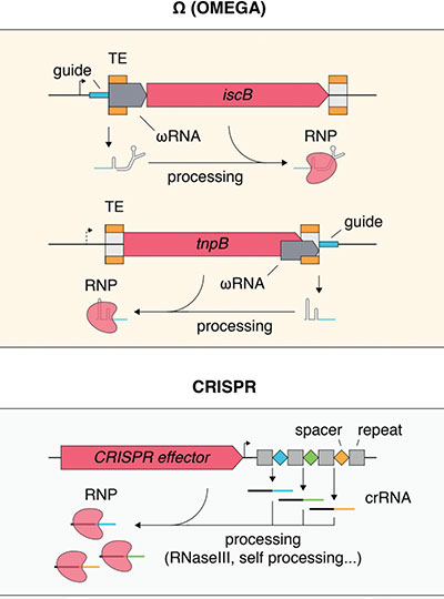
The first hints that OMEGA proteins might be directed by RNA came from the genes for proteins called IscBs. The IscBs are not involved in CRISPR immunity and were not known to associate with RNA, but they looked like small, DNA-cutting enzymes. The team discovered that each IscB had a small RNA encoded nearby and it directed IscB enzymes to cut specific DNA sequences. They named these RNAs “ωRNAs.”

The team’s experiments showed that two other classes of small proteins known as IsrBs and TnpBs, one of the most abundant genes in bacteria, also use ωRNAs that act as guides to direct the cleavage of DNA.

IscB, IsrB, and TnpB are found in mobile genetic elements called transposons. Graduate student Han Altae-Tran, co-first author on the paper, explains that each time these transposons move, they create a new guide RNA, allowing the enzyme they encode to cut somewhere else.

It’s not clear how bacteria benefit from this genomic shuffling—or whether they do at all.  Transposons are often thought of as selfish bits of DNA, concerned only with their own mobility and preservation, Kannan says. But if hosts can “co-opt” these systems and repurpose them, hosts may gain new abilities, as with CRISPR systems which confer adaptive immunity.

“A lot of the things that we have been thinking about may already exist naturally in some capacity,” says Altae-Tran.

IscBs and TnpBs appear to be predecessors of Cas9 and Cas12 CRISPR systems. The team suspects they, along with IsrB, likely gave rise to other RNA-guided enzymes, too—and they are eager to find them. They are curious about the range of functions that might be carried out in nature by RNA-guided enzymes, Kannan says, and suspect evolution likely already took advantage of OMEGA enzymes like IscBs and TnpBs to solve problems that biologists are keen to tackle.

Comparison of Ω (OMEGA) systems with other known RNA-guided systems. In contrast to CRISPR systems, which capture spacer sequences and store them in the locus within the CRISPR array, Ω systems may transpose their loci (or trans-acting loci) into target sequences, converting targets into ωRNA guides. Image courtesy of the researchers.

“A lot of the things that we have been thinking about may already exist naturally in some capacity,” says Altae-Tran. “Natural versions of these types of systems might be a good starting point to adapt for that particular task.”

The team is also interested in tracing the evolution of RNA-guided systems further into the past. “Finding all these new systems sheds light on how RNA-guided systems have evolved, but we don’t know where RNA-guided activity itself comes from,” Altae-Tran says. Understanding those origins, he says, could pave the way to developing even more classes of programmable tools.

This work was made possible with support from the Simons Center for the Social Brain at MIT; National Institutes of Health Intramural Research Program; National Institutes of Health grants 1R01-HG009761 and 1DP1-HL141201; Howard Hughes Medical Institute; Open Philanthropy; G. Harold and Leila Y. Mathers Charitable Foundation; Edward Mallinckrodt, Jr. Foundation; Poitras Center for Psychiatric Disorders Research at MIT; Hock E. Tan and K. Lisa Yang Center for Autism Research at MIT; Yang-Tan Center for Molecular Therapeutics at MIT; Lisa Yang; Phillips family; R. Metcalfe; and J. and P. Poitras.

RNA-targeting enzyme expands the CRISPR toolkit

Researchers at MIT’s McGovern Institute have discovered a bacterial enzyme that they say could expand scientists’ CRISPR toolkit, making it easy to cut and edit RNA with the kind of precision that, until now, has only been available for DNA editing. The enzyme, called Cas7-11, modifies RNA targets without harming cells, suggesting that in addition to being a valuable research tool, it provides a fertile platform for therapeutic applications.

“This new enzyme is like the Cas9 of RNA,” says McGovern Fellow Omar Abudayyeh, referring to the DNA-cutting CRISPR enzyme that has revolutionized modern biology by making DNA editing fast, inexpensive, and exact. “It creates two precise cuts and doesn’t destroy the cell in the process like other enzymes,” he adds.

Up until now, only one other family of RNA-targeting enzymes, Cas13, has extensively been developed for RNA targeting applications. However, when Cas13 recognizes its target, it shreds any RNAs in the cell, destroying the cell along the way. Like Cas9, Cas7-11 is part of a programmable system; it can be directed at specific RNA targets using a CRISPR guide. Abudayyeh, McGovern fellow Jonathan Gootenberg, and their colleagues discovered Cas7-11 through a deep exploration of the CRISPR systems found in the microbial world. Their findings are reported today in the journal Nature.

Exploring natural diversity

DNA tools in the CRISPR toolkit (red) are approaching capacity, but researchers are now beginning to find new tools to edit RNA (blue). Image: Steven Dixon

Like other CRISPR proteins, Cas7-11 is used by bacteria as a defense mechanism against viruses. After encountering a new virus, bacteria that employ the CRISPR system keep a record of the infection in the form of a small snippet of the pathogen’s genetic material. Should that virus reappear, the CRISPR system is activated, guided by a small piece of RNA to destroy the viral genome and eliminate the infection.

These ancient immune systems are widespread and diverse, with different bacteria deploying different proteins to counter their viral invaders.

“Some target DNA, some target RNA. Some are very efficient in cleaving the target but have some toxicity, and others do not. They introduce different types of cuts, they can differ in specificity—and so on,” says Eugene Koonin, an evolutionary biologist at the National Center for Biotechnology Information.

Abudayyeh, Gootenberg, and Koonin have been scouring genome sequences to learn about the natural diversity of CRISPR systems—and to mine them for potential tools. The idea, Abudayyeh says, is to take advantage of the work that evolution has already done in engineering protein machines.

“We don’t know what we’ll find,” Abudayyeh says, “but let’s just explore and see what’s out there.”

As the team was poring through public databases to examine the components of different bacterial defense systems, a protein from a bacterium that had been isolated from Tokyo Bay caught their attention. Its amino acid sequence indicated that it belonged to a class of CRISPR systems that use large, multiprotein machines to find and cleave their targets. But this protein appeared to have everything it needed to carry out the job on its own. Other known single-protein Cas enzymes, including the Cas9 protein that has been widely adopted for DNA editing, belong to a separate class of CRISPR systems—but Cas7-11 blurs the boundaries of the CRISPR classification system, Koonin says.

The enzyme, which the team eventually named Cas7-11, was attractive from an engineering perspective, because single proteins are easier to deliver to cells and make better tools than their complex counterparts. But its composition also signaled an unexpected evolutionary history. The team found evidence that through evolution, the components of a more complex Cas machine had fused together to make the Cas7-11 protein. Gootenberg equates this to discovering a bat when you had previously assumed that birds are the only animals that fly, thereby recognizing that there are multiple evolutionary paths to flight. “It totally changes the landscape of how these systems are thought about, both functionally and evolutionarily,” he says.

Precision editing

McGovern Fellows Jonathan Gootenberg and Omar Abudayyeh in their lab. Photo: Caitlin Cunningham

When Gootenberg and Abudayyeh produced the Cas7-11 protein in their lab and began experimenting with it, they realized this unusual enzyme offered a powerful means to manipulate and study RNA. When they introduced it into cells along with an RNA guide, it made remarkably precise cuts, snipping its targets while leaving other RNA undisturbed. This meant they could use Cas7-11 to change specific letters in the RNA code, correcting errors introduced by genetic mutations. They were also able to program Cas7-11 to either stabilize or destroy particular RNA molecules inside cells, which gave them the ability to adjust the levels of the proteins encoded by those RNAs.

Abudayyeh and Gootenberg also found that Cas7-11’s ability to cut RNA could be dampened by a protein that appeared likely to also be involved in triggering programmed cell death, suggesting a possible link between CRISPR defense and a more extreme response to infection.

The team showed that a gene therapy vector can deliver the complete Cas7-11 editing system to cells and that Cas7-11 does not compromise cells’ health. They hope that with further development, the enzyme might one day be used to edit disease-causing sequences out of a patient’s RNA so their cells can produce healthy proteins, or to dial down the level of a protein that is doing harm due to genetic disease.

“We think that the unique way that Cas7-11 cuts enables many interesting and diverse applications,” Gootenberg says, noting that no other CRISPR tool cuts RNA so precisely. “It’s yet another great example of how these basic-biology driven explorations can yield new tools for therapeutics and diagnostics,” he adds. “And we’re certainly still just scratching the surface of what’s out there in natural diversity.”

Scientists harness human protein to deliver molecular medicines to cells

Researchers from MIT, the McGovern Institute for Brain Research at MIT, the Howard Hughes Medical Institute, and the Broad Institute of MIT and Harvard have developed a new way to deliver molecular therapies to cells. The system, called SEND, can be programmed to encapsulate and deliver different RNA cargoes. SEND harnesses natural proteins in the body that form virus-like particles and bind RNA, and it may provoke less of an immune response than other delivery approaches.

The new delivery platform works efficiently in cell models, and, with further development, could open up a new class of delivery methods for a wide range of molecular medicines — including those for gene editing and gene replacement. Existing delivery vehicles for these therapeutics can be inefficient and randomly integrate into the genome of cells, and some can stimulate unwanted immune reactions. SEND has the promise to overcome these limitations, which could open up new opportunities to deploy molecular medicine.

“The biomedical community has been developing powerful molecular therapeutics, but delivering them to cells in a precise and efficient way is challenging,” said CRISPR pioneer Feng Zhang, senior author on the study, core institute member at the Broad Institute, investigator at the McGovern Institute, and the James and Patricia Poitras Professor of Neuroscience at MIT. “SEND has the potential to overcome these challenges.” Zhang is also an investigator at the Howard Hughes Medical Institute and a professor in MIT’s Departments of Brain and Cognitive Sciences and Biological Engineering.

SEND packages are introduced to diseased cells to deliver therapeutic mRNA and restore health. Image: McGovern Institute

Reporting in Science, the team describes how SEND (Selective Endogenous eNcapsidation for cellular Delivery) takes advantage of molecules made by human cells. At the center of SEND is a protein called PEG10, which normally binds to its own mRNA and forms a spherical protective capsule around it. In their study, the team engineered PEG10 to selectively package and deliver other RNA. The scientists used SEND to deliver the CRISPR-Cas9 gene editing system to mouse and human cells to edit targeted genes.

First author Michael Segel, a postdoctoral researcher in Zhang’s lab, and Blake Lash, second author and a graduate student in the lab, said PEG10 is not unique in its ability to transfer RNA. “That’s what’s so exciting,” said Segel. “This study shows that there are probably other RNA transfer systems in the human body that can also be harnessed for therapeutic purposes. It also raises some really fascinating questions about what the natural roles of these proteins might be.”

Inspiration from within

The PEG10 protein exists naturally in humans and is derived from a “retrotransposon” — a virus-like genetic element — that integrated itself into the genome of human ancestors millions of years ago. Over time, PEG10 has been co-opted by the body to become part of the repertoire of proteins important for life.

Four years ago, researchers showed that another retrotransposon-derived protein, ARC, forms virus-like structures and is involved in transferring RNA between cells. Although these studies suggested that it might be possible to engineer retrotransposon proteins as a delivery platform, scientists had not successfully harnessed these proteins to package and deliver specific RNA cargoes in mammalian cells.

Knowing that some retrotransposon-derived proteins are able to bind and package molecular cargo, Zhang’s team turned to these proteins as possible delivery vehicles. They systematically searched through these proteins in the human genome for ones that could form protective capsules. In their initial analysis, the team found 48 human genes encoding proteins that might have that ability. Of these, 19 candidate proteins were present in both mice and humans. In the cell line the team studied, PEG10 stood out as an efficient shuttle; the cells released significantly more PEG10 particles than any other protein tested. The PEG10 particles also mostly contained their own mRNA, suggesting that PEG10 might be able to package specific RNA molecules.

Developing a modular system

To develop the SEND technology, the team identified the molecular sequences, or “signals,” in PEG10’s mRNA that PEG10 recognizes and uses to package its mRNA. The researchers then used these signals to engineer both PEG10 and other RNA cargo so that PEG10 could selectively package those RNAs. Next, the team decorated the PEG10 capsules with additional proteins, called “fusogens,” that are found on the surface of cells and help them fuse together.

By engineering the fusogens on the PEG10 capsules, researchers should be able to target the capsule to a particular kind of cell, tissue, or organ. As a first step towards this goal, the team used two different fusogens, including one found in the human body, to enable delivery of SEND cargo.

“By mixing and matching different components in the SEND system, we believe that it will provide a modular platform for developing therapeutics for different diseases,” said Zhang.

Advancing gene therapy

SEND is composed of proteins that are produced naturally in the body, which means it may not trigger an immune response. If this is demonstrated in further studies, the researchers say SEND could open up opportunities to deliver gene therapies repeatedly with minimal side effects. “The SEND technology will complement viral delivery vectors and lipid nanoparticles to further expand the toolbox of ways to deliver gene and editing therapies to cells,” said Lash.

Next, the team will test SEND in animals and further engineer the system to deliver cargo to a variety of tissues and cells. They will also continue to probe the natural diversity of these systems in the human body to identify other components that can be added to the SEND platform.

“We’re excited to keep pushing this approach forward,” said Zhang. “The realization that we can use PEG10, and most likely other proteins, to engineer a delivery pathway in the human body to package and deliver new RNA and other potential therapies is a really powerful concept.”

This work was made possible with support from the Simons Center for the Social Brain at MIT; National Institutes of Health Intramural Research Program; National Institutes of Health grants 1R01-HG009761 and 1DP1-HL141201; Howard Hughes Medical Institute; Open Philanthropy; G. Harold and Leila Y. Mathers Charitable Foundation; Edward Mallinckrodt, Jr. Foundation; Poitras Center for Psychiatric Disorders Research at MIT; Hock E. Tan and K. Lisa Yang Center for Autism Research at MIT; Yang-Tan Center for Molecular Therapeutics at MIT; Lisa Yang; Phillips family; R. Metcalfe; and J. and P. Poitras.

Exploring the unknown

View the interactive version of this story in our Summer 2021 issue of BrainScan.


McGovern Investigator Ed Boyden.

McGovern Investigator Ed Boyden says his lab’s vision is clear.

“We want to understand how our brains take our sensory inputs, generate emotions and memories and decisions, and ultimately result in motor outputs. We want to be able to see the building blocks of life, and how they go into disarray in brain diseases. We want to be able to control the signals of the brain, so we can repair it,” Boyden says.

To get there, he and his team are exploring the brain’s complexity at every scale, from the function and architecture of its neural networks to the molecules that work together to process information.

And when they don’t have the tools to take them where they want to go, they create them, opening new frontiers for neuroscientists everywhere.

Open to discovery

Boyden’s team is highly interdisciplinary and collaborative. Its specialty, Boyden says, is problem solving. Creativity, adaptability, and deep curiosity are essential, because while many of neuroscience’s challenges are clear, the best way to address them is not. In its search for answers, Boyden’s lab is betting that an important path to discovery begins with finding new ways to explore.

They’ve made that possible with an innovative imaging approach called expansion microscopy (ExM). ExM physically enlarges biological samples so that minute details become visible under a standard laboratory microscope, enabling researchers everywhere to peer into spaces that once went unseen (see video below).

To use the technique, researchers permeate a biological sample with an absorbent gel, then add water, causing the components of the gel to spread apart and the tissue to expand.

This year, postdoctoral researcher Ruixuan Gao and graduate student Chih-Chieh (Jay) Yu made the method more precise, with a new material that anchors a sample’s molecules within a crystal-like lattice, better preserving structure during expansion than the irregular mesh-like composition of the original gel. The advance is an important step toward being able to image expanded samples with single-molecule precision, Gao says.

A revealing look

By opening space within the brain, ExM has let Boyden’s team venture into those spaces in new ways.

Areas of research and brain disorders page
Graduate student Oz Wassie examines expanded brain tissue. Photo: Justin Knight

In work led by Deblina Sarkar (who is now an assistant professor at MIT’s Media Lab), Jinyoung Kang, and Asmamaw (Oz) Wassie, they showed that they can pull apart proteins in densely packed regions like synapses so that it is easier to introduce fluorescent labels, illuminating proteins that were once too crowded to see. The process, called expansion revealing, has made it possible to visualize in intact brain tissue important structures such as ion channels that help transmit signals and fine-scale amyloid clusters in Alzheimer’s model mice.

Another reaction the lab has adapted to the expanded-brain context is RNA sequencing—an important tool for understanding cellular diversity. “Typically, the first thing you do in a sequencing project is you grind up the tissue, and you lose the spatial dimension,” explains Daniel Goodwin, a graduate student in Boyden’s lab. But when sequencing reactions are performed inside cells instead, new information is revealed.

Confocal image showing targeted ExSeq of a 34-panel gene set across a slice of mouse hippocampus. Green indicates YFP, magenta indicates reads identified with ExSeq, and white indicates reads localized within YFP-expressing cells. Image courtesy of the researchers.

Goodwin and fellow Boyden lab members Shahar Alon, Anubhav Sinha, Oz Wassie, and Fei Chen developed expansion sequencing (ExSeq), which copies RNA molecules, nucleotide by nucleotide, directly inside expanded tissue, using fluorescent labels that spell out the molecules’ codes just as they would in a sequencer.

The approach shows researchers which genes are turned on in which cells, as well as where those RNA molecules are—revealing, for example, which genes are active in the neuronal projections that carry out the brain’s communications. A next step, Sinha says, is to integrate expansion sequencing with other technologies to obtain even deeper insights.

That might include combining information revealed with ExSeq with a topographical map of the same cells’ genomes, using a method Boyden’s lab and collaborators Chen (who is now a core member of the Broad Institute) and Jason Buenrostro at Harvard have developed for DNA sequencing. That information is important because the shape of the genome varies across cells and circumstances, and that has consequences for how the genetic code is used.

Using similar techniques to those that make ExSeq possible, graduate students Andrew Payne, Zachary Chiang, and Paul Reginato figured out how to recreate the steps of commercial DNA sequencing within the genome’s natural environment.

By pinpointing the location of specific DNA sequences inside cells, the new method, called in situ genome sequencing (IGS) allows researchers to watch a genome reorganize itself in a developing embryo.

They haven’t yet performed this analysis inside expanded tissue, but Payne says integrating in situ genome sequencing (IGS) with ExM should open up new opportunities to study genomes’ structure.

Signaling clusters

Alongside these efforts, Boyden’s team is working to give researchers better tools to explore how molecules move, change, and interact, including a modular system that lets users assemble sets of sensors into clusters to simultaneously monitor multiple cellular activities.

Molecular sensors use fluorescence to report on certain changes inside cells, such as the calcium that surges into a neuron after it fires. But they come in a limited palette, so in most experiments only one or two things can be seen at once.

Graduate student Shannon Johnson and postdoctoral fellow Changyang Linghu solved this problem by putting different sensors at different points throughout a cell so they can report on different signals. Their technique, called spatial multiplexing, links sensors to molecular scaffolds designed to cling to their own kind. Sensors built on the same scaffold form islands inside cells, so when they light up their signals are distinct from those produced by other sensor islands.

Eventually, as new sensors and scaffolds become available, Johnson says the technique might be used to simultaneously follow dozens of molecular signals in living cells. The more precise information they can help people uncover, the better, Boyden says.

“The brain is so full of surprises, we don’t know where the next big discovery will come from,” he says. With new support from the recently established K. Lisa Yang and Hock E. Tan Center for Molecular Therapeutics in Neuroscience, the Boyden lab is positioned to make these big discoveries.

“My dream would be to image the signaling dynamics of the brain, and then perturb the dynamics, and then use expansion methods to make a map of the brain. If we can get those three data sets—the dynamics, the causality, and the molecular organization—I think stitching those together could potentially yield deep insights into how the brain works, and how we can repair it in disease states.”

New technique corrects disease-causing mutations

Gene editing, or purposefully changing a gene’s DNA sequence, is a powerful tool for studying how mutations cause disease, and for making changes in an individual’s DNA for therapeutic purposes. A novel method of gene editing that can be used for both purposes has now been developed by a team led by Guoping Feng, the James W. (1963) and Patricia T. Poitras Professor in Brain and Cognitive Sciences at MIT.

“This technical advance can accelerate the production of disease models in animals and, critically, opens up a brand-new methodology for correcting disease-causing mutations,” says Feng, who is also a member of the Broad Institute of Harvard and MIT and the associate director of the McGovern Institute for Brain Research at MIT. The new findings publish online May 26 and in print June 10 in the journal Cell.

Genetic models of disease

A major goal of the Feng lab is to precisely define what goes wrong in neurodevelopmental and neuropsychiatric disorders by engineering animal models that carry the gene mutations that cause these disorders in humans. New models can be generated by injecting embryos with gene editing tools, along with a piece of DNA carrying the desired mutation.

In one such method, the gene editing tool CRISPR is programmed to cut a targeted gene, thereby activating natural DNA mechanisms that “repair” the broken gene with the injected template DNA. The engineered cells are then used to generate offspring capable of passing the genetic change on to further generations, creating a stable genetic line in which the disease, and therapies, are tested.

Although CRISPR has accelerated the process of generating such disease models, the process can still take months or years. Reasons for the inefficiency are that many treated cells do not undergo the desired DNA sequence change at all, and the change only occurs on one of the two gene copies (for most genes, each cell contains two versions, one from the father and one from the mother).

In an effort to increase the efficiency of the gene editing process, the Feng lab team initially hypothesized that adding a DNA repair protein called RAD51 to a standard mixture of CRISPR gene editing tools would increase the chances that a cell (in this case a fertilized mouse egg, or one-cell embryo) would undergo the desired genetic change.

As a test case, they measured the rate at which they were able to insert (“knock-in”) a mutation in the gene Chd2 that is associated with autism.  The overall proportion of embryos that were correctly edited remained unchanged, but to their surprise, a significantly higher percentage carried the desired gene edit on both chromosomes. Tests with a different gene yielded the same unexpected outcome.

“Editing of both chromosomes simultaneously is normally very uncommon,” explains postdoctoral fellow Jonathan Wilde.  “The high rate of editing seen with RAD51 was really striking and what started as a simple attempt to make mutant Chd2 mice quickly turned into a much bigger project focused on RAD51 and its applications in genome editing,” said Wilde, who co-authored the Cell paper with research scientist Tomomi Aida.

A molecular copy machine

The Feng lab team next set out to understand the mechanism by which RAD51 enhances gene editing. They hypothesized that RAD51 engages a process called interhomolog repair (IHR), whereby a DNA break on one chromosome is repaired using the second copy of the chromosome (from the other parent) as the template.

To test this, they injected mouse embryos with RAD51 and CRISPR but left out the template DNA. They programmed CRISPR to cut only the gene sequence on one of the chromosomes, and then tested whether it was repaired to match the sequence on the uncut chromosome. For this experiment, they had to use mice in which the sequences on the maternal and paternal chromosomes were different.

They found that control embryos injected with CRISPR alone rarely showed IHR repair. However, addition of RAD51 significantly increased the number of embryos in which the CRISPR-targeted gene was edited to match the uncut chromosome.

“Previous studies of IHR found that it is incredibly inefficient in most cells,” says Wilde. “Our finding that it occurs much more readily in embryonic cells and can be enhanced by RAD51 suggest that a deeper understanding of what makes the embryo permissive to this type of DNA repair could help us design safer and more efficient gene therapies.”

A new way to correct disease-causing mutations          

Standard gene therapy strategies that rely on injecting a corrective piece of DNA to serve as a template for repairing the mutation engage a process called homology-directed repair (HDR).

“HDR-based strategies still suffer from low efficiency and carry the risk of unwanted integration of donor DNA throughout the genome,” explains Feng. “IHR has the potential to overcome these problems because it relies upon natural cellular pathways and the patient’s own normal chromosome for correction of the deleterious mutation.”

Feng’s team went on to identify additional DNA repair-associated proteins that can stimulate IHR, including several that not only promote high levels of IHR, but also repress errors in the DNA repair process. Additional experiments that allowed the team to examine the genomic features of IHR events gave deeper insight into the mechanism of IHR and suggested ways that the technique can be used to make gene therapies safer.

“While there is still a great deal to learn about this new application of IHR, our findings are the foundation for a new gene therapy approach that could help solve some of the big problems with current approaches,” says Aida.

This study was supported by the Hock E. Tan and K. Lisa Yang Center for Autism Research at MIT, the Poitras Center for Psychiatric Disorders Research at MIT, NIH/NIMH Conte Center Grant (P50 MH094271) and NIH Office of the Director (U24 OD026638).

A high-resolution glimpse of gene expression in cells

Using a novel technique for expanding tissue, MIT and Harvard Medical School researchers have devised a way to label individual molecules of messenger RNA within a tissue sample and then sequence the RNA.

This approach offers a unique snapshot of which genes are being expressed in different parts of a cell, and could allow scientists to learn much more about how gene expression is influenced by a cell’s location or its interactions with nearby cells. The technique could also be useful for mapping cells in the brain or other tissues and classifying them according to their function.

“Gene expression is one of the most fundamental processes in all of biology, and it plays roles in all biological processes, both healthy and disease-related. However, you need to know more than just whether a gene is on or off,” says Ed Boyden, the Y. Eva Tan Professor in Neurotechnology and a professor of biological engineering, media arts and sciences, and brain and cognitive sciences at MIT.

“You want to know where the gene products are located. You care what cell types they’re in, which individual cells they play roles in, and even which parts of cells they work in,” says Boyden.

In a study appearing today in Science, the researchers showed that they could use this technique to locate and then sequence thousands of different messenger RNA molecules within the mouse brain and in human tumor samples.

The senior authors of the study are Boyden, an investigator at the MIT McGovern Institute and the Howard Hughes Medical Institute; George Church, a professor of genetics at Harvard Medical School; and Adam Marblestone, a former MIT research scientist. The paper’s lead authors are Shahar Alon, a former MIT postdoc who is now a senior lecturer at Bar-Ilan University; Daniel Goodwin, an MIT graduate student; Anubhav Sinha ’14 MNG ’15, an MIT graduate student; Asmamaw Wassie ’12, PhD ’19; and Fei Chen PhD ’17, who is an assistant professor of stem cell and regenerative biology at Harvard University and a member of the Broad Institute of MIT and Harvard.

Tissue expansion

The new sequencing technique builds on a method that Boyden’s group devised in 2015 for expanding tissue samples and then imaging them. By embedding water-absorbent polymers into a tissue sample, researchers can swell the tissue sample while keeping its overall organization intact. Using this approach, tissues can be expanded by a factor of 100 or more, allowing scientists to obtain very high-resolution images of the brain or other tissues using a regular light microscope.

In 2014, Church’s lab developed an RNA sequencing technique known as FISSEQ (fluorescent in situ sequencing), which allows thousands of mRNA molecules to be located and sequenced within cells grown in a lab dish. The Boyden and Church labs decided to join forces to combine tissue expansion and in situ RNA sequencing, creating a new technique they call expansion sequencing (ExSeq).

Expanding the tissue before performing RNA sequencing has two main benefits: It offers a higher-resolution look at the RNA in cells, and it makes it easier to sequence those RNA molecules. “When you separate these molecules in the expanding sample, and move them away from each other, that gives you more room to actually perform the chemical reactions of in situ sequencing,” Marblestone says.

Once the tissue is expanded, the researchers can label and sequence thousands of RNA molecules in a sample, at a resolution that allows them to pinpoint the molecules’ locations not only within cells but within specific compartments such as dendrites — the tiny extensions of neurons that receive communications from other neurons.

“We know that the location of RNA in these small regions is important for learning and memory, but until now, we didn’t have any way to measure these locations because they are very small, on the order of nanometers,” Alon says.

Using an “untargeted” version of this technique, meaning that they are not looking for specific RNA sequences, the researchers can turn up thousands of different sequences. They estimate that in a given sample, they can sequence between 20 and 50 percent of all of the genes present.

In the mouse hippocampus, this technique yielded some surprising results. For one, the researchers found mRNA containing introns, which are sections of RNA that are normally edited out of mRNA in the nucleus, in dendrites. They also discovered mRNA molecules encoding transcription factors in the dendrites, which may help with novel forms of dendrite-to-nucleus communication.

“These are just examples of things that we never would have gone looking for intentionally, but now that we can sequence RNA exactly where it is in the neuron, we’re able to explore a lot more biology,” Goodwin says.

Cellular interactions

The researchers also showed that they could explore gene expression in a more targeted way, looking for a specific set of RNA sequences that correspond to genes of interest. In the visual cortex of the mouse, the researchers used this approach to classify neurons into different types based on an analysis of 42 different genes that they express.

In situ sequencing of physically expanded specimens enables multiplexed mapping of RNAs at nanoscale, subcellular resolution throughout intact tissues. Top: schematics of physical expansion and in situ sequencing (left), and image analysis (right). Bottom: characterization of nanoscale transcriptomic compartmentalization in mouse hippocampal neuron dendrites and spines (left, middle), and maps of cell types and states in a metastatic human breast cancer biopsy (right). Image courtesy of the researchers.

This technology could also be useful to analyze many other kinds of tissues, such as tumor biopsies. In this paper, the researchers studied breast cancer metastases, which contain many different cell types, including cancer cells and immune cells. The study revealed that these cell types can behave differently depending on their location within a tumor. For example, the researchers found that B cells that were near tumor cells expressed certain inflammatory genes at a higher level than B cells that were farther from tumor cells.

“The tumor microenvironment has been studied in many different contexts for a long time, but it’s been difficult to study it with any depth,” Sinha says. “A cancer biologist can give you a list of 20 or 30 marker genes that will identify most of the cell types in the tissue. Here, since we interrogated 297 different RNA transcripts in the sample, we can ask and answer more detailed questions about gene expression.”

The researchers now plan to further study the interactions between cancer cells and immune cells, as well as gene expression in the brain in healthy and disease states. They also plan to extend their techniques to allow them to map additional types of biomolecules, such as proteins, alongside RNA.

The research was funded, in part, by the National Institutes of Health and the National Science Foundation, as well as by Lisa Yang, John Doerr, the Open Philanthropy Project, Cancer Research UK, the Chan Zuckerberg Initiative Human Cell Atlas pilot program, and HHMI.