Nature: An unexpected source of innovative tools to study the brain

This story originally appeared in the Fall 2023 issue of BrainScan.

___

Scientist holds 3D printed phage over a natural background.
Genetic engineer Joseph Kreitz looks to the microscopic world for inspiration in Feng Zhang’s lab at the McGovern Institute. Photo: Steph Steve

In their quest to deepen their understanding of the brain, McGovern scientists take inspiration wherever it comes — and sometimes it comes from surprising sources. To develop new tools for research and innovative strategies for treating disease, they’ve drawn on proteins that organisms have been making for billions of years as well as sophisticated materials engineered for modern technology.

For McGovern investigator Feng Zhang, the natural world provides a rich source of molecules with remarkable and potentially useful functions.

Zhang is one of the pioneers of CRISPR, a programmable system for gene editing that is built from the components of a bacterial adaptive immune system. Scientists worldwide use CRISPR to modify genetic sequences in their labs, and many CRISPR-based therapies, which aim to treat disease through gene editing, are now in development. Meanwhile, Zhang and his team have continued to explore CRISPR-like systems beyond the bacteria in which they were originally discovered.

Turning to nature

This year, the search for evolutionarily related systems led Zhang’s team to a set of enzymes made by more complex organisms, including single-celled algae and hard-shell clams. Like the enzymes that power CRISPR, these newly discovered enzymes, called Fanzors, can be directed to cut DNA at specific sites by programming an RNA molecule as a guide.

Rhiannon Macrae, a scientific advisor in Zhang’s lab, says the discovery was surprising because Fanzors don’t seem to play the same role in immunity that CRISPR systems do. In fact, she says it’s not clear what Fanzors do at all. But as programmable gene editors, Fanzors might have an important advantage over current CRISPR tools — particularly for clinical applications. “Fanzor proteins are much smaller than the workhorse CRISPR tool, Cas9,” Macrae says. “This really matters when you actually want to be able to use one of these tools in a patient, because the bigger the tool, the harder it is to package and deliver to patients’ cells.”

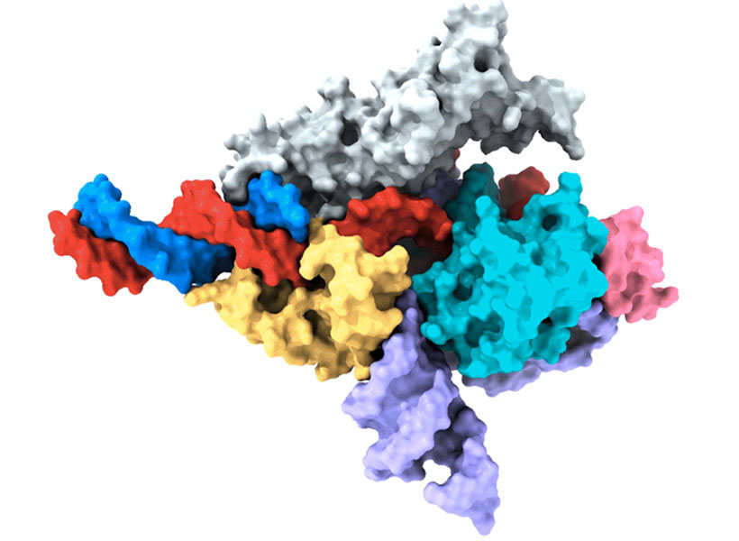
Cryo-EM map of a Fanzor protein (gray, yellow, light blue, and pink) in complex with ωRNA (purple) and its target DNA (red). Non-target DNA strand in blue. Image: Zhang lab

Zhang’s team has thought a lot about how to get therapies to patients’ cells, and size is only one consideration. They’ve also been looking for ways to direct drugs, gene-editing tools, or other therapies to specific cells and tissues in the body. One of the lab’s leading strategies comes from another unexpected natural source: a microscopic syringe produced by certain insect-infecting bacteria.

In their search for an efficient system for targeted drug delivery, Zhang and graduate student Joseph Kreitz first considered the injection systems of bacteria-infecting viruses: needle-like structures that pierce the outer membrane of their host to deliver their own genetic material. But these viral injection systems can’t easily be freed from the rest of the virus.

Then Zhang learned that some bacteria have injection systems of their own, which they release inside their hosts after packing them with toxins. They reengineered the bacterial syringe, devising a delivery system that works on human cells. Their current system can be programmed to inject proteins — including those used for gene editing — directly into specified cell types. With further development, Zhang hopes it will work with other types of therapies, as well.

Magnetic imaging

In McGovern Associate Investigator Alan Jasanoff’s lab, researchers are designing sensors that can track the activity of specific neurons or molecules in the brain, using magnetic resonance imaging (MRI) or related forms of non-invasive imaging. These tools are essential for understanding how the brain’s cells and circuits work together to process information. “We want to give MRI a suite of metaphorical colors: sensitivities that enable us to dissect the different kinds of mechanistically significant contributors to neural activity,” he explains.

Jasanoff can tick off a list of molecules with notable roles in biology and industry that his lab has repurposed to glean more information from brain imaging. These include manganese — a metal once used to tint ancient glass; nitric oxide synthase — the enzyme that causes blushing; and iron oxide nanoparticles — tiny magnets that enable compact data storage inside computers. But Jasanoff says none of these should be considered out of place in the imaging world. “Most are pretty logical choices,” he says. “They all do different things and we use them in pretty different ways, but they are either magnetic or interact with magnetic molecules to serve our purposes for brain imaging.”

Close-up picture of manganese metal
Manganese, a metal that interacts weakly with magnetic fields, is a key component in new MRI sensors being developed in Alan Jasanoff’s lab at the McGovern Institute.

The enzyme nitric oxide synthase, for example, plays an important role in most functional MRI scans. The enzyme produces nitric oxide, which causes blood vessels to expand. This can bring a blush to the cheeks, but in the brain, it increases blood flow to bring more oxygen to busy neurons. MRI can detect this change because it is sensitive to the magnetic properties of blood.

By using blood flow as a proxy for neural activity, functional MRI scans light up active regions of the brain, but they can’t pinpoint the activity of specific cells. So Jasanoff and his team devised a more informative MRI sensor by reengineering nitric oxide synthase. Their modified enzyme, which they call NOSTIC, can be introduced into a select group of cells, where it will produce nitric oxide in response to neural activity — triggering increased blood flow and strengthening the local MRI signal. Researchers can deliver it to specific kinds of brain cells, or they can deliver it exclusively to neurons that communicate directly with one another. Then they can watch for an elevated MRI signal when those cells fire. This lets them see how information flows through the brain and tie specific cells to particular tasks.

Miranda Dawson, a graduate student in Jasanoff’s lab, is using NOSTIC to study the brain circuits that fuel addiction. She’s interested in the involvement of a brain region called the insula, which may mediate the physical sensations that people with addiction experience during drug cravings or withdrawal. With NOSTIC, Dawson can follow how the insula communicates to other parts of the brain as a rat experiences these MITstages of addiction. “We give our sensor to the insula, and then it projects to anatomically connected brain regions,” she explains. “So we’re able to delineate what circuits are being activated at different points in the addiction cycle.”

Scientist with folded arms next to a picture of a brain
Miranda Dawson uses her lab’s novel MRI sensor, NOSTIC, to illuminate the brain circuits involved in fentanyl craving and withdrawal. Photo: Steph Stevens; MRI scan: Nan Li, Souparno Ghosh, Jasanoff lab

Mining biodiversity

McGovern investigators know that good ideas and useful tools can come from anywhere. Sometimes, the key to harnessing those tools is simply recognizing their potential. But there are also opportunities for a more deliberate approach to finding them.

McGovern Investigator Ed Boyden is leading a program that aims to accelerate the discovery of valuable natural products. Called the Biodiversity Network (BioNet), the project is collecting biospecimens from around the world and systematically analyzing them, looking for molecular tools that could be applied to major challenges in science and medicine, from brain research to organ preservation. “The idea behind BioNet,” Boyden explains, “is rather than wait for chance to give us these discoveries, can we go look for them on purpose?”

Researchers uncover new CRISPR-like system in animals that can edit the human genome

A team of researchers led by Feng Zhang at the McGovern Institute and the Broad Institute of MIT and Harvard has uncovered the first programmable RNA-guided system in eukaryotes — organisms that include fungi, plants, and animals.

In a study in Nature, the team describes how the system is based on a protein called Fanzor. They showed that Fanzor proteins use RNA as a guide to target DNA precisely, and that Fanzors can be reprogrammed to edit the genome of human cells. The compact Fanzor systems have the potential to be more easily delivered to cells and tissues as therapeutics than CRISPR/Cas systems, and further refinements to improve their targeting efficiency could make them a valuable new technology for human genome editing.

CRISPR/Cas was first discovered in prokaryotes (bacteria and other single-cell organisms that lack nuclei) and scientists including Zhang’s lab have long wondered whether similar systems exist in eukaryotes. The new study demonstrates that RNA-guided DNA-cutting mechanisms are present across all kingdoms of life.

“This new system is another way to make precise changes in human cells, complementing the genome editing tools we already have.” — Feng Zhang

“CRISPR-based systems are widely used and powerful because they can be easily reprogrammed to target different sites in the genome,” said Zhang, senior author on the study and a core institute member at the Broad, an investigator at MIT’s McGovern Institute, the James and Patricia Poitras Professor of Neuroscience at MIT, and a Howard Hughes Medical Institute investigator. “This new system is another way to make precise changes in human cells, complementing the genome editing tools we already have.”

Searching the domains of life

A major aim of the Zhang lab is to develop genetic medicines using systems that can modulate human cells by targeting specific genes and processes. “A number of years ago, we started to ask, ‘What is there beyond CRISPR, and are there other RNA-programmable systems out there in nature?’” said Zhang.

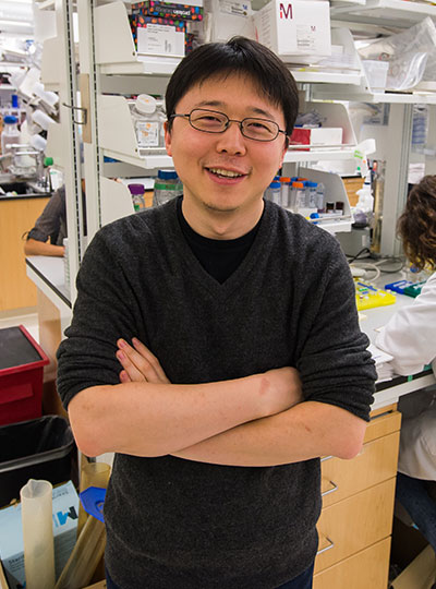
Feng Zhang with folded arms in lab
McGovern Investigator Feng Zhang in his lab.

Two years ago, Zhang lab members discovered a class of RNA-programmable systems in prokaryotes called OMEGAs, which are often linked with transposable elements, or “jumping genes”, in bacterial genomes and likely gave rise to CRISPR/Cas systems. That work also highlighted similarities between prokaryotic OMEGA systems and Fanzor proteins in eukaryotes, suggesting that the Fanzor enzymes might also use an RNA-guided mechanism to target and cut DNA.

In the new study, the researchers continued their study of RNA-guided systems by isolating Fanzors from fungi, algae, and amoeba species, in addition to a clam known as the Northern Quahog. Co-first author Makoto Saito of the Zhang lab led the biochemical characterization of the Fanzor proteins, showing that they are DNA-cutting endonuclease enzymes that use nearby non-coding RNAs known as ωRNAs to target particular sites in the genome. It is the first time this mechanism has been found in eukaryotes, such as animals.

Unlike CRISPR proteins, Fanzor enzymes are encoded in the eukaryotic genome within transposable elements and the team’s phylogenetic analysis suggests that the Fanzor genes have migrated from bacteria to eukaryotes through so-called horizontal gene transfer.

“These OMEGA systems are more ancestral to CRISPR and they are among the most abundant proteins on the planet, so it makes sense that they have been able to hop back and forth between prokaryotes and eukaryotes,” said Saito.

To explore Fanzor’s potential as a genome editing tool, the researchers demonstrated that it can generate insertions and deletions at targeted genome sites within human cells. The researchers found the Fanzor system to initially be less efficient at snipping DNA than CRISPR/Cas systems, but by systematic engineering, they introduced a combination of mutations into the protein that increased its activity 10-fold. Additionally, unlike some CRISPR systems and the OMEGA protein TnpB, the team found that a fungal-derived Fanzor protein did not exhibit “collateral activity,” where an RNA-guided enzyme cleaves its DNA target as well as degrading nearby DNA or RNA. The results suggest that Fanzors could potentially be developed as efficient genome editors.

Co-first author Peiyu Xu led an effort to analyze the molecular structure of the Fanzor/ωRNA complex and illustrate how it latches onto DNA to cut it. Fanzor shares structural similarities with its prokaryotic counterpart CRISPR-Cas12 protein, but the interaction between the ωRNA and the catalytic domains of Fanzor is more extensive, suggesting that the ωRNA might play a role in the catalytic reactions. “We are excited about these structural insights for helping us further engineer and optimize Fanzor for improved efficiency and precision as a genome editor,” said Xu.

Like CRISPR-based systems, the Fanzor system can be easily reprogrammed to target specific genome sites, and Zhang said it could one day be developed into a powerful new genome editing technology for research and therapeutic applications. The abundance of RNA-guided endonucleases like Fanzors further expands the number of OMEGA systems known across kingdoms of life and suggests that there are more yet to be found.

“Nature is amazing. There’s so much diversity,” said Zhang. “There are probably more RNA-programmable systems out there, and we’re continuing to explore and will hopefully discover more.”

The paper’s other authors include Guilhem Faure, Samantha Maguire, Soumya Kannan, Han Altae-Tran, Sam Vo, AnAn Desimone, and Rhiannon Macrae.

Support for this work was provided by the Howard Hughes Medical Institute; Poitras Center for Psychiatric Disorders Research at MIT; K. Lisa Yang and Hock E. Tan Molecular Therapeutics Center at MIT; Broad Institute Programmable Therapeutics Gift Donors; The Pershing Square Foundation, William Ackman, and Neri Oxman; James and Patricia Poitras; BT Charitable Foundation; Asness Family Foundation; Kenneth C. Griffin; the Phillips family; David Cheng; Robert Metcalfe; and Hugo Shong.


Season’s Greetings from the McGovern Institute

This year’s holiday video (shown above) was inspired by Ev Fedorenko’s July 2022 Nature Neuroscience paper, which found similar patterns of brain activation and language selectivity across speakers of 45 different languages.

Universal language network

Ev Fedorenko uses the widely translated book “Alice in Wonderland” to test brain responses to different languages. Photo: Caitlin Cunningham

Over several decades, neuroscientists have created a well-defined map of the brain’s “language network,” or the regions of the brain that are specialized for processing language. Found primarily in the left hemisphere, this network includes regions within Broca’s area, as well as in other parts of the frontal and temporal lobes. Although roughly 7,000 languages are currently spoken and signed across the globe, the vast majority of those mapping studies have been done in English speakers as they listened to or read English texts.

To truly understand the cognitive and neural mechanisms that allow us to learn and process such diverse languages, Fedorenko and her team scanned the brains of speakers of 45 different languages while they listened to Alice in Wonderland in their native language. The results show that the speakers’ language networks appear to be essentially the same as those of native English speakers — which suggests that the location and key properties of the language network appear to be universal.

The many languages of McGovern

English may be the primary language used by McGovern researchers, but more than 35 other languages are spoken by scientists and engineers at the McGovern Institute. Our holiday video features 30 of these researchers saying Happy New Year in their native (or learned) language. Below is the complete list of languages included in our video. Expand each accordion to learn more about the speaker of that particular language and the meaning behind their new year’s greeting.

New CRISPR-based tool inserts large DNA sequences at desired sites in cells

Building on the CRISPR gene-editing system, MIT researchers have designed a new tool that can snip out faulty genes and replace them with new ones, in a safer and more efficient way.

Using this system, the researchers showed that they could deliver genes as long as 36,000 DNA base pairs to several types of human cells, as well as to liver cells in mice. The new technique, known as PASTE, could hold promise for treating diseases that are caused by defective genes with a large number of mutations, such as cystic fibrosis.

“It’s a new genetic way of potentially targeting these really hard to treat diseases,” says Omar Abudayyeh, a McGovern Fellow at MIT’s McGovern Institute for Brain Research. “We wanted to work toward what gene therapy was supposed to do at its original inception, which is to replace genes, not just correct individual mutations.”

The new tool combines the precise targeting of CRISPR-Cas9, a set of molecules originally derived from bacterial defense systems, with enzymes called integrases, which viruses use to insert their own genetic material into a bacterial genome.

“Just like CRISPR, these integrases come from the ongoing battle between bacteria and the viruses that infect them,” says Jonathan Gootenberg, also a McGovern Fellow. “It speaks to how we can keep finding an abundance of interesting and useful new tools from these natural systems.”

Gootenberg and Abudayyeh are the senior authors of the new study, which appears today in Nature Biotechnology. The lead authors of the study are MIT technical associates Matthew Yarnall and Rohan Krajeski, former MIT graduate student Eleonora Ioannidi, and MIT graduate student Cian Schmitt-Ulms.

DNA insertion

The CRISPR-Cas9 gene editing system consists of a DNA-cutting enzyme called Cas9 and a short RNA strand that guides the enzyme to a specific area of the genome, directing Cas9 where to make its cut. When Cas9 and the guide RNA targeting a disease gene are delivered into cells, a specific cut is made in the genome, and the cells’ DNA repair processes glue the cut back together, often deleting a small portion of the genome.

If a DNA template is also delivered, the cells can incorporate a corrected copy into their genomes during the repair process. However, this process requires cells to make double-stranded breaks in their DNA, which can cause chromosomal deletions or rearrangements that are harmful to cells. Another limitation is that it only works in cells that are dividing, as nondividing cells don’t have active DNA repair processes.

The MIT team wanted to develop a tool that could cut out a defective gene and replace it with a new one without inducing any double-stranded DNA breaks. To achieve this goal, they turned to a family of enzymes called integrases, which viruses called bacteriophages use to insert themselves into bacterial genomes.

For this study, the researchers focused on serine integrases, which can insert huge chunks of DNA, as large as 50,000 base pairs. These enzymes target specific genome sequences known as attachment sites, which function as “landing pads.” When they find the correct landing pad in the host genome, they bind to it and integrate their DNA payload.

In past work, scientists have found it challenging to develop these enzymes for human therapy because the landing pads are very specific, and it’s difficult to reprogram integrases to target other sites. The MIT team realized that combining these enzymes with a CRISPR-Cas9 system that inserts the correct landing site would enable easy reprogramming of the powerful insertion system.

The new tool, PASTE (Programmable Addition via Site-specific Targeting Elements), includes a Cas9 enzyme that cuts at a specific genomic site, guided by a strand of RNA that binds to that site. This allows them to target any site in the genome for insertion of the landing site, which contains 46 DNA base pairs. This insertion can be done without introducing any double-stranded breaks by adding one DNA strand first via a fused reverse transcriptase, then its complementary strand.

Once the landing site is incorporated, the integrase can come along and insert its much larger DNA payload into the genome at that site.

“We think that this is a large step toward achieving the dream of programmable insertion of DNA,” Gootenberg says. “It’s a technique that can be easily tailored both to the site that we want to integrate as well as the cargo.”

Gene replacement

In this study, the researchers showed that they could use PASTE to insert genes into several types of human cells, including liver cells, T cells, and lymphoblasts (immature white blood cells). They tested the delivery system with 13 different payload genes, including some that could be therapeutically useful, and were able to insert them into nine different locations in the genome.

In these cells, the researchers were able to insert genes with a success rate ranging from 5 to 60 percent. This approach also yielded very few unwanted “indels” (insertions or deletions) at the sites of gene integration.

“We see very few indels, and because we’re not making double-stranded breaks, you don’t have to worry about chromosomal rearrangements or large-scale chromosome arm deletions,” Abudayyeh says.

The researchers also demonstrated that they could insert genes in “humanized” livers in mice. Livers in these mice consist of about 70 percent human hepatocytes, and PASTE successfully integrated new genes into about 2.5 percent of these cells.

The DNA sequences that the researchers inserted in this study were up to 36,000 base pairs long, but they believe even longer sequences could also be used. A human gene can range from a few hundred to more than 2 million base pairs, although for therapeutic purposes only the coding sequence of the protein needs to be used, drastically reducing the size of the DNA segment that needs to be inserted into the genome.

“The ability to site-specifically make large genomic integrations is of huge value to both basic science and biotechnology studies. This toolset will, I anticipate, be very enabling for the research community,” says Prashant Mali, a professor of bioengineering at the University of California at San Diego, who was not involved in the study.

The researchers are now further exploring the possibility of using this tool as a possible way to replace the defective cystic fibrosis gene. This technique could also be useful for treating blood diseases caused by faulty genes, such as hemophilia and G6PD deficiency, or Huntington’s disease, a neurological disorder caused by a defective gene that has too many gene repeats.

The researchers have also made their genetic constructs available online for other scientists to use.

“One of the fantastic things about engineering these molecular technologies is that people can build on them, develop and apply them in ways that maybe we didn’t think of or hadn’t considered,” Gootenberg says. “It’s really great to be part of that emerging community.”

The research was funded by a Swiss National Science Foundation Postdoc Mobility Fellowship, the U.S. National Institutes of Health, the McGovern Institute Neurotechnology Program, the K. Lisa Yang and Hock E. Tan Center for Molecular Therapeutics in Neuroscience, the G. Harold and Leila Y. Mathers Charitable Foundation, the MIT John W. Jarve Seed Fund for Science Innovation, Impetus Grants, a Cystic Fibrosis Foundation Pioneer Grant, Google Ventures, Fast Grants, the Harvey Family Foundation, and the McGovern Institute.

RNA-activated protein cutter protects bacteria from infection

Our growing understanding of the ways bacteria defend themselves against viruses continues to change the way scientists work and offer new opportunities to improve human health. Ancient immune systems known as CRISPR systems have already been widely adopted as powerful genome editing tools, and the CRISPR toolkit is continuing to expand. Now, scientists at MIT’s McGovern Institute have uncovered an unexpected and potentially useful tool that some bacteria use to respond to infection: an RNA-activated protein-cutting enzyme.

McGovern Fellows Jonathan Gootenberg and Omar Abudayyeh in their lab. Photo: Caitlin Cunningham

The enzyme is part of a CRISPR system discovered last year by McGovern Fellows Omar Abudayyeh and Jonathan Gootenberg. The system, found in bacteria from Tokyo Bay, originally caught their interest because of the precision with which its RNA-activated enzyme cuts RNA. That enzyme, Cas7-11, is considered a promising tool for editing RNA for both research and potential therapeutics. Now, the same researchers have taken a closer look at this bacterial immune system and found that once Cas7-11 has been activated by the right RNA, it also turns on an enzyme that snips apart a particular bacterial protein.

That makes the Cas7-11 system notably more complex than better-studied CRISPR systems, which protect bacteria simply by chopping up the genetic material of an invading virus. “This is a much more elegant and complex signaling mechanism to really defend the bacteria,” Abudayyeh says. A team led by Abudayyeh, Gootenberg, and collaborator Hiroshi Nishimasu at the University of Tokyo report these findings in the November 3, 2022, issue of the journal Science.

Protease programming

The team’s experiments reveal that in bacteria, activation of the protein-cutting enzyme, known as a protease, triggers a series of events that ultimately slow the organism’s growth. But the components of the CRISPR system can be engineered to achieve different outcomes. Gootenberg and Abudayyeh have already programmed the RNA-activated protease to report on the presence of specific RNAs in mammalian cells. With further adaptations, they say it might one day be used to diagnose or treat disease.

The discovery grew out of the researchers’ curiosity about how bacteria protect themselves from infection using Cas7-11. They knew that the enzyme was capable of cutting viral RNA, but there were hints that something more might be going on. They wondered whether a set of genes that clustered near the Cas7-11 gene might also be involved in the bacteria’s infection response, and when graduate students Cian Schmitt-Ulms and Kaiyi Jiang began experimenting with those proteins, they discovered that they worked with Cas7-11 to execute a surprisingly elaborate response to a target RNA.

One of those proteins was the protease Csx29. In the team’s test tube experiments, Csx29 and Cas7-11 couldn’t cut anything on their own—but in the presence of a target RNA, Cas7-11 switched it on. Even then, when the researchers mixed the protease with Cas7-11 and its RNA target and allowed them to mingle with other proteins, most of the proteins remained intact. But one, a protein called Csx30, was reliably snipped apart by the protein-cutting enzyme.

Their experiments had uncovered an enzyme that cut a specific protein, but only in the presence of its particular target RNA. It was unusual—and potentially useful. “That was when we knew we were onto something,” Abudayyeh says.

As the team continued to explore the system, they found that the Csx29’s RNA-activated cut frees a fragment of Csx30 that then works with other bacterial proteins to execute a key aspect of the bacteria’s response to infection—slowing down growth. “Our growth experiments suggest that the cleavage is modulating the bacteria’s stress response in some way,” Gootenberg says.

The scientists quickly recognized that this RNA-activated protease could have uses beyond its natural role in antiviral defense. They have shown that the system can be adapted so that when the protease cuts Csx30 in the presence of its target RNA, it generates an easy to detect fluorescent signal. Because Cas7-11 can be directed to recognize any target RNA, researchers can program the system to detect and report on any RNA of interest. And even though the original system evolved in bacteria, this RNA sensor works well in mammalian cells.

Gootenberg and Abudayyeh say understanding this surprisingly elaborate CRISPR system opens new possibilities by adding to scientists’ growing toolkit of RNA-guided enzymes. “We’re excited to see how people use these tools and how they innovate on them,” Gootenberg says. It’s easy to imagine both diagnostic and therapeutic applications, they say. For example, an RNA sensor could detect signatures of disease in patient samples or to limit delivery of a potential therapy to specific types of cells, enabling that drug to carry out its work without side effects.

In addition to Gootenberg, Abudayyeh, Schmitt-Ulms, and Jiang, Abudayyeh-Gootenberg lab postdoc Nathan Wenyuan Zhou contributed to the project. This work was supported by NIH grants 1R21-AI149694, R01-EB031957, and R56-HG011857, the McGovern Institute Neurotechnology (MINT) program, the K. Lisa Yang and Hock E. Tan Center for Molecular Therapeutics in Neuroscience, the G. Harold & Leila Y. Mathers Charitable Foundation, the MIT John W. Jarve (1978) Seed Fund for Science Innovation, the Cystic Fibrosis Foundation, Google Ventures, Impetus Grants, the NHGRI/TDCC Opportunity Fund, and the McGovern Institute.

RNA-sensing system controls protein expression in cells based on specific cell states

Researchers at the Broad Institute of MIT and Harvard and the McGovern Institute for Brain Research at MIT have developed a system that can detect a particular RNA sequence in live cells and produce a protein of interest in response. Using the technology, the team showed how they could identify specific cell types, detect and measure changes in the expression of individual genes, track transcriptional states, and control the production of proteins encoded by synthetic mRNA.

The platform, called Reprogrammable ADAR Sensors, or RADARS, even allowed the team to target and kill a specific cell type. The team said RADARS could one day help researchers detect and selectively kill tumor cells, or edit the genome in specific cells. The study appears today in Nature Biotechnology and was led by co-first authors Kaiyi Jiang (MIT), Jeremy Koob (Broad), Xi Chen (Broad), Rohan Krajeski (MIT), and Yifan Zhang (Broad).

“One of the revolutions in genomics has been the ability to sequence the transcriptomes of cells,” said Fei Chen, a core institute member at the Broad, Merkin Fellow, assistant professor at Harvard University, and co-corresponding author on the study. “That has really allowed us to learn about cell types and states. But, often, we haven’t been able to manipulate those cells specifically. RADARS is a big step in that direction.”

“Right now, the tools that we have to leverage cell markers are hard to develop and engineer,” added Omar Abudayyeh, a McGovern Institute Fellow and co-corresponding author on the study. “We really wanted to make a programmable way of sensing and responding to a cell state.”

Jonathan Gootenberg, who is also a McGovern Institute Fellow and co-corresponding author, says that their team was eager to build a tool to take advantage of all the data provided by single-cell RNA sequencing, which has revealed a vast array of cell types and cell states in the body.

“We wanted to ask how we could manipulate cellular identities in a way that was as easy as editing the genome with CRISPR,” he said. “And we’re excited to see what the field does with it.” 

Omar Abudayyeh, Jonathan Gootenberg and Fei Chen at the Broad Institute
Study authors (from left to right) Omar Abudayyeh, Jonathan Gootenberg, and Fei Chen. Photo: Namrita Sengupta

Repurposing RNA editing

The RADARS platform generates a desired protein when it detects a specific RNA by taking advantage of RNA editing that occurs naturally in cells.

The system consists of an RNA containing two components: a guide region, which binds to the target RNA sequence that scientists want to sense in cells, and a payload region, which encodes the protein of interest, such as a fluorescent signal or a cell-killing enzyme. When the guide RNA binds to the target RNA, this generates a short double-stranded RNA sequence containing a mismatch between two bases in the sequence — adenosine (A) and cytosine (C). This mismatch attracts a naturally occurring family of RNA-editing proteins called adenosine deaminases acting on RNA (ADARs).

In RADARS, the A-C mismatch appears within a “stop signal” in the guide RNA, which prevents the production of the desired payload protein. The ADARs edit and inactivate the stop signal, allowing for the translation of that protein. The order of these molecular events is key to RADARS’s function as a sensor; the protein of interest is produced only after the guide RNA binds to the target RNA and the ADARs disable the stop signal.

The team tested RADARS in different cell types and with different target sequences and protein products. They found that RADARS distinguished between kidney, uterine, and liver cells, and could produce different fluorescent signals as well as a caspase, an enzyme that kills cells. RADARS also measured gene expression over a large dynamic range, demonstrating their utility as sensors.

Most systems successfully detected target sequences using the cell’s native ADAR proteins, but the team found that supplementing the cells with additional ADAR proteins increased the strength of the signal. Abudayyeh says both of these cases are potentially useful; taking advantage of the cell’s native editing proteins would minimize the chance of off-target editing in therapeutic applications, but supplementing them could help produce stronger effects when RADARS are used as a research tool in the lab.

On the radar

Abudayyeh, Chen, and Gootenberg say that because both the guide RNA and payload RNA are modifiable, others can easily redesign RADARS to target different cell types and produce different signals or payloads. They also engineered more complex RADARS, in which cells produced a protein if they sensed two RNA sequences and another if they sensed either one RNA or another. The team adds that similar RADARS could help scientists detect more than one cell type at the same time, as well as complex cell states that can’t be defined by a single RNA transcript.

Ultimately, the researchers hope to develop a set of design rules so that others can more easily develop RADARS for their own experiments. They suggest other scientists could use RADARS to manipulate immune cell states, track neuronal activity in response to stimuli, or deliver therapeutic mRNA to specific tissues.

“We think this is a really interesting paradigm for controlling gene expression,” said Chen. “We can’t even anticipate what the best applications will be. That really comes from the combination of people with interesting biology and the tools you develop.”

This work was supported by the The McGovern Institute Neurotechnology (MINT) program, the K. Lisa Yang and Hock E. Tan Center for Molecular Therapeutics in Neuroscience, the G. Harold & Leila Y. Mathers Charitable Foundation, Massachusetts Institute of Technology, Impetus Grants, the Cystic Fibrosis Foundation, Google Ventures, FastGrants, the McGovern Institute, National Institutes of Health, the Burroughs Wellcome Fund, the Searle Scholars Foundation, the Harvard Stem Cell Institute, and the Merkin Institute.

MIT scientists discover new antiviral defense system in bacteria

Bacteria use a variety of defense strategies to fight off viral infection, and some of these systems have led to groundbreaking technologies, such as CRISPR-based gene-editing. Scientists predict there are many more antiviral weapons yet to be found in the microbial world.

A team led by researchers at the Broad Institute of MIT and Harvard and the McGovern Institute for Brain Research at MIT has discovered and characterized one of these unexplored microbial defense systems. They found that certain proteins in bacteria and archaea (together known as prokaryotes) detect viruses in surprisingly direct ways, recognizing key parts of the viruses and causing the single-celled organisms to commit suicide to quell the infection within a microbial community. The study is the first time this mechanism has been seen in prokaryotes and shows that organisms across all three domains of life — bacteria, archaea, and eukaryotes (which includes plants and animals) — use pattern recognition of conserved viral proteins to defend against pathogens.

The study appears in Science.

“This work demonstrates a remarkable unity in how pattern recognition occurs across very different organisms,” said senior author Feng Zhang, who is a core institute member at the Broad, the James and Patricia Poitras Professor of Neuroscience at MIT, a professor of brain and cognitive sciences and biological engineering at MIT, and an investigator at MIT’s McGovern Institute and the Howard Hughes Medical Institute. “It’s been very exciting to integrate genetics, bioinformatics, biochemistry, and structural biology approaches in one study to understand this fascinating molecular system.”

Microbial armory

In an earlier study, the researchers scanned data on the DNA sequences of hundreds of thousands of bacteria and archaea, which revealed several thousand genes harboring signatures of microbial defense. In the new study, they homed in on a handful of these genes encoding enzymes that are members of the STAND ATPase family of proteins, which in eukaryotes are involved in the innate immune response.

In humans and plants, the STAND ATPase proteins fight infection by recognizing patterns in a pathogen itself or in the cell’s response to infection. In the new study, the researchers wanted to know if the proteins work the same way in prokaryotes to defend against infection. The team chose a few STAND ATPase genes from the earlier study, delivered them to bacterial cells, and challenged those cells with bacteriophage viruses. The cells underwent a dramatic defensive response and survived.

The scientists next wondered which part of the bacteriophage triggers that response, so they delivered viral genes to the bacteria one at a time. Two viral proteins elicited an immune response: the portal, a part of the virus’s capsid shell, which contains viral DNA; and the terminase, the molecular motor that helps assemble the virus by pushing the viral DNA into the capsid. Each of these viral proteins activated a different STAND ATPase to protect the cell.

The finding was striking and unprecedented. Most known bacterial defense systems work by sensing viral DNA or RNA, or cellular stress due to the infection. These bacterial proteins were instead directly sensing key parts of the virus.

The team next showed that bacterial STAND ATPase proteins could recognize diverse portal and terminase proteins from different phages. “It’s surprising that bacteria have these highly versatile sensors that can recognize all sorts of different phage threats that they might encounter,” said co-first author Linyi Gao, a junior fellow in the Harvard Society of Fellows and a former graduate student in the Zhang lab.

Structural analysis

For a detailed look at how the microbial STAND ATPases detect the viral proteins, the researchers used cryo-electron microscopy to examine their molecular structure when bound to the viral proteins. “By analyzing the structure, we were able to precisely answer a lot of the questions about how these things actually work,” said co-first author Max Wilkinson, a postdoctoral researcher in the Zhang lab.

The team saw that the portal or terminase protein from the virus fits within a pocket in the STAND ATPase protein, with each STAND ATPase protein grasping one viral protein. The STAND ATPase proteins then group together in sets of four known as tetramers, which brings together key parts of the bacterial proteins called effector domains. This activates the proteins’ endonuclease function, shredding cellular DNA and killing the cell.

The tetramers bound viral proteins from other bacteriophages just as tightly, demonstrating that the STAND ATPases sense the viral proteins’ three-dimensional shape, rather than their sequence. This helps explain how one STAND ATPase can recognize dozens of different viral proteins. “Regardless of sequence, they all fit like a hand in a glove,” said Wilkinson.

STAND ATPases in humans and plants also work by forming multi-unit complexes that activate specific functions in the cell. “That’s the most exciting part of this work,” said Strecker. “To see this across the domains of life is unprecedented.”

The research was funded in part by the National Institutes of Health, the Howard Hughes Medical Institute, Open Philanthropy, the Edward Mallinckrodt, Jr. Foundation, the Poitras Center for Psychiatric Disorders Research, the Hock E. Tan and K. Lisa Yang Center for Autism Research, the K. Lisa Yang and Hock E. Tan Center for Molecular Therapeutics in Neuroscience, the Phillips family, J. and P. Poitras, and the BT Charitable Foundation.

McGovern Fellows recognized with life sciences innovation award

McGovern Institute Fellows Omar Abudayyeh and Jonathan Gootenberg have been named the inaugural recipients of the Termeer Scholars Awards, which recognize “emerging biomedical researchers that represent the future of the biotechnology industry.” The Termeer Foundation is a nonprofit organization focused on connecting life science innovators and catalyzing the creation of new medicines.

“The Termeer Foundation is committed to championing emerging biotechnology leaders and finding people who want to solve the biggest problems in human health,” said Belinda Termeer, president of the Termeer Foundation. “By supporting researchers like Omar and Jonathan, we plant the seeds for future success in individuals who are preparing to make significant contributions in academia and industry.”

The Abudayyeh-Gootenberg lab is developing a suite of new tools to enable next-generation cellular engineering, with uses in basic research, therapeutics and diagnostics. Building off the revolutionary biology of natural biological systems, including mobile genetic elements and CRISPR systems, the team develops new approaches for understanding and manipulating genomes, transcriptomes and cellular fate. The technologies have broad applications, including in oncology, aging and genetic disease.

These tools have been adopted by researchers over the world and formed the basis for four companies that Abudayyeh and Gootenberg have co-founded. They will receive a $50,000 grant to support professional development, knowledge advancement and/or stakeholder engagement and will become part of The Termeer Foundation’s signature Network of Termeer Fellows (first-time CEOs and entrepreneurs) and Mentors (experienced industry leaders).

“The Termeer Foundation is working to improve the long odds of biotechnology by identifying and supporting future biotech leaders; if we help them succeed as leaders, we can help their innovations reach patients,” said Alan Waltws, co-founder of the Termeer Foundation. “While our Termeer Fellows program has supported first time CEOs and entrepreneurs for the past five years, our new Termeer Scholars program will provide much needed support to the researchers whose innovative ideas represent the future of the biotechnology industry – researchers like Omar and Jonathan.”

Abudayyeh and Gootenberg were honored at the Termeer Foundation’s annual dinner in Boston on June 16, 2022.

Convenience-sized RNA editing

Last year, researchers at MIT’s McGovern Institute discovered and characterized Cas7-11, the first CRISPR enzyme capable of making precise, guided cuts to strands of RNA without harming cells in the process. Now, working with collaborators at the University of Tokyo, the same team has revealed that Cas7-11 can be shrunk to a more compact version, making it an even more viable option for editing the RNA inside living cells. The new, compact Cas7-11 was described today in the journal Cell along with a detailed structural analysis of the original enzyme.

“When we looked at the structure, it was clear there were some pieces that weren’t needed which we could actually remove,” says McGovern Fellow Omar Abudayyeh, who led the new work with McGovern Fellow Jonathan Gootenberg and collaborator Hiroshi Nishimasu from the University of Tokyo. “This makes the enzyme small enough that it fits into a single viral vector for therapeutic applications.”

The authors, who also include postdoctoral researcher Nathan Zhou from the McGovern Institute and Kazuki Kato from the University Tokyo, see the new three-dimensional structure of Cas7-11 as a rich resource toanswer questions about the basic biology of the enzymes and reveal other ways to tweak its function in the future.

Targeting RNA

McGovern Fellows Jonathan Gootenberg and Omar Abudayyeh in their lab. Photo: Caitlin Cunningham

Over the past decade, the CRISPR-Cas9 genome editing technology has given researchers the ability to modify the genes inside human cells—a boon for both basic research and the development of therapeutics to reverse disease-causing genetic mutations. But CRISPR-Cas9 only works to alter DNA, and for some research and clinical purposes, editing RNA is more effective or useful.

A cell retains its DNA for life, and passes an identical copy to daughter cells as it duplicates, so any changes to DNA are relatively permanent. However, RNA is a more transient molecule, transcribed from DNA and degraded not long after.

“There are lots of positives about being able to permanently change DNA, especially when it comes to treating an inherited genetic disease,” Gootenberg says. “But for an infection, an injury or some other temporary disease, being able to temporarily modify a gene through RNA targeting makes more sense.”

Until Abudayyeh, Gootenberg and their colleagues discovered and characterized Cas7-11, the only enzyme that could target RNA had a messy side effect; when it recognized a particular gene, the enzyme—Cas13—began cutting up all the RNA around it. This property makes Cas13 effective for diagnostic tests, where it is used to detect the presence of a piece of RNA, but not very useful for therapeutics, where targeted cuts are required.

The discovery of Cas7-11 opened the doors to a more precise form of RNA editing, analogous to the Cas9 enzyme for DNA. However, the massive Cas7-11 protein was too big to fit inside a single viral vector—the empty shell of a virus that researchers typically use to deliver gene editing machinery into patient’s cells.

Structural insight

To determine the overall structure of Cas7-11, Abudayyeh, Gootenberg and Nishimasu used cryo-electron microscopy, which shines beams of electrons on frozen protein samples and measures how the beams are transmitted. The researchers knew that Cas7-11 was like an amalgamation of five separate Cas enzymes, fused into one single gene, but were not sure exactly how those parts folded and fit together.

“The really fascinating thing about Cas7-11, from a fundamental biology perspective, is that it should be all these separate pieces that come together, but instead you have a fusion into one gene,” Gootenberg says. “We really didn’t know what that would look like.”

The structure of Cas7-11, caught in the act of binding both its target tRNA strand and the guide RNA, which directs that binding, revealed how the pieces assembled and which parts of the protein were critical to recognizing and cutting RNA. This kind of structural insight is critical to figuring out how to make Cas7-11 carry out targeted jobs inside human cells.

The structure also illuminated a section of the protein that wasn’t serving any apparent functional role. This finding suggested the researchers could remove it, re-engineering Cas7-11 to make it smaller without taking away its ability to target RNA. Abudayyeh and Gootenberg tested the impact of removing different bits of this section, resulting in a new compact version of the protein, dubbed Cas7-11S. With Cas7-11S in hand, they packaged the system inside a single viral vector, delivered it into mammalian cells and efficiently targeted RNA.

The team is now planning future studies on other proteins that interact with Cas7-11 in the bacteria that it originates from, and also hopes to continue working towards the use of Cas7-11 for therapeutic applications.

“Imagine you could have an RNA gene therapy, and when you take it, it modifies your RNA, but when you stop taking it, that modification stops,” Abudayyeh says. “This is really just the beginning of enabling that tool set.”

This research was funded, in part, by the McGovern Institute Neurotechnology Program, K. Lisa Yang and Hock E. Tan Center for Molecular Therapeutics in Neuroscience, G. Harold & Leila Y. Mathers Charitable Foundation, MIT John W. Jarve (1978) Seed Fund for Science Innovation, FastGrants, Basis for Supporting Innovative Drug Discovery and Life Science Research Program, JSPS KAKENHI, Takeda Medical Research Foundation, and Inamori Research Institute for Science.

New research center focused on brain-body relationship established at MIT

The inextricable link between our brains and our bodies has been gaining increasing recognition among researchers and clinicians over recent years. Studies have shown that the brain-body pathway is bidirectional — meaning that our mental state can influence our physical health and vice versa. But exactly how the two interact is less clear.

A new research center at MIT, funded by a $38 million gift to the McGovern Institute for Brain Research from philanthropist K. Lisa Yang, aims to unlock this mystery by creating and applying novel tools to explore the multidirectional, multilevel interplay between the brain and other body organ systems. This gift expands Yang’s exceptional philanthropic support of human health and basic science research at MIT over the past five years.

“Lisa Yang’s visionary gift enables MIT scientists and engineers to pioneer revolutionary technologies and undertake rigorous investigations into the brain’s complex relationship with other organ systems,” says MIT President L. Rafael Reif.  “Lisa’s tremendous generosity empowers MIT scientists to make pivotal breakthroughs in brain and biomedical research and, collectively, improve human health on a grand scale.”

The K. Lisa Yang Brain-Body Center will be directed by Polina Anikeeva, professor of materials science and engineering and brain and cognitive sciences at MIT and an associate investigator at the McGovern Institute. The center will harness the power of MIT’s collaborative, interdisciplinary life sciences research and engineering community to focus on complex conditions and diseases affecting both the body and brain, with a goal of unearthing knowledge of biological mechanisms that will lead to promising therapeutic options.

“Under Professor Anikeeva’s brilliant leadership, this wellspring of resources will encourage the very best work of MIT faculty, graduate fellows, and research — and ultimately make a real impact on the lives of many,” Reif adds.

microscope image of gut
Mouse small intestine stained to reveal cell nucleii (blue) and peripheral nerve fibers (red).
Image: Polina Anikeeva, Marie Manthey, Kareena Villalobos

Center goals  

Initial projects in the center will focus on four major lines of research:

  • Gut-Brain: Anikeeva’s group will expand a toolbox of new technologies and apply these tools to examine major neurobiological questions about gut-brain pathways and connections in the context of autism spectrum disorders, Parkinson’s disease, and affective disorders.
  • Aging: CRISPR pioneer Feng Zhang, the James and Patricia Poitras Professor of Neuroscience at MIT and investigator at the McGovern Institute, will lead a group in developing molecular tools for precision epigenomic editing and erasing accumulated “errors” of time, injury, or disease in various types of cells and tissues.
  • Pain: The lab of Fan Wang, investigator at the McGovern Institute and professor of brain and cognitive sciences, will design new tools and imaging methods to study autonomic responses, sympathetic-parasympathetic system balance, and brain-autonomic nervous system interactions, including how pain influences these interactions.
  • Acupuncture: Wang will also collaborate with Hilda (“Scooter”) Holcombe, a veterinarian in MIT’s Division of Comparative Medicine, to advance techniques for documenting changes in brain and peripheral tissues induced by acupuncture in mouse models. If successful, these techniques could lay the groundwork for deeper understandings of the mechanisms of acupuncture, specifically how the treatment stimulates the nervous system and restores function.

A key component of the K. Lisa Yang Brain-Body Center will be a focus on educating and training the brightest young minds who aspire to make true breakthroughs for individuals living with complex and often devastating diseases. A portion of center funding will endow the new K. Lisa Yang Brain-Body Fellows Program, which will support four annual fellowships for MIT graduate students and postdocs working to advance understanding of conditions that affect both the body and brain.

Mens sana in corpore sano

“A phrase I remember reading in secondary school has always stuck with me: ‘mens sana in corpore sano’ ‘a healthy mind in a healthy body,’” says Lisa Yang, a former investment banker committed to advocacy for individuals with visible and invisible disabilities. “When we look at how stress, nutrition, pain, immunity, and other complex factors impact our health, we truly see how inextricably linked our brains and bodies are. I am eager to help MIT scientists and engineers decode these links and make real headway in creating therapeutic strategies that result in longer, healthier lives.”

“This center marks a once-in-a-lifetime opportunity for labs like mine to conduct bold and risky studies into the complexities of brain-body connections,” says Anikeeva, who works at the intersection of materials science, electronics, and neurobiology. “The K. Lisa Yang Brain-Body Center will offer a pathbreaking, holistic approach that bridges multiple fields of study. I have no doubt that the center will result in revolutionary strides in our understanding of the inextricable bonds between the brain and the body’s peripheral organ systems, and a bold new way of thinking in how we approach human health overall.”