Reevaluating an approach to functional brain imaging

A new way of imaging the brain with magnetic resonance imaging (MRI) does not directly detect neural activity as originally reported, according to scientists at MIT’s McGovern Institute. The method, first described in 2022, generated excitement within the neuroscience community as a potentially transformative approach. But a study from the lab of McGovern Associate Investigator Alan Jasanoff, reported March 27, 2024, in the journal Science Advances, demonstrates that MRI signals produced by the new method are generated in large part by the imaging process itself, not neuronal activity.

A man stands with his arms crossed in front of a board with mathematical equations written on it.
Alan Jasanoff, associate member of the McGovern Institute, and a professor of brain and cognitive sciences, biological engineering, and nuclear science and engineering at MIT. Photo: Justin Knight

Jasanoff explains that having a noninvasive means of seeing neuronal activity in the brain is a long-sought goal for neuroscientists. The functional MRI methods that researchers currently use to monitor brain activity don’t actually detect neural signaling. Instead, they use blood flow changes triggered by brain activity as a proxy. This reveals which parts of the brain are engaged during imaging, but it cannot pinpoint neural activity to precise locations, and it is too slow to truly track neurons’ rapid-fire communications.

So when a team of scientists reported in Science a new MRI method called DIANA, for “direct imaging of neuronal activity,” neuroscientists paid attention. The authors claimed that DIANA detected MRI signals in the brain that corresponded to the electrical signals of neurons, and that it acquired signals far faster than the methods now used for functional MRI.

“Everyone wants this,” Jasanoff says. “If we could look at the whole brain and follow its activity with millisecond precision and know that all the signals that we’re seeing have to do with cellular activity, this would be just wonderful. It could tell us all kinds of things about how the brain works and what goes wrong in disease.”

Jasanoff adds that from the initial report, it was not clear what brain changes DIANA was detecting to produce such a rapid readout of neural activity. Curious, he and his team began to experiment with the method. “We wanted to reproduce it, and we wanted to understand how it worked,” he says.

Decoding DIANA

Recreating the MRI procedure reported by DIANA’s developers, postdoctoral researcher Valerie Doan Phi Van imaged the brain of a rat as an electric stimulus was delivered to one paw. Phi Van says she was excited to see an MRI signal appear in the brain’s sensory cortex, exactly when and where neurons were expected to respond to the sensation on the paw. “I was able to reproduce it,” she says. “I could see the signal.”

With further tests of the system, however, her enthusiasm waned. To investigate the source of the signal, she disconnected the device used to stimulate the animal’s paw, then repeated the imaging. Again, signals showed up in the sensory processing part of the brain. But this time, there was no reason for neurons in that area to be activated. In fact, Phi Van found, the MRI produced the same kinds of signals when the animal inside the scanner was replaced with a tube of water. It was clear DIANA’s functional signals were not arising from neural activity.

Phi Van traced the source of the specious signals to the pulse program that directs DIANA’s imaging process, detailing the sequence of steps the MRI scanner uses to collect data. Embedded within DIANA’s pulse program was a trigger for the device that delivers sensory input to the animal inside the scanner. That synchronizes the two processes, so the stimulation occurs at a precise moment during data acquisition. That trigger appeared to be causing signals that DIANA’s developers had concluded indicated neural activity.

It was clear DIANA’s functional signals were not arising from neural activity.

Phi Van altered the pulse program, changing the way the stimulator was triggered. Using the updated program, the MRI scanner detected no functional signal in the brain in response to the same paw stimulation that had produced a signal before. “If you take this part of the code out, then the signal will also be gone. So that means the signal we see is an artifact of the trigger,” she says.

Jasanoff and Phi Van went on to find reasons why other researchers have struggled to reproduce the results of the original DIANA report, noting that the trigger-generated signals can disappear with slight variations in the imaging process. With their postdoctoral colleague Sajal Sen, they also found evidence that cellular changes that DIANA’s developers had proposed might give rise to a functional MRI signal were not related to neuronal activity.

Jasanoff and Phi Van say it was important to share their findings with the research community, particularly as efforts continue to develop new neuroimaging methods. “If people want to try to repeat any part of the study or implement any kind of approach like this, they have to avoid falling into these pits,” Jasanoff says. He adds that they admire the authors of the original study for their ambition: “The community needs scientists who are willing to take risks to move the field ahead.”

Beyond the brain

Like many people, graduate student Guillermo Herrera-Arcos found himself working from home in the spring of 2020. Surrounded by equipment he’d hastily borrowed from the lab, he began testing electrical components he would need to control muscles in a new way. If it worked, he and colleagues in Hugh Herr’s lab might have found a promising strategy for restoring movement when signals from the brain fail to reach the muscles, such as after a spinal cord injury or stroke.

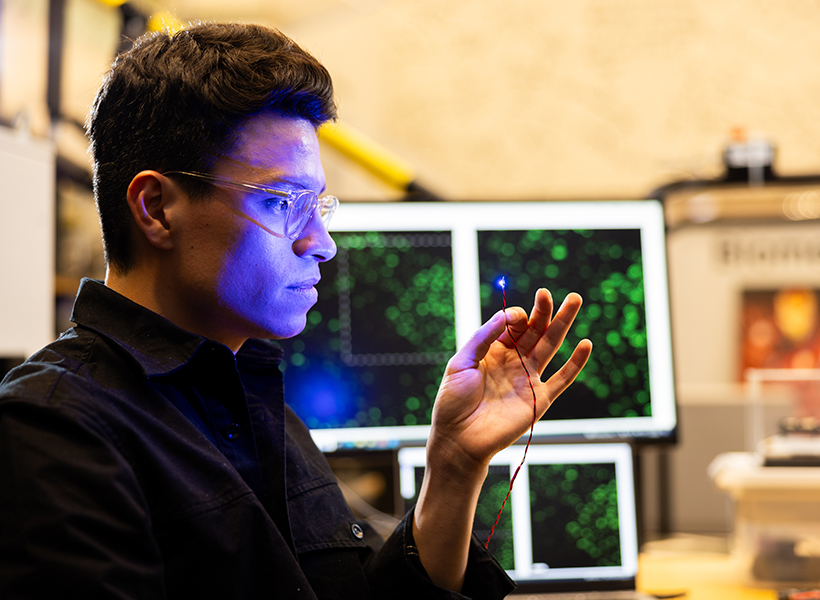
Man holds a fiber that is illuminated with blue light at its tip.
Guillermo Herrera-Arcos, a graduate student in Hugh Herr’s lab, is developing an optical technology with the potential to restore movement in people with spinal cord injury or stroke. Photo: Steph Stevens

Herrera-Arcos and Herr’s work is one way McGovern neuroscientists are working at the interface of brain and machine. Such work aims to enable better ways of understanding and treating injury and disease, offering scientists tools to manipulate neural signaling as well as to replace its function when it is lost.

Restoring movement

The system Herrera-Arcos and Herr were developing wouldn’t be the first to bypass the brain to move muscles. Neuroprosthetic devices that use electricity to stimulate muscle-activating motor neurons are sometimes used during rehabilitation from an injury, helping patients maintain muscle mass when they can’t use their muscles on their own. But existing neuroprostheses lack the precision of the body’s natural movement system. They send all-or-nothing signals that quickly tire muscles out.

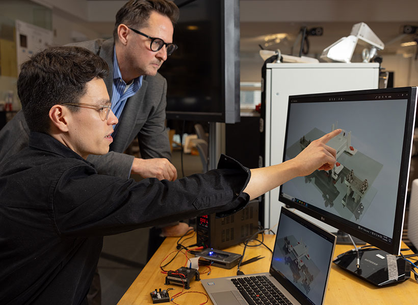
TWo men looking at a computer screen, one points to the image on the screen.
Hugh Herr (left) and graduate student Guillermo Herrera-Arco at work in the lab. Photo: Steph Stevens

Researchers attribute that fatigue to an unnatural recruitment of neurons and muscle fibers. Electrical signals go straight to the largest, most powerful components of the system, even when smaller units could do the job. “You turn up the stimulus and you get no force, and then suddenly, you get too much force. And then fatigue, a lack of controllability, and so on,” Herr explains. The nervous system, in contrast, calls first on small motor units and recruits larger ones only when needed to generate more force.

Optical solution

In hopes of recreating this strategic pattern of muscle activation, Herr and Herrera-Arcos turned to a technique pioneered by McGovern Investigator Edward Boyden that has become common research: controlling neural activity with light. To put neurons under their control, researchers equip them with light-sensitive proteins. The cells can then be switched on or off within milliseconds using an optic fiber.

When a return to the lab enabled Herr and Herrera-Arcos to test their idea, they were thrilled with the results. Using light to switch on motor neurons and stimulate a single muscle in mice, they recreated the nervous system’s natural muscle activation pattern. Consequently, fatigue did not set in nearly as quickly as it would with an electrically-activated system. Herrera-Arcos says he set out to measure the force generated by the muscle and how long it took to fatigue, and he had to keep extending his experiments: After an hour of light stimulation, it was still going strong.

To optimize the force generated by the system, the researchers used feedback from the muscle to modulate the intensity of the neuron-activating light. Their success suggests this type of closed-loop system could enable fatigue-resistant neuroprostheses for muscle control.

“The field has been struggling for many decades with the challenge of how to control living muscle tissue,” Herr says. “So the idea that this could be solved is very, very exciting.”

There’s work to be done to translate what the team has learned into practical neuroprosthetics for people who need them. To use light to stimulate human motor neurons, light-sensitive proteins will need to be delivered to those cells. Figuring out how to do that safely is a high priority at the K. Lisa Yang Center for Bionics, which Herr co-directs with Boyden, and might lead to better ways of obtaining tactile and proprioceptive feedback from prosthetic limbs, as well as to control muscles for the restoration of natural movements after spinal cord injury. “It would be a game changer for a number of conditions,” Herr says.

Gut-brain connection

While Herr’s team works where the nervous system meets the muscle, researchers in Polina Anikeeva’s lab are exploring the brain’s relationship with an often-overlooked part of the nervous system — the hundreds of millions of neurons in the gut.

“Classically, when we think of brain function in neuroscience, it is always studied in the framework of how the brain interacts with the surrounding environment and how it integrates different stimuli,” says Atharva Sahasrabudhe, a graduate student in the group. “But the brain does not function in a vacuum. It’s constantly getting and integrating signals from the peripheral organs.”

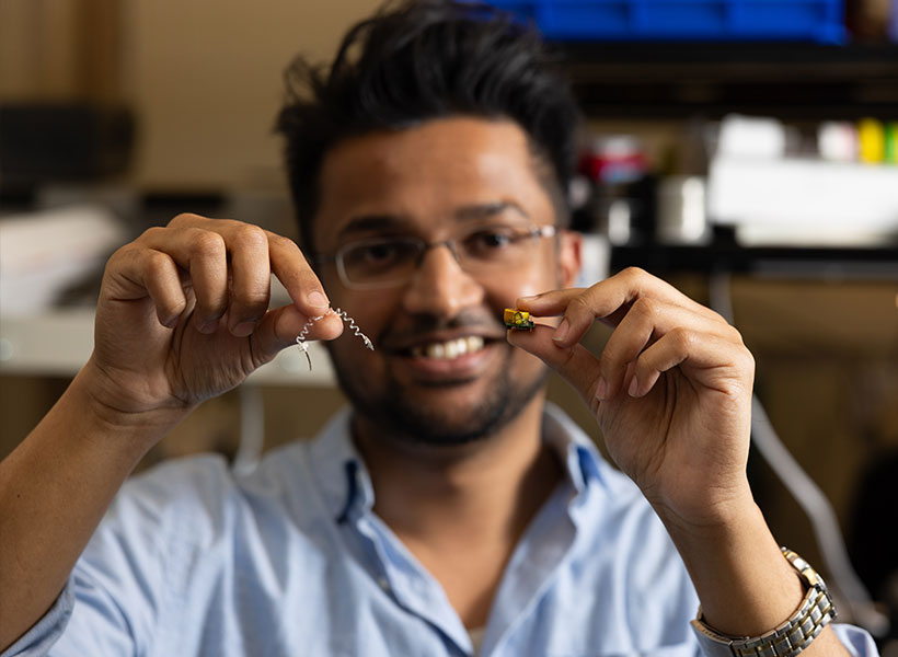
Man smiles at camera while holding up tiny devices.
Atharva Sahasrabudhe holds some of the fiber technology he developed in the Anikeeva lab. Photo: Steph Stevens

The nervous system has a particularly pronounced presence in the gut. Neurons embedded within the walls of the gastrointestinal (GI) tract monitor local conditions and relay information to the brain. This mind-body connection may help explain the GI symptoms associated with some brain-related conditions, including Parkinson’s disease, mood disorders, and autism. Researchers have yet to untangle whether GI symptoms help drive these conditions, are a consequence of them, or are coincidental. Either way, Anikeeva says, “if there is a GI connection, maybe we can tap into this connection to improve the quality of life of affected individuals.”

Flexible fibers

At the K. Lisa Yang Brain-Body Center that Anikeeva directs, studying how the gut communicates with the brain is a high priority. But most of neuroscientists’ tools are designed specifically to investigate the brain. To explore new territory, Sahasrabudhe devised a device that is compatible with the long and twisty GI tract of a mouse.

The new tool is a slender, flexible fiber equipped with light emitters for activating subsets of cells and tiny channels for delivering nutrients or drugs. To access neurons dispersed throughout the GI tract, its wirelessly controlled components are embedded along its length. A more rigid probe at one end of the device is designed to monitor and manipulate neural activity in the brain, so researchers can follow the nervous system’s swift communications across the gut-brain axis.

Scientists on Anikeeva’s team are deploying the device to investigate how gut-brain communications contribute to several conditions. Postdoctoral researcher Sharmelee Selvaraji is focused on Parkinson’s disease. Like many scientists, she wonders whether the neurodegenerative movement disorder might actually start in the gut. There’s a molecular link: the misshapen protein that sickens brain cells in patients with Parkinson’s disease has been found aggregating in the gut, too. And the constipation and other GI problems that are common complaints for people with Parkinson’s disease usually start decades before the onset of motor symptoms. She hopes that by investigating gut-brain communications in a mouse model of the disease, she will uncover important clues about its origins and progression.

“We’re trying to observe the effects of Parkinson’s in the gut, and then eventually, we may be able to intervene at an earlier stage to slow down the disease progression, or even cure it,” says Selvaraji.

Meanwhile, colleagues in the lab are exploring related questions about gut-brain communications in mouse models of autism, anxiety disorders, and addiction. Others continue to focus on technology development, adding new capabilities to the gut-brain probe or applying similar engineering principles to new problems.

“We are realizing that the brain is very much connected to the rest of the body,” Anikeeva says. “There is now a lot of effort in the lab to create technology suitable for a variety of really interesting organs that will help us study brain-body connections.”

A new way to see the activity inside a living cell

Living cells are bombarded with many kinds of incoming molecular signal that influence their behavior. Being able to measure those signals and how cells respond to them through downstream molecular signaling networks could help scientists learn much more about how cells work, including what happens as they age or become diseased.

Right now, this kind of comprehensive study is not possible because current techniques for imaging cells are limited to just a handful of different molecule types within a cell at one time. However, MIT researchers have developed an alternative method that allows them to observe up to seven different molecules at a time, and potentially even more than that.

“There are many examples in biology where an event triggers a long downstream cascade of events, which then causes a specific cellular function,” says Edward Boyden, the Y. Eva Tan Professor in Neurotechnology. “How does that occur? It’s arguably one of the fundamental problems of biology, and so we wondered, could you simply watch it happen?”

It’s arguably one of the fundamental problems of biology, and so we wondered, could you simply watch it happen? – Ed Boyden

The new approach makes use of green or red fluorescent molecules that flicker on and off at different rates. By imaging a cell over several seconds, minutes, or hours, and then extracting each of the fluorescent signals using a computational algorithm, the amount of each target protein can be tracked as it changes over time.

Boyden, who is also a professor of biological engineering and of brain and cognitive sciences at MIT, a Howard Hughes Medical Institute investigator, and a member of MIT’s McGovern Institute for Brain Research and Koch Institute for Integrative Cancer Research, as well as the co-director of the K. Lisa Yang Center for Bionics, is the senior author of the study, which appears today in Cell. MIT postdoc Yong Qian is the lead author of the paper.

Fluorescent signals

Labeling molecules inside cells with fluorescent proteins has allowed researchers to learn a great deal about the functions of many cellular molecules. This type of study is often done with green fluorescent protein (GFP), which was first deployed for imaging in the 1990s. Since then, several fluorescent proteins that glow in other colors have been developed for experimental use.

However, a typical light microscope can only distinguish two or three of these colors, allowing researchers only a tiny glimpse of the overall activity that is happening inside a cell. If they could track a greater number of labeled molecules, researchers could measure a brain cell’s response to different neurotransmitters during learning, for example, or investigate the signals that prompt a cancer cell to metastasize.

“Ideally, you would be able to watch the signals in a cell as they fluctuate in real time, and then you could understand how they relate to each other. That would tell you how the cell computes,” Boyden says. “The problem is that you can’t watch very many things at the same time.”

In 2020, Boyden’s lab developed a way to simultaneously image up to five different molecules within a cell, by targeting glowing reporters to distinct locations inside the cell. This approach, known as “spatial multiplexing,” allows researchers to distinguish signals for different molecules even though they may all be fluorescing the same color.

In the new study, the researchers took a different approach: Instead of distinguishing signals based on their physical location, they created fluorescent signals that vary over time. The technique relies on “switchable fluorophores” — fluorescent proteins that turn on and off at a specific rate. For this study, Boyden and his group members identified four green switchable fluorophores, and then engineered two more, all of which turn on and off at different rates. They also identified two red fluorescent proteins that switch at different rates, and engineered one additional red fluorophore.

Using four switchable fluorophores, MIT researchers were able to label and image four different kinases inside these cells (top four rows). In the bottom row, the cell nuclei are labeled in blue.
Image: Courtesy of the researchers

Each of these switchable fluorophores can be used to label a different type of molecule within a living cell, such an enzyme, signaling protein, or part of the cell cytoskeleton. After imaging the cell for several minutes, hours, or even days, the researchers use a computational algorithm to pick out the specific signal from each fluorophore, analogous to how the human ear can pick out different frequencies of sound.

“In a symphony orchestra, you have high-pitched instruments, like the flute, and low-pitched instruments, like a tuba. And in the middle are instruments like the trumpet. They all have different sounds, and our ear sorts them out,” Boyden says.

The mathematical technique that the researchers used to analyze the fluorophore signals is known as linear unmixing. This method can extract different fluorophore signals, similar to how the human ear uses a mathematical model known as a Fourier transform to extract different pitches from a piece of music.

Once this analysis is complete, the researchers can see when and where each of the fluorescently labeled molecules were found in the cell during the entire imaging period. The imaging itself can be done with a simple light microscope, with no specialized equipment required.

Biological phenomena

In this study, the researchers demonstrated their approach by labeling six different molecules involved in the cell division cycle, in mammalian cells. This allowed them to identify patterns in how the levels of enzymes called cyclin-dependent kinases change as a cell progresses through the cell cycle.

The researchers also showed that they could label other types of kinases, which are involved in nearly every aspect of cell signaling, as well as cell structures and organelles such as the cytoskeleton and mitochondria. In addition to their experiments using mammalian cells grown in a lab dish, the researchers showed that this technique could work in the brains of zebrafish larvae.

This method could be useful for observing how cells respond to any kind of input, such as nutrients, immune system factors, hormones, or neurotransmitters, according to the researchers. It could also be used to study how cells respond to changes in gene expression or genetic mutations. All of these factors play important roles in biological phenomena such as growth, aging, cancer, neurodegeneration, and memory formation.

“You could consider all of these phenomena to represent a general class of biological problem, where some short-term event — like eating a nutrient, learning something, or getting an infection — generates a long-term change,” Boyden says.

In addition to pursuing those types of studies, Boyden’s lab is also working on expanding the repertoire of switchable fluorophores so that they can study even more signals within a cell. They also hope to adapt the system so that it could be used in mouse models.

The research was funded by an Alana Fellowship, K. Lisa Yang, John Doerr, Jed McCaleb, James Fickel, Ashar Aziz, the K. Lisa Yang and Hock E. Tan Center for Molecular Therapeutics at MIT, the Howard Hughes Medical Institute, and the National Institutes of Health.

A multifunctional tool for cognitive neuroscience

A team of researchers at MIT’s McGovern and Picower Institutes has advanced the clinical potential of a thin, flexible fiber designed to simultaneously monitor and manipulate neural activity at targeted sites in the brain. The collaborative team improved upon an earlier model of the multifunctional fiber, developed in the lab of McGovern Institute Associate Investigator Polina Anikeeva, to explore dynamic changes to neural signaling as large animals engage in a working memory task. The results appear Oct. 6 in Science Advances.

The new device, developed by Indie Garwood, who recently received her PhD in the Harvard-MIT Program in Health Sciences and Technology, includes four microelectrodes for detecting neural activity and two microfluidic channels through which drugs can be delivered. This means scientists can deliver a drug that alters neural signaling within a particular part of the brain, then monitor the consequences for local brain activity. This technology was a collaborative effort between Anikeeva, who is also the Matoula S. Salapatas Professor in Materials Science and Engineering and a professor of brain and cognitive sciences, and Picower Institute Investigators Emery Brown and Earl Miller, who jointly supervised Garwood to develop a multifunctional neurotechnology for larger and translational animal models, which are necessary to investigate the neural circuits that underlie high-level cognitive functions.  With further development and testing, similar devices might one day be deployed to diagnose or treat brain disorders in human patients.

Brown is the Edward Hood Taplin Professor of Medical Engineering and Computational Neuroscience in the Picower Institute, the Institute for Medical Engineering and Science, and the Department of Brain and Cognitive Sciences, as well as an anesthesiologist at Massachusetts General Hospital and Harvard Medical School. Miller is the Picower Professor of Neuroscience and a professor of brain and cognitive sciences at MIT.

The new multifunctional fiber is not the first produced by Anikeeva and her team. An earlier model engineered in their lab has already reached the neuroscience community, whose members use it to simultaneously monitor and manipulate neural activity in the brains of mice and rats. But for studies in larger animals, the existing tools for delivering drugs to the brains were rigid, bulky devices, which were both fragile and prone to causing tissue damage. A better tool was needed, both to advance cognitive neuroscience research and to set the stage for developing devices that can deliver drugs directly to the brains of patients and monitor the effects.

Like the devices that Anikeeva’s team designed for rodent studies, the new tool is created by first assembling a larger version of the fiber—a preform cylinder with multiple channels that is then heated and stretched until it is thin and long. As the channels narrow, microelectrodes are incorporated into to the fiber. The final step is to link the electrodes in the fiber to a connector that will relay data collected inside the brain to a unit in the lab.

The final device is long enough to access areas deep in the brain of a large animal. It is built to withstand rigorous sterilization procedures and to stay in place even in an active animal. And it integrates directly with experimental systems that cognitive neuroscientists already use in their labs. “We really wanted this to be something that we could easily hand somebody and they’re going to know how to implement it in their system,” says Garwood, who led development of the device as a graduate student in Anikeeva’s lab.

Once the new device was developed, Garwood and colleagues in the Miller and Brown labs put it to work.  They used the tool to study changes in neural activity as an animal completed a task requiring working memory. The fluid channels in the fiber were used to deliver small amounts of GABA, a neurotransmitter that dampens neuronal activity, to the animal’s premotor cortex, a part of the brain that helps plan movement. At the same time, the device recorded electrical activity from individual neurons, as well as broader patterns of activity in this part of the brain. By monitoring these signals over time, the team learned how neural circuits adapted to the local inhibition they had applied. In another experiment, the team used the device to record neural activity from the putamen, a region deep in the brain involved in reward processing and motivation.

The data collected by the device was extensive and complex, tracking changes that unfolded in the brain over seconds to hours. Interpreting those data required the team to devise new methods of data analysis, which Garwood worked on closely with the Brown lab. Garwood says these methods will be shared with users of the new devices, providing “a roadmap for extracting all of these rich dynamics that you can get out of them.”

These successes, the researchers say, are an important step toward the development of tools to modulate and manipulate neuronal activity in the human brain to benefit patients. For example, they say, a multifunctional fiber might one day be used to more accurately pinpoint the origin of seizures in people with epilepsy, by testing the effects of activating or inhibiting specific brain cells.


Soft optical fibers block pain while moving and stretching with the body

Scientists have a new tool to precisely illuminate the roots of nerve pain.

Engineers at MIT have developed soft and implantable fibers that can deliver light to major nerves through the body. When these nerves are genetically manipulated to respond to light, the fibers can send pulses of light to the nerves to inhibit pain. The optical fibers are flexible and stretch with the body.

The new fibers are meant as an experimental tool that can be used by scientists to explore the causes and potential treatments for peripheral nerve disorders in animal models. Peripheral nerve pain can occur when nerves outside the brain and spinal cord are damaged, resulting in tingling, numbness, and pain in affected limbs. Peripheral neuropathy is estimated to affect more than 20 million people in the United States.

“Current devices used to study nerve disorders are made of stiff materials that constrain movement, so that we can’t really study spinal cord injury and recovery if pain is involved,” says Siyuan Rao, assistant professor of biomedical engineering at the University of Massachusetts at Amherst, who carried out part of the work as a postdoc at MIT. “Our fibers can adapt to natural motion and do their work while not limiting the motion of the subject. That can give us more precise information.”

“Now, people have a tool to study the diseases related to the peripheral nervous system, in very dynamic, natural, and unconstrained conditions,” adds Xinyue Liu PhD ’22, who is now an assistant professor at Michigan State University (MSU).

Details of their team’s new fibers are reported today in a study appearing in Nature Methods. Rao’s and Liu’s MIT co-authors include Atharva Sahasrabudhe, a graduate student in chemistry; Xuanhe Zhao, professor of mechanical engineering and civil and environmental engineering; and Polina Anikeeva, professor of materials science and engineering, along with others at MSU, UMass-Amherst, Harvard Medical School, and the National Institutes of Health.

Beyond the brain

The new study grew out of the team’s desire to expand the use of optogenetics beyond the brain. Optogenetics is a technique by which nerves are genetically engineered to respond to light. Exposure to that light can then either activate or inhibit the nerve, which can give scientists information about how the nerve works and interacts with its surroundings.

Neuroscientists have applied optogenetics in animals to precisely trace the neural pathways underlying a range of brain disorders, including addiction, Parkinson’s disease, and mood and sleep disorders — information that has led to targeted therapies for these conditions.

To date, optogenetics has been primarily employed in the brain, an area that lacks pain receptors, which allows for the relatively painless implantation of rigid devices. However, the rigid devices can still damage neural tissues. The MIT team wondered whether the technique could be expanded to nerves outside the brain. Just as with the brain and spinal cord, nerves in the peripheral system can experience a range of impairment, including sciatica, motor neuron disease, and general numbness and pain.

Optogenetics could help neuroscientists identify specific causes of peripheral nerve conditions as well as test therapies to alleviate them. But the main hurdle to implementing the technique beyond the brain is motion. Peripheral nerves experience constant pushing and pulling from the surrounding muscles and tissues. If rigid silicon devices were used in the periphery, they would constrain an animal’s natural movement and potentially cause tissue damage.

Crystals and light

The researchers looked to develop an alternative that could work and move with the body. Their new design is a soft, stretchable, transparent fiber made from hydrogel — a rubbery, biocompatible mix of polymers and water, the ratio of which they tuned to create tiny, nanoscale crystals of polymers scattered throughout a more Jell-O-like solution.

The fiber embodies two layers — a core and an outer shell or “cladding.” The team mixed the solutions of each layer to generate a specific crystal arrangement. This arrangement gave each layer a specific, different refractive index, and together the layers kept any light traveling through the fiber from escaping or scattering away.

The team tested the optical fibers in mice whose nerves were genetically modified to respond to blue light that would excite neural activity or yellow light that would inhibit their activity. They found that even with the implanted fiber in place, mice were able to run freely on a wheel. After two months of wheel exercises, amounting to some 30,000 cycles, the researchers found the fiber was still robust and resistant to fatigue, and could also transmit light efficiently to trigger muscle contraction.

The team then turned on a yellow laser and ran it through the implanted fiber. Using standard laboratory procedures for assessing pain inhibition, they observed that the mice were much less sensitive to pain than rodents that were not stimulated with light. The fibers were able to significantly inhibit sciatic pain in those light-stimulated mice.

The researchers see the fibers as a new tool that can help scientists identify the roots of pain and other peripheral nerve disorders.

“We are focusing on the fiber as a new neuroscience technology,” Liu says. “We hope to help dissect mechanisms underlying pain in the peripheral nervous system. With time, our technology may help identify novel mechanistic therapies for chronic pain and other debilitating conditions such as nerve degeneration or injury.”

This research was supported, in part, by the National Institutes of Health, the National Science Foundation, the U.S. Army Research Office, the McGovern Institute for Brain Research, the Hock E. Tan and K. Lisa Yang Center for Autism Research, the K. Lisa Yang Brain-Body Center, and the Brain and Behavior Research Foundation.

Nature: An unexpected source of innovative tools to study the brain

This story originally appeared in the Fall 2023 issue of BrainScan.

___

Scientist holds 3D printed phage over a natural background.
Genetic engineer Joseph Kreitz looks to the microscopic world for inspiration in Feng Zhang’s lab at the McGovern Institute. Photo: Steph Steve

In their quest to deepen their understanding of the brain, McGovern scientists take inspiration wherever it comes — and sometimes it comes from surprising sources. To develop new tools for research and innovative strategies for treating disease, they’ve drawn on proteins that organisms have been making for billions of years as well as sophisticated materials engineered for modern technology.

For McGovern investigator Feng Zhang, the natural world provides a rich source of molecules with remarkable and potentially useful functions.

Zhang is one of the pioneers of CRISPR, a programmable system for gene editing that is built from the components of a bacterial adaptive immune system. Scientists worldwide use CRISPR to modify genetic sequences in their labs, and many CRISPR-based therapies, which aim to treat disease through gene editing, are now in development. Meanwhile, Zhang and his team have continued to explore CRISPR-like systems beyond the bacteria in which they were originally discovered.

Turning to nature

This year, the search for evolutionarily related systems led Zhang’s team to a set of enzymes made by more complex organisms, including single-celled algae and hard-shell clams. Like the enzymes that power CRISPR, these newly discovered enzymes, called Fanzors, can be directed to cut DNA at specific sites by programming an RNA molecule as a guide.

Rhiannon Macrae, a scientific advisor in Zhang’s lab, says the discovery was surprising because Fanzors don’t seem to play the same role in immunity that CRISPR systems do. In fact, she says it’s not clear what Fanzors do at all. But as programmable gene editors, Fanzors might have an important advantage over current CRISPR tools — particularly for clinical applications. “Fanzor proteins are much smaller than the workhorse CRISPR tool, Cas9,” Macrae says. “This really matters when you actually want to be able to use one of these tools in a patient, because the bigger the tool, the harder it is to package and deliver to patients’ cells.”

Cryo-EM map of a Fanzor protein (gray, yellow, light blue, and pink) in complex with ωRNA (purple) and its target DNA (red). Non-target DNA strand in blue. Image: Zhang lab

Zhang’s team has thought a lot about how to get therapies to patients’ cells, and size is only one consideration. They’ve also been looking for ways to direct drugs, gene-editing tools, or other therapies to specific cells and tissues in the body. One of the lab’s leading strategies comes from another unexpected natural source: a microscopic syringe produced by certain insect-infecting bacteria.

In their search for an efficient system for targeted drug delivery, Zhang and graduate student Joseph Kreitz first considered the injection systems of bacteria-infecting viruses: needle-like structures that pierce the outer membrane of their host to deliver their own genetic material. But these viral injection systems can’t easily be freed from the rest of the virus.

Then Zhang learned that some bacteria have injection systems of their own, which they release inside their hosts after packing them with toxins. They reengineered the bacterial syringe, devising a delivery system that works on human cells. Their current system can be programmed to inject proteins — including those used for gene editing — directly into specified cell types. With further development, Zhang hopes it will work with other types of therapies, as well.

Magnetic imaging

In McGovern Associate Investigator Alan Jasanoff’s lab, researchers are designing sensors that can track the activity of specific neurons or molecules in the brain, using magnetic resonance imaging (MRI) or related forms of non-invasive imaging. These tools are essential for understanding how the brain’s cells and circuits work together to process information. “We want to give MRI a suite of metaphorical colors: sensitivities that enable us to dissect the different kinds of mechanistically significant contributors to neural activity,” he explains.

Jasanoff can tick off a list of molecules with notable roles in biology and industry that his lab has repurposed to glean more information from brain imaging. These include manganese — a metal once used to tint ancient glass; nitric oxide synthase — the enzyme that causes blushing; and iron oxide nanoparticles — tiny magnets that enable compact data storage inside computers. But Jasanoff says none of these should be considered out of place in the imaging world. “Most are pretty logical choices,” he says. “They all do different things and we use them in pretty different ways, but they are either magnetic or interact with magnetic molecules to serve our purposes for brain imaging.”

Close-up picture of manganese metal
Manganese, a metal that interacts weakly with magnetic fields, is a key component in new MRI sensors being developed in Alan Jasanoff’s lab at the McGovern Institute.

The enzyme nitric oxide synthase, for example, plays an important role in most functional MRI scans. The enzyme produces nitric oxide, which causes blood vessels to expand. This can bring a blush to the cheeks, but in the brain, it increases blood flow to bring more oxygen to busy neurons. MRI can detect this change because it is sensitive to the magnetic properties of blood.

By using blood flow as a proxy for neural activity, functional MRI scans light up active regions of the brain, but they can’t pinpoint the activity of specific cells. So Jasanoff and his team devised a more informative MRI sensor by reengineering nitric oxide synthase. Their modified enzyme, which they call NOSTIC, can be introduced into a select group of cells, where it will produce nitric oxide in response to neural activity — triggering increased blood flow and strengthening the local MRI signal. Researchers can deliver it to specific kinds of brain cells, or they can deliver it exclusively to neurons that communicate directly with one another. Then they can watch for an elevated MRI signal when those cells fire. This lets them see how information flows through the brain and tie specific cells to particular tasks.

Miranda Dawson, a graduate student in Jasanoff’s lab, is using NOSTIC to study the brain circuits that fuel addiction. She’s interested in the involvement of a brain region called the insula, which may mediate the physical sensations that people with addiction experience during drug cravings or withdrawal. With NOSTIC, Dawson can follow how the insula communicates to other parts of the brain as a rat experiences these MITstages of addiction. “We give our sensor to the insula, and then it projects to anatomically connected brain regions,” she explains. “So we’re able to delineate what circuits are being activated at different points in the addiction cycle.”

Scientist with folded arms next to a picture of a brain
Miranda Dawson uses her lab’s novel MRI sensor, NOSTIC, to illuminate the brain circuits involved in fentanyl craving and withdrawal. Photo: Steph Stevens; MRI scan: Nan Li, Souparno Ghosh, Jasanoff lab

Mining biodiversity

McGovern investigators know that good ideas and useful tools can come from anywhere. Sometimes, the key to harnessing those tools is simply recognizing their potential. But there are also opportunities for a more deliberate approach to finding them.

McGovern Investigator Ed Boyden is leading a program that aims to accelerate the discovery of valuable natural products. Called the Biodiversity Network (BioNet), the project is collecting biospecimens from around the world and systematically analyzing them, looking for molecular tools that could be applied to major challenges in science and medicine, from brain research to organ preservation. “The idea behind BioNet,” Boyden explains, “is rather than wait for chance to give us these discoveries, can we go look for them on purpose?”

Making invisible therapy targets visible

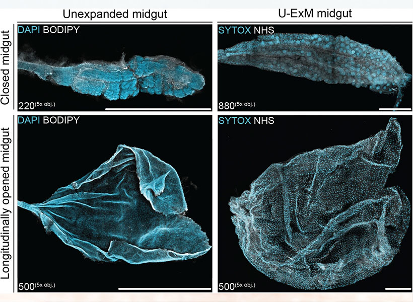
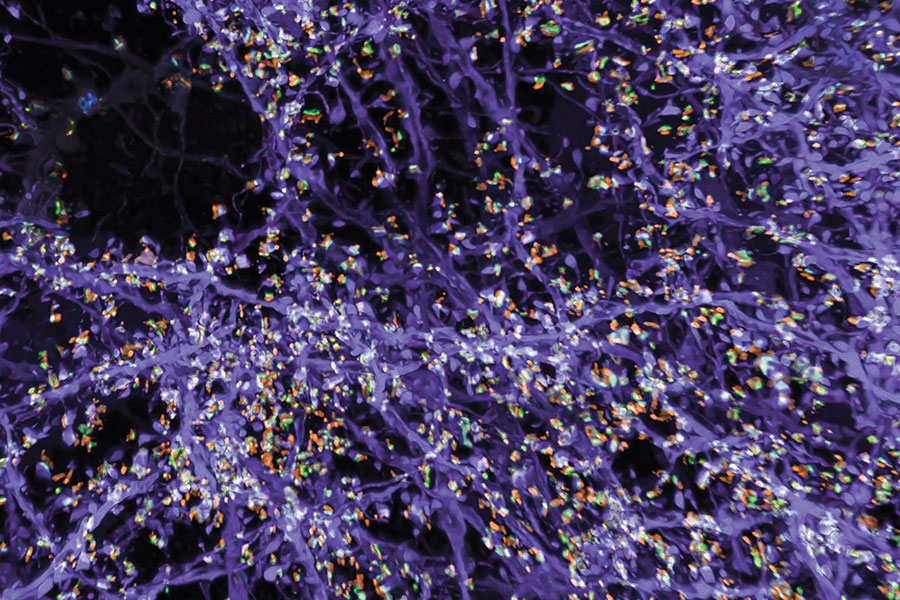
The lab of Edward Boyden, the Y. Eva Tan Professor in Neurotechnology, has developed a powerful technology called Expansion Revealing (ExR) that makes visible molecular structures that were previously too hidden to be seen with even the most powerful microscopes. It “reveals” the nanoscale alterations in synapses, neural wiring, and other molecular assemblies using ordinary lab microscopes. It does so this way: Inside a cell, proteins and other molecules are often tightly packed together. These dense clusters can be difficult to image because the fluorescent labels used to make them visible can’t wedge themselves between the molecules. ExR “de-crowds” the molecules by expanding the cell using a chemical process, making the molecules accessible to fluorescent tags.

Jinyoung Kang is a J. Douglas Tan Postdoctoral Fellow in the Boyden and Feng labs. Photo: Steph Stevens

“This technology can be used to answer a lot of biological questions about dysfunction in synaptic proteins, which are involved in neurodegenerative diseases,” says Jinyoung Kang, a J. Douglas Tan Postdoctoral Fellow in the labs of Boyden and Guoping Feng, the James W. (1963) and Patricia T. Poitras Professor of Brain and Cognitive Sciences. “Until now, there has been no tool to visualize synapses very well at nanoscale.”

Over the past year, the Boyden team has been using ExR to explore the underlying mechanisms of brain disorders, including autism spectrum disorder (ASD) and Alzheimer’s disease. Since the method can be applied iteratively, Boyden imagines it may one day succeed in creating a 100-fold magnification of molecular structures.

“Using earlier technology, researchers may be missing entire categories of molecular phenomena, both functional and dysfunctional,” says Boyden. “It’s critical to bring these nanostructures into view so that we can identify potential targets for new therapeutics that can restore functional molecular arrangements.”

The team is applying ExR to the study of mutant-animal-model brain slices to expose complex synapse 3D nanoarchitecture and configuration. Among their questions: How do synapses differ when mutations that cause autism and other neurological conditions are present?

Using the new technology, Kang and her collaborator Menglong Zeng characterized the molecular architecture of excitatory synapses on parvalbumin interneurons, cells that drastically influence the downstream effects of neuronal signaling and ultimately change cognitive behaviors. They discovered condensed AMPAR clustering in parvalbumin interneurons is essential for normal brain function. The next step is to explore their role in the function of parvalbumin interneurons, which are vulnerable to stressors and have been implicated in brain disorders including autism and Alzheimer’s disease.

The researchers are now investigating whether ExR can reveal abnormal protein nanostructures in SHANK3 knockout mice and marmosets. Mutations in the SHANK3 gene lead to one of the most severe types of ASD, Phelan-McDermid syndrome, which accounts for about 2 percent of all ASD patients with intellectual disability.

Unraveling connections between the brain and gut

The brain and the digestive tract are in constant communication, relaying signals that help to control feeding and other behaviors. This extensive communication network also influences our mental state and has been implicated in many neurological disorders.

MIT engineers have designed a new technology for probing those connections. Using fibers embedded with a variety of sensors, as well as light sources for optogenetic stimulation, the researchers have shown that they can control neural circuits connecting the gut and the brain, in mice.

In a new study, the researchers demonstrated that they could induce feelings of fullness or reward-seeking behavior in mice by manipulating cells of the intestine. In future work, they hope to explore some of the correlations that have been observed between digestive health and neurological conditions such as autism and Parkinson’s disease.

“The exciting thing here is that we now have technology that can drive gut function and behaviors such as feeding. More importantly, we have the ability to start accessing the crosstalk between the gut and the brain with the millisecond precision of optogenetics, and we can do it in behaving animals,” says Polina Anikeeva, the Matoula S. Salapatas Professor in Materials Science and Engineering, a professor of brain and cognitive sciences, director of the K. Lisa Yang Brain-Body Center, associate director of MIT’s Research Laboratory of Electronics, and a member of MIT’s McGovern Institute for Brain Research.

Portait of MIT scientist Polina Anikeeva
McGovern Institute Associate Investigator Polina Anikeeva in her lab. Photo: Steph Stevens

Anikeeva is the senior author of the new study, which appears today in Nature Biotechnology. The paper’s lead authors are MIT graduate student Atharva Sahasrabudhe, Duke University postdoc Laura Rupprecht, MIT postdoc Sirma Orguc, and former MIT postdoc Tural Khudiyev.

The brain-body connection

Last year, the McGovern Institute launched the K. Lisa Yang Brain-Body Center to study the interplay between the brain and other organs of the body. Research at the center focuses on illuminating how these interactions help to shape behavior and overall health, with a goal of developing future therapies for a variety of diseases.

“There’s continuous, bidirectional crosstalk between the body and the brain,” Anikeeva says. “For a long time, we thought the brain is a tyrant that sends output into the organs and controls everything. But now we know there’s a lot of feedback back into the brain, and this feedback potentially controls some of the functions that we have previously attributed exclusively to the central neural control.”

As part of the center’s work, Anikeeva set out to probe the signals that pass between the brain and the nervous system of the gut, also called the enteric nervous system. Sensory cells in the gut influence hunger and satiety via both the neuronal communication and hormone release.

Untangling those hormonal and neural effects has been difficult because there hasn’t been a good way to rapidly measure the neuronal signals, which occur within milliseconds.

“We needed a device that didn’t exist. So, we decided to make it,” says Atharva Sahasrabudhe.

“To be able to perform gut optogenetics and then measure the effects on brain function and behavior, which requires millisecond precision, we needed a device that didn’t exist. So, we decided to make it,” says Sahasrabudhe, who led the development of the gut and brain probes.

The electronic interface that the researchers designed consists of flexible fibers that can carry out a variety of functions and can be inserted into the organs of interest. To create the fibers, Sahasrabudhe used a technique called thermal drawing, which allowed him to create polymer filaments, about as thin as a human hair, that can be embedded with electrodes and temperature sensors.

The filaments also carry microscale light-emitting devices that can be used to optogenetically stimulate cells, and microfluidic channels that can be used to deliver drugs.

The mechanical properties of the fibers can be tailored for use in different parts of the body. For the brain, the researchers created stiffer fibers that could be threaded deep into the brain. For digestive organs such as the intestine, they designed more delicate rubbery fibers that do not damage the lining of the organs but are still sturdy enough to withstand the harsh environment of the digestive tract.

“To study the interaction between the brain and the body, it is necessary to develop technologies that can interface with organs of interest as well as the brain at the same time, while recording physiological signals with high signal-to-noise ratio,” Sahasrabudhe says. “We also need to be able to selectively stimulate different cell types in both organs in mice so that we can test their behaviors and perform causal analyses of these circuits.”

The fibers are also designed so that they can be controlled wirelessly, using an external control circuit that can be temporarily affixed to the animal during an experiment. This wireless control circuit was developed by Orguc, a Schmidt Science Fellow, and Harrison Allen ’20, MEng ’22, who were co-advised between the Anikeeva lab and the lab of Anantha Chandrakasan, dean of MIT’s School of Engineering and the Vannevar Bush Professor of Electrical Engineering and Computer Science.

Driving behavior

Using this interface, the researchers performed a series of experiments to show that they could influence behavior through manipulation of the gut as well as the brain.

First, they used the fibers to deliver optogenetic stimulation to a part of the brain called the ventral tegmental area (VTA), which releases dopamine. They placed mice in a cage with three chambers, and when the mice entered one particular chamber, the researchers activated the dopamine neurons. The resulting dopamine burst made the mice more likely to return to that chamber in search of the dopamine reward.

Then, the researchers tried to see if they could also induce that reward-seeking behavior by influencing the gut. To do that, they used fibers in the gut to release sucrose, which also activated dopamine release in the brain and prompted the animals to seek out the chamber they were in when sucrose was delivered.

Next, working with colleagues from Duke University, the researchers found they could induce the same reward-seeking behavior by skipping the sucrose and optogenetically stimulating nerve endings in the gut that provide input to the vagus nerve, which controls digestion and other bodily functions.

Three scientists holding a fiber in a lab.
Duke University postdoc Laura Rupprecht, MIT graduate student Atharva Sahasrabudhe, and MIT postdoc Sirma Orguc holding their engineered flexible fiber in Polina Anikeeva’s lab at MIT. Photo: Courtesy of the researchers

“Again, we got this place preference behavior that people have previously seen with stimulation in the brain, but now we are not touching the brain. We are just stimulating the gut, and we are observing control of central function from the periphery,” Anikeeva says.

Sahasrabudhe worked closely with Rupprecht, a postdoc in Professor Diego Bohorquez’ group at Duke, to test the fibers’ ability to control feeding behaviors. They found that the devices could optogenetically stimulate cells that produce cholecystokinin, a hormone that promotes satiety. When this hormone release was activated, the animals’ appetites were suppressed, even though they had been fasting for several hours. The researchers also demonstrated a similar effect when they stimulated cells that produce a peptide called PYY, which normally curbs appetite after very rich foods are consumed.

The researchers now plan to use this interface to study neurological conditions that are believed to have a gut-brain connection. For instance, studies have shown that autistic children are far more likely than their peers to be diagnosed with GI dysfunction, while anxiety and irritable bowel syndrome share genetic risks.

“We can now begin asking, are those coincidences, or is there a connection between the gut and the brain? And maybe there is an opportunity for us to tap into those gut-brain circuits to begin managing some of those conditions by manipulating the peripheral circuits in a way that does not directly ‘touch’ the brain and is less invasive,” Anikeeva says.

The research was funded, in part, by the Hock E. Tan and K. Lisa Yang Center for Autism Research and the K. Lisa Yang Brain-Body Center, the National Institute of Neurological Disorders and Stroke, the National Science Foundation (NSF) Center for Materials Science and Engineering, the NSF Center for Neurotechnology, the National Center for Complementary and Integrative Health, a National Institutes of Health Director’s Pioneer Award, the National Institute of Mental Health, and the National Institute of Diabetes and Digestive and Kidney Diseases.

Magnetic robots walk, crawl, and swim

MIT scientists have developed tiny, soft-bodied robots that can be controlled with a weak magnet. The robots, formed from rubbery magnetic spirals, can be programmed to walk, crawl, swim—all in response to a simple, easy-to-apply magnetic field.

“This is the first time this has been done, to be able to control three-dimensional locomotion of robots with a one-dimensional magnetic field,” says McGovern associate investigator Polina Anikeeva, whose team reported on the magnetic robots June 3, 2023, in the journal Advanced Materials. “And because they are predominantly composed of polymer and polymers are soft, you don’t need a very large magnetic field to activate them. It’s actually a really tiny magnetic field that drives these robots,” says Anikeeva, who is also the Matoula S. Salapatas Professor in Materials Science and Engineering and a professor of brain and cognitive sciences at MIT, as well as the associate director of MIT’s Research Laboratory of Electronics and director of MIT’s K. Lisa Yang Brain-Body Center.

Portait of MIT scientist Polina Anikeeva
McGovern Institute Associate Investigator Polina Anikeeva in her lab. Photo: Steph Stevens

The new robots are well suited to transport cargo through confined spaces and their rubber bodies are gentle on fragile environments, opening the possibility that the technology could be developed for biomedical applications. Anikeeva and her team have made their robots millimeters long, but she says the same approach could be used to produce much smaller robots.

Engineering magnetic robots

Anikeeva says that until now, magnetic robots have moved in response to moving magnetic fields. She explains that for these models, “if you want your robot to walk, your magnet walks with it. If you want it to rotate, you rotate your magnet.” That limits the settings in which such robots might be deployed. “If you are trying to operate in a really constrained environment, a moving magnet may not be the safest solution. You want to be able to have a stationary instrument that just applies magnetic field to the whole sample,” she explains.

Youngbin Lee, a former graduate student in Anikeeva’s lab, engineered a solution to this problem. The robots he developed in Anikeeva’s lab are not uniformly magnetized. Instead, they are strategically magnetized in different zones and directions so a single magnetic field can enable a movement-driving profile of magnetic forces.

Before they are magnetized, however, the flexible, lightweight bodies of the robots must be fabricated. Lee starts this process with two kinds of rubber, each with a different stiffness. These are sandwiched together, then heated and stretched into a long, thin fiber. Because of the two materials’ different properties, one of the rubbers retains its elasticity through this stretching process, but the other deforms and cannot return to its original size. So when the strain is released, one layer of the fiber contracts, tugging on the other side and pulling the whole thing into a tight coil. Anikeeva says the helical fiber is modeled after the twisty tendrils of a cucumber plant, which spiral when one layer of cells loses water and contracts faster than a second layer.

A third material—one whose particles have the potential to become magnetic—is incorporated in a channel that runs through the rubbery fiber. So once the spiral has been made, a magnetization pattern that enables a particular type of movement can be introduced.

“Youngbin thought very carefully about how to magnetize our robots to make them able to move just as he programmed them to move,” Anikeeva says. “He made calculations to determine how to establish such a profile of forces on it when we apply a magnetic field that it will actually start walking or crawling.”

To form a caterpillar-like crawling robot, for example, the helical fiber is shaped into gentle undulations, and then the body, head, and tail are magnetized so that a magnetic field applied perpendicular to the robot’s plane of motion will cause the body to compress. When the field is reduced to zero, the compression is released, and the crawling robot stretches. Together, these movements propel the robot forward. Another robot in which two foot-like helical fibers are connected with a joint is magnetized in a pattern that enables a movement more like walking.

Biomedical potential

This precise magnetization process generates a program for each robot and ensures that that once the robots are made, they are simple to control. A weak magnetic field activates each robot’s program and drives its particular type of movement. A single magnetic field can even send multiple robots moving in opposite directions, if they have been programmed to do so. The team found that one minor manipulation of the magnetic field has a useful effect: With the flip of a switch to reverse the field, a cargo-carrying robot can be made to gently shake and release its payload.

Anikeeva says she can imagine these soft-bodied robots—whose straightforward production will be easy to scale up—delivering materials through narrow pipes or even inside the human body. For example, they might carry a drug through narrow blood vessels, releasing it exactly where it is needed. She says the magnetically-actuated devices have biomedical potential beyond robots as well, and might one day be incorporated into artificial muscles or materials that support tissue regeneration.

Francesca Riccio-Ackerman works to improve access to prosthetics

This story originally appeared in the Spring 2023 issue of Spectrum.

___

In Sierra Leone, war and illness have left up to 40,000 people requiring orthotics and prosthetics services, but there is a profound lack of access to specialized care, says Francesca Riccio-Ackerman, a biomedical engineer and PhD student studying health equity and health systems. There is just one fully certified prosthetist available for the thousands of patients in the African nation who are living with amputation, she notes. The ideal number is one for every 250, according to the World Health Organization and the International Society of Orthotics and Prosthetics.

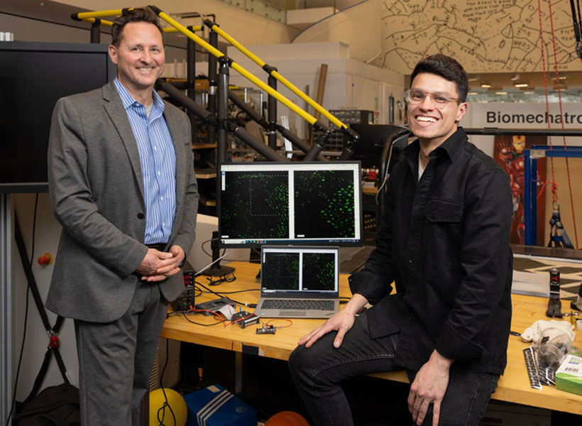
The data point is significant for Riccio-Ackerman, who conducts research in the MIT Media Lab’s Biomechatronics Group and in the K. Lisa Yang Center for Bionics, both of which aim to improve translation of assistive technologies to people with disabilities. “We’re really focused on improving and augmenting human mobility,” she says. For Riccio-Ackerman, part of the quest to improve human mobility means ensuring that the people who need access to prosthetic care can get it—for the duration of their lives.

“We’re really focused on improving and augmenting human mobility,” says Riccio-Ackerman.

In September 2021, the Yang Center provided funding for Riccio-Ackerman to travel to Sierra Leone, where she witnessed the lingering physical effects of a brutal decade-long civil war that ended in 2002. Prosthetic and orthotic care in the country, where a vast number of patients are also disabled by untreated polio or diabetes, has become more elusive, she says, as global media attention on the war’s aftermath has subsided. “People with amputation need low-level, consistent care for years. There really needs to be a long-term investment in improving this.”

Through the Yang Center and supported by a fellowship from the new MIT Morningside Academy for Design, Riccio-Ackerman is designing and building a sustainable care and delivery model in Sierra Leone that aims to multiply the production of prosthetic limbs and strengthen the country’s prosthetic sector. “[We’re working] to improve access to orthotic and prosthetic services,” she says.

She is also helping to establish a supply chain for prosthetic limb and orthotic brace parts and equipping clinics with machines and infrastructure to serve more patients. In January 2023, her team launched a four-year collaboration with the Sierra Leone Ministry of Health and Sanitation. One of the goals of the joint effort is to enable Sierra Leoneans to obtain professional prosthetics training, so they can care for their own community without leaving home.

From engineering to economics

Riccio-Ackerman was drawn to issues around human mobility after witnessing her aunt suffer from rheumatoid arthritis. “My aunt was young, but she looked like she was 80 or 90. She was sick, in pain, in a wheelchair— a young spirit in an old body,” she says.

As a biomedical engineering undergraduate student at Florida International University, Riccio-Ackerman worked on clinical trials for neural-enabled myoelectric arms controlled by nerves in the body. She says that the technology was thrilling yet heartbreaking. She would often have to explain to patients who participated in testing that they couldn’t take the devices home and that they may never be covered by insurance.

Riccio-Ackerman began asking questions: “What factors determine who gets an amputation? Why are we making devices that are so expensive and inaccessible?” This sense of injustice inspired her to pivot away from device design and toward a master’s degree in health economics and policy at the SDA Bocconi School of Management in Milan.

She began work as a research specialist with Hugh Herr SM ’93, professor of arts and sciences at the MIT Media Lab and codirector of the Yang Center, helping to study communities that were medically neglected in prosthetic care. “I knew that the devices weren’t getting to the people who need them, and I didn’t know if the best way to solve it was through engineering,” Riccio-Ackerman explains.

While Riccio-Ackerman’s PhD should be finished within three years, she’s only at the beginning of her health care equity work. “We’re forging ahead in Sierra Leone and thinking about translating our strategy and methodologies to other communities around the globe that could benefit,” she says. “We hope to be able to do this in many, many countries in the future.”