Controlling attention with brain waves

Having trouble paying attention? MIT neuroscientists may have a solution for you: Turn down your alpha brain waves. In a new study, the researchers found that people can enhance their attention by controlling their own alpha brain waves based on neurofeedback they receive as they perform a particular task.

The study found that when subjects learned to suppress alpha waves in one hemisphere of their parietal cortex, they were able to pay better attention to objects that appeared on the opposite side of their visual field. This is the first time that this cause-and-effect relationship has been seen, and it suggests that it may be possible for people to learn to improve their attention through neurofeedback.

Desimone lab study shows that people can boost attention by manipulating their own alpha brain waves with neurofeedback training.

“There’s a lot of interest in using neurofeedback to try to help people with various brain disorders and behavioral problems,” says Robert Desimone, director of MIT’s McGovern Institute for Brain Research. “It’s a completely noninvasive way of controlling and testing the role of different types of brain activity.”

It’s unknown how long these effects might last and whether this kind of control could be achieved with other types of brain waves, such as beta waves, which are linked to Parkinson’s disease. The researchers are now planning additional studies of whether this type of neurofeedback training might help people suffering from attentional or other neurological disorders.

Desimone is the senior author of the paper, which appears in Neuron on Dec. 4. McGovern Institute postdoc Yasaman Bagherzadeh is the lead author of the study. Daniel Baldauf, a former McGovern Institute research scientist, and Dimitrios Pantazis, a McGovern Institute principal research scientist, are also authors of the paper.

Alpha and attention

There are billions of neurons in the brain, and their combined electrical signals generate oscillations known as brain waves. Alpha waves, which oscillate in the frequency of 8 to 12 hertz, are believed to play a role in filtering out distracting sensory information.

Previous studies have shown a strong correlation between attention and alpha brain waves, particularly in the parietal cortex. In humans and in animal studies, a decrease in alpha waves has been linked to enhanced attention. However, it was unclear if alpha waves control attention or are just a byproduct of some other process that governs attention, Desimone says.

To test whether alpha waves actually regulate attention, the researchers designed an experiment in which people were given real-time feedback on their alpha waves as they performed a task. Subjects were asked to look at a grating pattern in the center of a screen, and told to use mental effort to increase the contrast of the pattern as they looked at it, making it more visible.

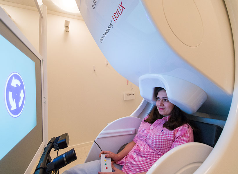
During the task, subjects were scanned using magnetoencephalography (MEG), which reveals brain activity with millisecond precision. The researchers measured alpha levels in both the left and right hemispheres of the parietal cortex and calculated the degree of asymmetry between the two levels. As the asymmetry between the two hemispheres grew, the grating pattern became more visible, offering the participants real-time feedback.

McGovern postdoc Yasaman sits in a magnetoencephalography (MEG) scanner. Photo: Justin Knight

Although subjects were not told anything about what was happening, after about 20 trials (which took about 10 minutes), they were able to increase the contrast of the pattern. The MEG results indicated they had done so by controlling the asymmetry of their alpha waves.

“After the experiment, the subjects said they knew that they were controlling the contrast, but they didn’t know how they did it,” Bagherzadeh says. “We think the basis is conditional learning — whenever you do a behavior and you receive a reward, you’re reinforcing that behavior. People usually don’t have any feedback on their brain activity, but when we provide it to them and reward them, they learn by practicing.”

Although the subjects were not consciously aware of how they were manipulating their brain waves, they were able to do it, and this success translated into enhanced attention on the opposite side of the visual field. As the subjects looked at the pattern in the center of the screen, the researchers flashed dots of light on either side of the screen. The participants had been told to ignore these flashes, but the researchers measured how their visual cortex responded to them.

One group of participants was trained to suppress alpha waves in the left side of the brain, while the other was trained to suppress the right side. In those who had reduced alpha on the left side, their visual cortex showed a larger response to flashes of light on the right side of the screen, while those with reduced alpha on the right side responded more to flashes seen on the left side.

“Alpha manipulation really was controlling people’s attention, even though they didn’t have any clear understanding of how they were doing it,” Desimone says.

Persistent effect

After the neurofeedback training session ended, the researchers asked subjects to perform two additional tasks that involve attention, and found that the enhanced attention persisted. In one experiment, subjects were asked to watch for a grating pattern, similar to what they had seen during the neurofeedback task, to appear. In some of the trials, they were told in advance to pay attention to one side of the visual field, but in others, they were not given any direction.

When the subjects were told to pay attention to one side, that instruction was the dominant factor in where they looked. But if they were not given any cue in advance, they tended to pay more attention to the side that had been favored during their neurofeedback training.

In another task, participants were asked to look at an image such as a natural outdoor scene, urban scene, or computer-generated fractal shape. By tracking subjects’ eye movements, the researchers found that people spent more time looking at the side that their alpha waves had trained them to pay attention to.

“It is promising that the effects did seem to persist afterwards,” says Desimone, though more study is needed to determine how long these effects might last.

The research was funded by the McGovern Institute.

Dimitrios Pantazis

Holistic Imagery

The most widely used imaging method, functional magnetic resonance imaging (fMRI) provides precise information about where in the brain activity occurs, but it cannot detect with the same degree of precision when these events occur in the brain. This kind of temporal precision can be accomplished with magnetoencephalography (MEG), a tool developed at MIT and found in the Martinos Imaging Center at MIT.  Dimitrios Pantazis’ research helps to bridge the gap between spatial and temporal brain imaging data. Director of the MEG lab, Pantazis develops new methods for extracting neural representations from MEG data, and the development of multimodal imaging techniques that give more holistic information about brain function. Using such approaches, he gets insight into processes such as how the brain handles information in the ventral visual stream.

MEG matters

Somewhere nearby, most likely, sits a coffee mug. Give it a glance. An image of that mug travels from desktop to retina and into the brain, where it is processed, categorized and recognized, within a fraction of a second.

All this feels effortless to us, but programming a computer to do the same reveals just how complex that process is. Computers can handle simple objects in expected positions, such as an upright mug. But tilt that cup on its side? “That messes up a lot of standard computer vision algorithms,” says Leyla Isik, a graduate student in Tomaso Poggio’s lab at the McGovern Institute.

For her thesis research, Isik is working to build better computer vision models, inspired by how human brains recognize objects. But to track this process, she needed an imaging tool that could keep up with the brain’s astonishing speed. In 2011, soon after Isik arrived at MIT, the McGovern Institute opened its magnetoencephalography (MEG) lab, one of only a few dozens in the entire country. MEG operates on the same timescale as the human brain. Now, with easy access to a MEG facility dedicated to brain research, neuroscientists at McGovern and across MIT—even those like Isik who had never scanned human subjects—are delving into human neural processing in ways never possible before.

The making of…

MEG was developed at MIT in the early 1970s by physicist David Cohen. He was searching for the tiny magnetic fields that were predicted to arise within electrically active tissues such as the brain. Magnetic fields can travel unimpeded through the skull, so Cohen hoped it might be possible to detect them noninvasively. Because the signals are so small—a billion times weaker than the magnetic field of the Earth—Cohen experimented with a newly invented device called a SQUID (short for superconducting quantum interference device), a highly sensitive magnetometer. In 1972, he succeeded in recording alpha waves, brain rhythms that occur when the eyes close. The recording scratched out on yellow graph paper with notes scrawled in the margins, led to a seminal paper that launched a new field. Cohen’s prototype has now evolved into a sophisticated machine with an array of 306 SQUID detectors contained within a helmet that sits over the subject’s head like a giant hairdryer.

As MEG technology advanced, neuroscientists watched with growing interest. Animal studies were revealing the importance of high-frequency electrical oscillations such as gamma waves, which appear to have a key role in the communication between different brain regions. But apart from occasional neurosurgery patients, it was very difficult to study these signals in the human brain or to understand how they might contribute to human cognition. The most widely used imaging method, functional magnetic resonance imaging (fMRI) could provide precise spatial localization, but it could not detect events on the necessary millisecond timescale. “We needed to bridge that gap,” says Robert Desimone, director of the McGovern Institute.

Desimone decided to make MEG a priority, and with support from donors including Thomas F. Peterson, Jr., Edward and Kay Poitras, and the Simons Foundation, the institute was able to purchase a Triux scanner from Elekta, the newest model on the market and the first to be installed in North America.

One challenge was the high level of magnetic background noise from the surrounding environment, and so the new scanner was installed in a 13-ton shielded room that deflects interference away from the scanner. “We have a challenging location, but we were able to work with it and to get clear signals,” says Desimone.

“An engineer might have picked a different site, but we cannot overstate the importance of having MEG right here, next to the MRI scanners and easily accessible for our researchers.”

To run the new lab, Desimone recruited Dimitrios Pantazis, an expert in MEG signal processing from the University of Southern California. Pantazis knew a lot about MEG data analysis, but he had never actually scanned human subjects himself. In March 2011, he watched in anticipation as Elekta engineers uncrated the new system. Within a few months, he had the lab up and running.

Computer vision quest

When the MEG lab opened, Isik attended a training session. Like Pantazis, she had no previous experience scanning human subjects, but MEG seemed an ideal tool for teasing out the complexities of human object recognition.

She recorded the brain activity of volunteers as they viewed images of objects in various orientations. She also asked them to track the color of a cross on each image, partly to keep their eyes on the screen and partly to keep them alert. “It’s a dark and quiet room and a comfy chair,” she says. “You have to give them something to do to keep them awake.”

To process the data, Isik used a computational tool called a machine learning classifier, which learns to recognize patterns of brain activity evoked by different stimuli. By comparing responses to different types of objects, or similar objects from different viewpoints (such as a cup lying on its side), she was able to show that the human visual system processes objects in stages, starting with the specific view and then generalizing to features that are independent of the size and position of the object.

Isik is now working to develop a computer model that simulates this step-wise processing. “Having this data to work with helps ground my models,” she says. Meanwhile, Pantazis was impressed by the power of machine learning classifiers to make sense of the huge quantities of data produced by MEG studies. With support from the National Science Foundation, he is working to incorporate them into a software analysis package that is widely used by the MEG community.

Mixology

Because fMRI and MEG provide complementary information, it was natural that researchers would want to combine them. This is a computationally challenging task, but MIT research scientist Aude Oliva and postdoc Radoslaw Cichy, in collaboration with Pantazis, have developed a new way to do so. They presented 92 images to volunteers subjects, once in the MEG scanner, and then again in the MRI scanner across the hall. For each data set, they looked for patterns of similarity between responses to different stimuli. Then, by aligning the two ‘similarity maps,’ they could determine which MEG signals correspond to which fMRI signals, providing information about the location and timing of brain activity that could not be revealed by either method in isolation. “We could see how visual information flows from the rear of the brain to the more anterior regions where objects are recognized and categorized,” says Pantazis. “It all happens within a few hundred milliseconds. You could not see this level of detail without the combination of fMRI and MEG.”

Another study combining fMRI and MEG data focused on attention, a longstanding research interest for Desimone. Daniel Baldauf, a postdoc in Desimone’s lab, shares that fascination. “Our visual experience is amazingly rich,” says Baldauf. “Most mysteries about how we deal with all this information boil down to attention.”

Baldauf set out to study how the brain switches attention between two well-studied object categories, faces and houses. These stimuli are known to be processed by different brain areas, and Baldauf wanted to understand how signals might be routed to one area or the other during shifts of attention. By scanning subjects with MEG and fMRI, Baldauf identified a brain region, the inferior frontal junction (IFJ), that synchronizes its gamma oscillations with either the face or house areas depending on which stimulus the subject was attending to—akin to tuning a radio to a particular station.

Having found a way to trace attention within the brain, Desimone and his colleagues are now testing whether MEG can be used to improve attention. Together with Baldauf and two visiting students, Yasaman Bagherzadeh and Ben Lu, he has rigged the scanner so that subjects can be given feedback on their own activity on a screen in real time as it is being recorded. “By concentrating on a task, participants can learn to steer their own brain activity,” says Baldauf, who hopes to determine whether these exercises can help people perform better on everyday tasks that require attention.

Comfort zone

In addition to exploring basic questions about brain function, MEG is also a valuable tool for studying brain disorders such as autism. Margaret Kjelgaard, a clinical researcher at Massachusetts General Hospital, is collaborating with MIT faculty member Pawan Sinha to understand why people with autism often have trouble tolerating sounds, smells, and lights. This is difficult to study using fMRI, because subjects are often unable to tolerate the noise of the scanner, whereas they find MEG much more comfortable.

“Big things are probably going to happen here.”
— David Cohen, inventor of MEG technology

In the scanner, subjects listened to brief repetitive sounds as their brain responses were recorded. In healthy controls, the responses became weaker with repetition as the subjects adapted to the sounds. Those with autism, however, did not adapt. The results are still preliminary and as-yet unpublished, but Kjelgaard hopes that the work will lead to a biomarker for autism, and perhaps eventually for other disorders. In 2012, the McGovern Institute organized a symposium to mark the opening of the new lab. Cohen, who had invented MEG forty years earlier, spoke at the event and made a prediction: “Big things are probably going to happen here.” Two years on, researchers have pioneered new MEG data analysis techniques, invented novel ways to combine MEG and fMRI, and begun to explore the neural underpinnings of autism. Odds are, there are more big things to come.

Expanding our view of vision

Every time you open your eyes, visual information flows into your brain, which interprets what you’re seeing. Now, for the first time, MIT neuroscientists have noninvasively mapped this flow of information in the human brain with unique accuracy, using a novel brain-scanning technique.

This technique, which combines two existing technologies, allows researchers to identify precisely both the location and timing of human brain activity. Using this new approach, the MIT researchers scanned individuals’ brains as they looked at different images and were able to pinpoint, to the millisecond, when the brain recognizes and categorizes an object, and where these processes occur.

“This method gives you a visualization of ‘when’ and ‘where’ at the same time. It’s a window into processes happening at the millisecond and millimeter scale,” says Aude Oliva, a principal research scientist in MIT’s Computer Science and Artificial Intelligence Laboratory (CSAIL).

Oliva is the senior author of a paper describing the findings in the Jan. 26 issue of Nature Neuroscience. Lead author of the paper is CSAIL postdoc Radoslaw Cichy. Dimitrios Pantazis, a research scientist at MIT’s McGovern Institute for Brain Research, is also an author of the paper.

When and where

Until now, scientists have been able to observe the location or timing of human brain activity at high resolution, but not both, because different imaging techniques are not easily combined. The most commonly used type of brain scan, functional magnetic resonance imaging (fMRI), measures changes in blood flow, revealing which parts of the brain are involved in a particular task. However, it works too slowly to keep up with the brain’s millisecond-by-millisecond dynamics.

Another imaging technique, known as magnetoencephalography (MEG), uses an array of hundreds of sensors encircling the head to measure magnetic fields produced by neuronal activity in the brain. These sensors offer a dynamic portrait of brain activity over time, down to the millisecond, but do not tell the precise location of the signals.

To combine the time and location information generated by these two scanners, the researchers used a computational technique called representational similarity analysis, which relies on the fact that two similar objects (such as two human faces) that provoke similar signals in fMRI will also produce similar signals in MEG. This method has been used before to link fMRI with recordings of neuronal electrical activity in monkeys, but the MIT researchers are the first to use it to link fMRI and MEG data from human subjects.

In the study, the researchers scanned 16 human volunteers as they looked at a series of 92 images, including faces, animals, and natural and manmade objects. Each image was shown for half a second.

“We wanted to measure how visual information flows through the brain. It’s just pure automatic machinery that starts every time you open your eyes, and it’s incredibly fast,” Cichy says. “This is a very complex process, and we have not yet looked at higher cognitive processes that come later, such as recalling thoughts and memories when you are watching objects.”

Each subject underwent the test multiple times — twice in an fMRI scanner and twice in an MEG scanner — giving the researchers a huge set of data on the timing and location of brain activity. All of the scanning was done at the Athinoula A. Martinos Imaging Center at the McGovern Institute.

Millisecond by millisecond

By analyzing this data, the researchers produced a timeline of the brain’s object-recognition pathway that is very similar to results previously obtained by recording electrical signals in the visual cortex of monkeys, a technique that is extremely accurate but too invasive to use in humans.

About 50 milliseconds after subjects saw an image, visual information entered a part of the brain called the primary visual cortex, or V1, which recognizes basic elements of a shape, such as whether it is round or elongated. The information then flowed to the inferotemporal cortex, where the brain identified the object as early as 120 milliseconds. Within 160 milliseconds, all objects had been classified into categories such as plant or animal.

The MIT team’s strategy “provides a rich new source of evidence on this highly dynamic process,” says Nikolaus Kriegeskorte, a principal investigator in cognition and brain sciences at Cambridge University.

“The combination of MEG and fMRI in humans is no surrogate for invasive animal studies with techniques that simultaneously have high spatial and temporal precision, but Cichy et al. come closer to characterizing the dynamic emergence of representational geometries across stages of processing in humans than any previous work. The approach will be useful for future studies elucidating other perceptual and cognitive processes,” says Kriegeskorte, who was not part of the research team.

The MIT researchers are now using representational similarity analysis to study the accuracy of computer models of vision by comparing brain scan data with the models’ predictions of how vision works.

Using this approach, scientists should also be able to study how the human brain analyzes other types of information such as motor, verbal, or sensory signals, the researchers say. It could also shed light on processes that underlie conditions such as memory disorders or dyslexia, and could benefit patients suffering from paralysis or neurodegenerative diseases.

“This is the first time that MEG and fMRI have been connected in this way, giving us a unique perspective,” Pantazis says. “We now have the tools to precisely map brain function both in space and time, opening up tremendous possibilities to study the human brain.”

The research was funded by the National Eye Institute, the National Science Foundation, and a Feodor Lynen Research Fellowship from the Humboldt Foundation.

Detecting the brain’s magnetic signals with MEG

Magnetoencephalography (MEG) is a noninvasive technique for measuring neuronal activity in the human brain. Electrical currents flowing through neurons generate weak magnetic fields that can be recorded at the surface of the head using very sensitive magnetic detectors known as superconducting quantum interference devices (SQUIDs).

MEG is a purely passive method that relies on detection of signals that are produced naturally by the brain. It does not involve exposure to radiation or strong magnetic fields, and there are no known hazards associated with MEG.

MEG was developed at MIT in the early 1970s by physicist David Cohen. Photo: David Cohen

Magnetic signals from the brain are very small compared to the magnetic fluctuations that are produced by interfering sources such as nearby electrical equipment or moving metal objects. Therefore MEG scans are typically performed within a special magnetically shielded room that blocks this external interference.

It is fitting that MIT should have a state-of-the-art MEG scanner, since the MEG technology was pioneered by David Cohen in the early 1970s while he was a member of MIT’s Francis Bitter Magnet Laboratory.

MEG can detect the timing of magnetic signals with millisecond precision. This is the timescale on which neurons communicate, and MEG is thus well suited to measuring the rapid signals that reflect communication between different parts of the human brain.

MEG is complementary to other brain imaging modalities such as functional magnetic resonance imaging (fMRI) and positron emission tomography (PET), which depend on changes in blood flow, and which have higher spatial resolution but much lower temporal resolution than MEG.

Our MEG scanner, an Elekta Neuromag Triux with 306 channels plus 128 channels for EEG, was installed in 2011 and is the first of its kind in North America. It is housed within a magnetically shielded room to reduce background noise.

The MEG lab is part of the Martinos Imaging Center at MIT, operating as a core facility, and accessible to all members of the local research community. Potential users should contact Dimitrios Pantazis for more information.

The MEG Lab was made possible through a grant from the National Science Foundation and through the generous support of the following donors: Thomas F. Peterson, Jr. ’57; Edward and Kay Poitras; The Simons Foundation; and an anonymous donor.