Empowering faculty partnerships across the globe

MIT faculty share their creative and technical talent on campus as well as across the globe, compounding the Institute’s impact through strong international partnerships. Thanks to the MIT Global Seed Funds (GSF) program, managed by the MIT International Science and Technology Initiatives (MISTI), more of these faculty members will be able to build on these relationships to develop ideas and create new projects.

“This MISTI fund was extremely helpful in consolidating our collaboration and has been the start of a long-term interaction between the two teams,” says 2017 GSF awardee Mehrdad Jazayeri, associate professor of brain and cognitive sciences and investigator at the McGovern Institute for Brain Research. “We have already submitted multiple abstracts to conferences together, mapped out several ongoing projects, and secured international funding thanks to the preliminary progress this seed fund enabled.”

This year, the 28 funds that comprise MISTI GSF received 232 MIT applications. Over $2.3 million was awarded to 107 projects from 23 departments across the entire Institute. This brings the amount awarded to $22 million over the 12-year life of the program. Besides supporting faculty, these funds also provide meaningful educational opportunities for students. The majority of GSF teams include students from MIT and international collaborators, bolstering both their research portfolios and global experience.

“This project has had important impact on my grad student’s education and development. She was able to apply techniques she has learned to a new and challenging system, mentor an international student, participate in a major international meeting, and visit CEA,” says Professor of Chemistry Elizabeth Nolan, a 2017 GSF awardee.

On top of these academic and research goals, students are actively broadening their cultural experience and scope. “The environment at CEA differs enormously from MIT because it is a national lab and because lab structure and graduate education in France is markedly different than at MIT,” Nolan continues. “At CEA, she had the opportunity to present research to distinguished international colleagues.”

These impactful partnerships unite faculty teams behind common goals to tackle worldwide challenges, helping to develop solutions that would not be possible without international collaboration. 2017 GSF winner Emilio Bizzi, professor emeritus of brain and cognitive sciences and emeritus investigator at the McGovern Institute, articulated the advantage of combining these individual skills within a high-level team. “The collaboration among researchers was valuable in sharing knowledge, experience, skills and techniques … as well as offering the probability of future development of systems to aid in rehabilitation of patients suffering TBI.”

The research opportunities that grow from these seed funds often lead to published papers and additional funding leveraged from early results. The next call for proposals will be in mid-May.

MISTI creates applied international learning opportunities for MIT students that increase their ability to understand and address real-world problems. MISTI collaborates with partners at MIT and beyond, serving as a vital nexus of international activity and bolstering the Institute’s research mission by promoting collaborations between MIT faculty members and their counterparts abroad.

Emilio Bizzi

Controlling Actions

A fundamental job of the brain is to produce actions. Emeritus Professor Emilio Bizzi examined how the brain handles the enormous complexity involved in making even the simplest movement. One of his key discoveries was that groups of muscles are activated synergistically by circuits of neurons in the spinal cord. He argued that these synergies represent fundamental building blocks for assembling repertoires of complex movements and might be used to restore limb movements compromised by stroke or muscle injury.

Controlling movement with light

For the first time, MIT neuroscientists have shown they can control muscle movement by applying optogenetics — a technique that allows scientists to control neurons’ electrical impulses with light — to the spinal cords of animals that are awake and alert.

Led by MIT Institute Professor Emilio Bizzi, the researchers studied mice in which a light-sensitive protein that promotes neural activity was inserted into a subset of spinal neurons. When the researchers shone blue light on the animals’ spinal cords, their hind legs were completely but reversibly immobilized. The findings, described in the June 25 issue of PLoS One, offer a new approach to studying the complex spinal circuits that coordinate movement and sensory processing, the researchers say.

In this study, Bizzi and Vittorio Caggiano, a postdoc at MIT’s McGovern Institute for Brain Research, used optogenetics to explore the function of inhibitory interneurons, which form circuits with many other neurons in the spinal cord. These circuits execute commands from the brain, with additional input from sensory information from the limbs.

Previously, neuroscientists have used electrical stimulation or pharmacological intervention to control neurons’ activity and try to tease out their function. Those approaches have revealed a great deal of information about spinal control, but they do not offer precise enough control to study specific subsets of neurons.

Optogenetics, on the other hand, allows scientists to control specific types of neurons by genetically programming them to express light-sensitive proteins. These proteins, called opsins, act as ion channels or pumps that regulate neurons’ electrical activity. Some opsins suppress activity when light shines on them, while others stimulate it.

“With optogenetics, you are attacking a system of cells that have certain characteristics similar to each other. It’s a big shift in terms of our ability to understand how the system works,” says Bizzi, who is a member of MIT’s McGovern Institute.

Muscle control

Inhibitory neurons in the spinal cord suppress muscle contractions, which is critical for maintaining balance and for coordinating movement. For example, when you raise an apple to your mouth, the biceps contract while the triceps relax. Inhibitory neurons are also thought to be involved in the state of muscle inhibition that occurs during the rapid eye movement (REM) stage of sleep.

To study the function of inhibitory neurons in more detail, the researchers used mice developed by Guoping Feng, the Poitras Professor of Neuroscience at MIT, in which all inhibitory spinal neurons were engineered to express an opsin called channelrhodopsin 2. This opsin stimulates neural activity when exposed to blue light. They then shone light at different points along the spine to observe the effects of neuron activation.

When inhibitory neurons in a small section of the thoracic spine were activated in freely moving mice, all hind-leg movement ceased. This suggests that inhibitory neurons in the thoracic spine relay the inhibition all the way to the end of the spine, Caggiano says. The researchers also found that activating inhibitory neurons had no effect on the transmission of sensory information from the limbs to the brain, or on normal reflexes.

“The spinal location where we found this complete suppression was completely new,” Caggiano says. “It has not been shown by any other scientists that there is this front-to-back suppression that affects only motor behavior without affecting sensory behavior.”

“It’s a compelling use of optogenetics that raises a lot of very interesting questions,” says Simon Giszter, a professor of neurobiology and anatomy at Drexel University who was not part of the research team. Among those questions is whether this mechanism behaves as a global “kill switch,” or if the inhibitory neurons form modules that allow for more selective suppression of movement patterns.

Now that they have demonstrated the usefulness of optogenetics for this type of study, the MIT team hopes to explore the roles of other types of spinal cord neurons. They also plan to investigate how input from the brain influences these spinal circuits.

“There’s huge interest in trying to extend these studies and dissect these circuits because we tackled only the inhibitory system in a very global way,” Caggiano says. “Further studies will highlight the contribution of single populations of neurons in the spinal cord for the control of limbs and control of movement.”

The research was funded by the Human Frontier Science Program and the National Science Foundation. Mriganka Sur, the Paul E. and Lilah Newton Professor of Neuroscience at MIT, is also an author of the paper.

Brain balances learning new skills, retaining old skills

To learn new motor skills, the brain must be plastic: able to rapidly change the strengths of connections between neurons, forming new patterns that accomplish a particular task. However, if the brain were too plastic, previously learned skills would be lost too easily.

A new computational model developed by MIT neuroscientists explains how the brain maintains the balance between plasticity and stability, and how it can learn very similar tasks without interference between them.

The key, the researchers say, is that neurons are constantly changing their connections with other neurons. However, not all of the changes are functionally relevant — they simply allow the brain to explore many possible ways to execute a certain skill, such as a new tennis stroke.

“Your brain is always trying to find the configurations that balance everything so you can do two tasks, or three tasks, or however many you’re learning,” says Robert Ajemian, a research scientist in MIT’s McGovern Institute for Brain Research and lead author of a paper describing the findings in the Proceedings of the National Academy of Sciences the week of Dec. 9. “There are many ways to solve a task, and you’re exploring all the different ways.”

As the brain explores different solutions, neurons can become specialized for specific tasks, according to this theory.

Noisy circuits

As the brain learns a new motor skill, neurons form circuits that can produce the desired output — a command that will activate the body’s muscles to perform a task such as swinging a tennis racket. Perfection is usually not achieved on the first try, so feedback from each effort helps the brain to find better solutions.

This works well for learning one skill, but complications arise when the brain is trying to learn many different skills at once. Because the same distributed network controls related motor tasks, new modifications to existing patterns can interfere with previously learned skills.

“This is particularly tricky when you’re learning very similar things,” such as two different tennis strokes, says Institute Professor Emilio Bizzi, the paper’s senior author and a member of the McGovern Institute.

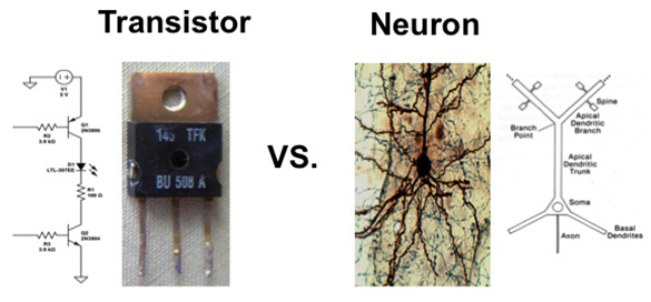
The Bizzi lab shows how the brain utilizes the operating characteristics of neurons to form sensorimotor memories in a way that differs profoundly from computer memory.

In a serial network such as a computer chip, this would be no problem — instructions for each task would be stored in a different location on the chip. However, the brain is not organized like a computer chip. Instead, it is massively parallel and highly connected — each neuron connects to, on average, about 10,000 other neurons.

That connectivity offers an advantage, however, because it allows the brain to test out so many possible solutions to achieve combinations of tasks. The constant changes in these connections, which the researchers call hyperplasticity, is balanced by another inherent trait of neurons — they have a very low signal to noise ratio, meaning that they receive about as much useless information as useful input from their neighbors.

Most models of neural activity don’t include noise, but the MIT team says noise is a critical element of the brain’s learning ability. “Most people don’t want to deal with noise because it’s a nuisance,” Ajemian says. “We set out to try to determine if noise can be used in a beneficial way, and we found that it allows the brain to explore many solutions, but it can only be utilized if the network is hyperplastic.”

This model helps to explain how the brain can learn new things without unlearning previously acquired skills, says Ferdinando Mussa-Ivaldi, a professor of physiology at Northwestern University.

“What the paper shows is that, counterintuitively, if you have neural networks and they have a high level of random noise, that actually helps instead of hindering the stability problem,” says Mussa-Ivaldi, who was not part of the research team.

Without noise, the brain’s hyperplasticity would overwrite existing memories too easily. Conversely, low plasticity would not allow any new skills to be learned, because the tiny changes in connectivity would be drowned out by all of the inherent noise.

The model is supported by anatomical evidence showing that neurons exhibit a great deal of plasticity even when learning is not taking place, as measured by the growth and formation of connections of dendrites — the tiny extensions that neurons use to communicate with each other.

Like riding a bike

The constantly changing connections explain why skills can be forgotten unless they are practiced often, especially if they overlap with other routinely performed tasks.

“That’s why an expert tennis player has to warm up for an hour before a match,” Ajemian says. The warm-up is not for their muscles, instead, the players need to recalibrate the neural networks that control different tennis strokes that are stored in the brain’s motor cortex.

However, skills such as riding a bicycle, which is not very similar to other common skills, are retained more easily. “Once you’ve learned something, if it doesn’t overlap or intersect with other skills, you will forget it but so slowly that it’s essentially permanent,” Ajemian says.

The researchers are now investigating whether this type of model could also explain how the brain forms memories of events, as well as motor skills.

The research was funded by the National Science Foundation.

Stroke disrupts how brain controls muscle synergies

The simple act of picking up a pencil requires the coordination of dozens of muscles: The eyes and head must turn toward the object as the hand reaches forward and the fingers grasp it. To make this job more manageable, the brain’s motor cortex has implemented a system of shortcuts. Instead of controlling each muscle independently, the cortex is believed to activate muscles in groups, known as “muscle synergies.” These synergies can be combined in different ways to achieve a wide range of movements.

A new study from MIT, Harvard Medical School and the San Camillo Hospital in Venice finds that after a stroke, these muscle synergies are activated in altered ways. Furthermore, those disruptions follow specific patterns depending on the severity of the stroke and the amount of time that has passed since the stroke.

The findings, published this week in the Proceedings of the National Academy of Sciences, could lead to improved rehabilitation for stroke patients, as well as a better understanding of how the motor cortex coordinates movements, says Emilio Bizzi, an Institute Professor at MIT and senior author of the paper.

“The cortex is responsible for motor learning and for controlling movement, so we want to understand what’s going on there,” says Bizzi, who is a member of the McGovern Institute for Brain Research at MIT. “How does the cortex translate an idea to move into a series of commands to accomplish a task?”

Coordinated control

One way to explore motor cortical functions is to study how motor patterns are disrupted in stroke patients who suffered damage to the motor areas.

In 2009, Bizzi and his colleagues first identified muscle synergies in the arms of people who had suffered mild strokes by measuring electrical activity in each muscle as the patients moved. Then, by utilizing a specially designed factorization algorithm, the researchers identified characteristic muscle synergies in both the stroke-affected and unaffected arms.

“To control, precisely, each muscle needed for the task would be very hard. What we have proven is that the central nervous system, when it programs the movement, makes use of these modules,” Bizzi says. “Instead of activating simultaneously 50 muscles for a single action, you will combine a few synergies to achieve that goal.”

In the 2009 study, and again in the new paper, the researchers showed that synergies in the affected arms of patients who suffered mild strokes in the cortex are very similar to those seen in their unaffected arms even though the muscle activation patterns are different. This shows that muscle synergies are structured within the spinal cord, and that cortical stroke alters the ability of the brain to activate these synergies in the appropriate combinations.

However, the new study found a much different pattern in patients who suffered more severe strokes. In those patients, synergies in the affected arm merged to form a smaller number of larger synergies. And in a third group of patients, who had suffered their stroke many years earlier, the muscle synergies of the affected arm split into fragments of the synergies seen in the unaffected arm.

This phenomenon, known as fractionation, does not restore the synergies to what they would have looked like before the stroke. “These fractionations appear to be something totally new,” says Vincent Cheung, a research scientist at the McGovern Institute and lead author of the new PNAS paper. “The conjecture would be that these fragments could be a way that the nervous system tries to adapt to the injury, but we have to do further studies to confirm that.”

This is the first time that fractionation of muscle synergies identified by factorization has been seen in chronic stroke patients, says Simon Giszter, a professor of neurobiology and anatomy at Drexel University. “It raises the question of how this occurs and if it’s a compensatory process. If it is, we can use this measurement to study how the recovery process can be accelerated,” says Giszter, who was not involved in this study.

Toward better rehabilitation

The researchers believe that these patterns of synergies, which are determined by both the severity of the deficit and the time since the stroke occurred, could be used as markers to more fully describe individual patients’ impaired status. “In some of the patients, we see a mixture of these patterns. So you can have severe but chronic patients, for instance, who show both merging and fractionation,” Cheung says.

The findings could also help doctors design better rehabilitation programs. The MIT team is now working with several hospitals to establish new therapeutic protocols based on the discovered markers.

About 700,000 people suffer strokes in the United States every year, and many different rehabilitation programs exist to treat them. Choosing one is currently more of an art than a science, Bizzi says. “There is a great deal of need to sharpen current procedures for rehabilitation by turning to principles derived from the most advanced brain research,” he says. “It is very likely that different strategies of rehabilitation will have to be used in patients who have one type of marker versus another.”

The research was funded by the National Institutes of Health and the Italian Ministry of Health.