Thousands of programmable DNA-cutters found in algae, snails, and other organisms

A diverse set of species, from snails to algae to amoebas, make programmable DNA-cutting enzymes called Fanzors—and a new study from scientists at MIT’s McGovern Institute has identified thousands of them. Fanzors are RNA-guided enzymes that can be programmed to cut DNA at specific sites, much like the bacterial enzymes that power the widely used gene-editing system known as CRISPR. The newly recognized diversity of natural Fanzor enzymes, reported September 27, 2023, in the journal Science Advances, gives scientists an extensive set of programmable enzymes that might be adapted into new tools for research or medicine.

“RNA-guided biology is what lets you make programmable tools that are really easy to use. So the more we can find, the better,” says McGovern fellow Omar Abudayyeh, who led the research with McGovern fellow Jonathan Gootenberg.

CRISPR, an ancient bacterial defense system, has made it clear how useful RNA-guided enzymes can be when they are adapted for use in the lab. CRISPR-based genome editing tools developed by McGovern investigator Feng Zhang, Abudayyeh, Gootenberg and others have changed the way scientists modify DNA, accelerating research and enabling the development of many experimental gene therapies.

Researchers have since uncovered other RNA-guide enzymes throughout the bacterial world, many with features that make them valuable in the lab. The discovery of Fanzors, whose ability to cut DNA in an RNA-guided manner was reported by Zhang’s group earlier this year, opens a new frontier of RNA-guided biology. Fanzors were the first such enzymes to be found in eukaryotic organisms—a wide group of lifeforms, including plants, animals, and fungi, defined by the membrane-bound nucleus that holds each cell’s genetic material. (Bacteria, which lack nuclei, belong to a group known as prokaryotes.)

Structural illustration of Fanzors.
Predicted structural image of Fanzors. Image: Jonathan Gootenberg and Omar Abudayyeh

“People have been searching for interesting tools in prokaryotic systems for a long time, and I think that that has been incredibly fruitful,” says Gootenberg. “Eukaryotic systems are really just a whole new kind of playground to work in.”

One hope, Abudayyeh and Gootenberg say, is that enzymes that naturally evolved in eukaryotic organisms might be better suited to function safely and efficiently in the cells of other eukaryotic organisms, including humans. Zhang’s group has shown that Fanzor enzymes can be engineered to precisely cut specific DNA sequences in human cells. In the new work, Abudayyeh and Gootenberg discovered that some Fanzors can target DNA sequences in human cells even without optimization. “The fact that they work quite efficiently in mammalian cells was really fantastic to see,” Gootenberg says.

Prior to the current study, hundreds of Fanzors had been found among eukaryotic organisms. Through an extensive search of genetic databases led by lab member Justin Lim, Gootenberg and Abudayyeh’s team has now expanded the known diversity of these enzymes by an order of magnitude.

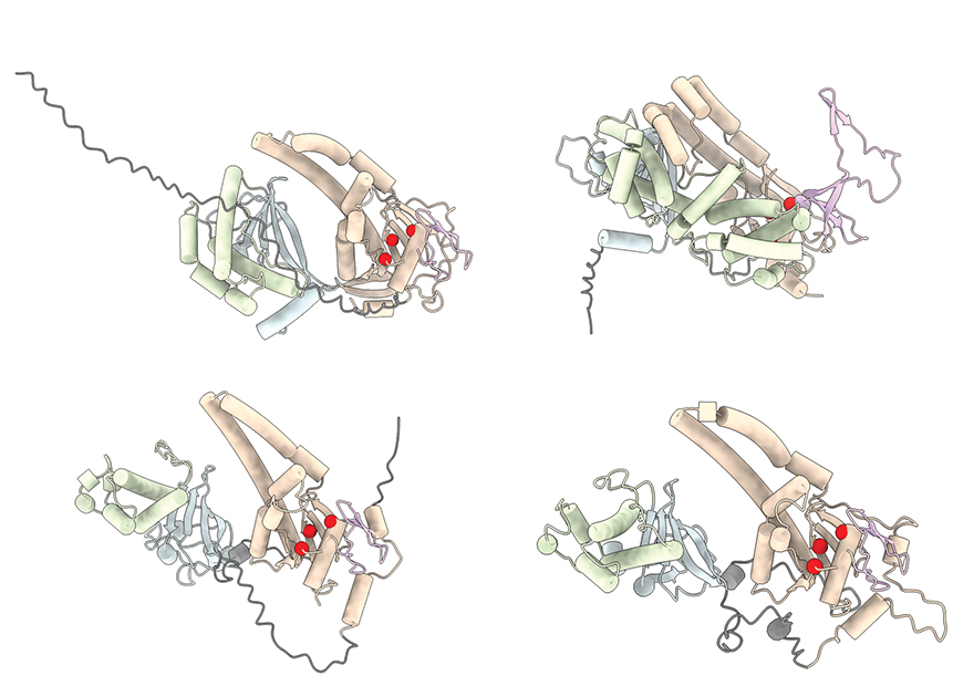
Among the more than 3,600 Fanzors that the team found in eukaryotes and the viruses that infect them, the researchers were able to identify five different families of the enzymes. By comparing these enzymes’ precise makeup, they found evidence of a long evolutionary history.

Fanzors likely evolved from RNA-guided DNA-cutting bacterial enzymes called TnpBs. In fact, it was Fanzors’ genetic similarities to these bacterial enzymes that first caught the attention of both Zhang’s group and Gootenberg and Abudayyeh’s team.

The evolutionary connections that Gootenberg and Abudayyeh traced suggest that these bacterial predecessors of Fanzors probably entered eukaryotic cells, initiating their evolution, more than once. Some were likely transmitted by viruses, while others may have been introduced by symbiotic bacteria. The research also suggests that after they were taken up by eukaryotes, the enzymes evolved features suited to their new environment, such as a signal that allows them to enter a cell nucleus, where they have access to DNA.

Through genetic and biochemical experiments led by graduate student Kaiyi Jiang, the team determined that Fanzors have evolved a DNA-cutting active site that is distinct from that of their bacterial predecessors. This seems to allow the enzyme to cut its target sequence more precisely the ancestors of TnpB, when targeted to a sequence of DNA in a test tube, become activated and cut other sequences in the tube; Fanzors lack this promiscuous activity. When they used an RNA guide to direct the enzymes to cut specific sites in the genome of human cells, they found that certain Fanzors were able to cut these target sequences with about 10 to 20 percent efficiency.

With further research, Abudayyeh and Gootenberg hope that a variety of sophisticated genome editing tools can be developed from Fanzors. “It’s a new platform, and they have many capabilities,” says Gootenberg. “Opening up the whole eukaryotic world to these types of RNA-guided systems is going to give us a lot to work on,” Abudayyeh adds.

New CRISPR-based tool inserts large DNA sequences at desired sites in cells

Building on the CRISPR gene-editing system, MIT researchers have designed a new tool that can snip out faulty genes and replace them with new ones, in a safer and more efficient way.

Using this system, the researchers showed that they could deliver genes as long as 36,000 DNA base pairs to several types of human cells, as well as to liver cells in mice. The new technique, known as PASTE, could hold promise for treating diseases that are caused by defective genes with a large number of mutations, such as cystic fibrosis.

“It’s a new genetic way of potentially targeting these really hard to treat diseases,” says Omar Abudayyeh, a McGovern Fellow at MIT’s McGovern Institute for Brain Research. “We wanted to work toward what gene therapy was supposed to do at its original inception, which is to replace genes, not just correct individual mutations.”

The new tool combines the precise targeting of CRISPR-Cas9, a set of molecules originally derived from bacterial defense systems, with enzymes called integrases, which viruses use to insert their own genetic material into a bacterial genome.

“Just like CRISPR, these integrases come from the ongoing battle between bacteria and the viruses that infect them,” says Jonathan Gootenberg, also a McGovern Fellow. “It speaks to how we can keep finding an abundance of interesting and useful new tools from these natural systems.”

Gootenberg and Abudayyeh are the senior authors of the new study, which appears today in Nature Biotechnology. The lead authors of the study are MIT technical associates Matthew Yarnall and Rohan Krajeski, former MIT graduate student Eleonora Ioannidi, and MIT graduate student Cian Schmitt-Ulms.

DNA insertion

The CRISPR-Cas9 gene editing system consists of a DNA-cutting enzyme called Cas9 and a short RNA strand that guides the enzyme to a specific area of the genome, directing Cas9 where to make its cut. When Cas9 and the guide RNA targeting a disease gene are delivered into cells, a specific cut is made in the genome, and the cells’ DNA repair processes glue the cut back together, often deleting a small portion of the genome.

If a DNA template is also delivered, the cells can incorporate a corrected copy into their genomes during the repair process. However, this process requires cells to make double-stranded breaks in their DNA, which can cause chromosomal deletions or rearrangements that are harmful to cells. Another limitation is that it only works in cells that are dividing, as nondividing cells don’t have active DNA repair processes.

The MIT team wanted to develop a tool that could cut out a defective gene and replace it with a new one without inducing any double-stranded DNA breaks. To achieve this goal, they turned to a family of enzymes called integrases, which viruses called bacteriophages use to insert themselves into bacterial genomes.

For this study, the researchers focused on serine integrases, which can insert huge chunks of DNA, as large as 50,000 base pairs. These enzymes target specific genome sequences known as attachment sites, which function as “landing pads.” When they find the correct landing pad in the host genome, they bind to it and integrate their DNA payload.

In past work, scientists have found it challenging to develop these enzymes for human therapy because the landing pads are very specific, and it’s difficult to reprogram integrases to target other sites. The MIT team realized that combining these enzymes with a CRISPR-Cas9 system that inserts the correct landing site would enable easy reprogramming of the powerful insertion system.

The new tool, PASTE (Programmable Addition via Site-specific Targeting Elements), includes a Cas9 enzyme that cuts at a specific genomic site, guided by a strand of RNA that binds to that site. This allows them to target any site in the genome for insertion of the landing site, which contains 46 DNA base pairs. This insertion can be done without introducing any double-stranded breaks by adding one DNA strand first via a fused reverse transcriptase, then its complementary strand.

Once the landing site is incorporated, the integrase can come along and insert its much larger DNA payload into the genome at that site.

“We think that this is a large step toward achieving the dream of programmable insertion of DNA,” Gootenberg says. “It’s a technique that can be easily tailored both to the site that we want to integrate as well as the cargo.”

Gene replacement

In this study, the researchers showed that they could use PASTE to insert genes into several types of human cells, including liver cells, T cells, and lymphoblasts (immature white blood cells). They tested the delivery system with 13 different payload genes, including some that could be therapeutically useful, and were able to insert them into nine different locations in the genome.

In these cells, the researchers were able to insert genes with a success rate ranging from 5 to 60 percent. This approach also yielded very few unwanted “indels” (insertions or deletions) at the sites of gene integration.

“We see very few indels, and because we’re not making double-stranded breaks, you don’t have to worry about chromosomal rearrangements or large-scale chromosome arm deletions,” Abudayyeh says.

The researchers also demonstrated that they could insert genes in “humanized” livers in mice. Livers in these mice consist of about 70 percent human hepatocytes, and PASTE successfully integrated new genes into about 2.5 percent of these cells.

The DNA sequences that the researchers inserted in this study were up to 36,000 base pairs long, but they believe even longer sequences could also be used. A human gene can range from a few hundred to more than 2 million base pairs, although for therapeutic purposes only the coding sequence of the protein needs to be used, drastically reducing the size of the DNA segment that needs to be inserted into the genome.

“The ability to site-specifically make large genomic integrations is of huge value to both basic science and biotechnology studies. This toolset will, I anticipate, be very enabling for the research community,” says Prashant Mali, a professor of bioengineering at the University of California at San Diego, who was not involved in the study.

The researchers are now further exploring the possibility of using this tool as a possible way to replace the defective cystic fibrosis gene. This technique could also be useful for treating blood diseases caused by faulty genes, such as hemophilia and G6PD deficiency, or Huntington’s disease, a neurological disorder caused by a defective gene that has too many gene repeats.

The researchers have also made their genetic constructs available online for other scientists to use.

“One of the fantastic things about engineering these molecular technologies is that people can build on them, develop and apply them in ways that maybe we didn’t think of or hadn’t considered,” Gootenberg says. “It’s really great to be part of that emerging community.”

The research was funded by a Swiss National Science Foundation Postdoc Mobility Fellowship, the U.S. National Institutes of Health, the McGovern Institute Neurotechnology Program, the K. Lisa Yang and Hock E. Tan Center for Molecular Therapeutics in Neuroscience, the G. Harold and Leila Y. Mathers Charitable Foundation, the MIT John W. Jarve Seed Fund for Science Innovation, Impetus Grants, a Cystic Fibrosis Foundation Pioneer Grant, Google Ventures, Fast Grants, the Harvey Family Foundation, and the McGovern Institute.

RNA-activated protein cutter protects bacteria from infection

Our growing understanding of the ways bacteria defend themselves against viruses continues to change the way scientists work and offer new opportunities to improve human health. Ancient immune systems known as CRISPR systems have already been widely adopted as powerful genome editing tools, and the CRISPR toolkit is continuing to expand. Now, scientists at MIT’s McGovern Institute have uncovered an unexpected and potentially useful tool that some bacteria use to respond to infection: an RNA-activated protein-cutting enzyme.

McGovern Fellows Jonathan Gootenberg and Omar Abudayyeh in their lab. Photo: Caitlin Cunningham

The enzyme is part of a CRISPR system discovered last year by McGovern Fellows Omar Abudayyeh and Jonathan Gootenberg. The system, found in bacteria from Tokyo Bay, originally caught their interest because of the precision with which its RNA-activated enzyme cuts RNA. That enzyme, Cas7-11, is considered a promising tool for editing RNA for both research and potential therapeutics. Now, the same researchers have taken a closer look at this bacterial immune system and found that once Cas7-11 has been activated by the right RNA, it also turns on an enzyme that snips apart a particular bacterial protein.

That makes the Cas7-11 system notably more complex than better-studied CRISPR systems, which protect bacteria simply by chopping up the genetic material of an invading virus. “This is a much more elegant and complex signaling mechanism to really defend the bacteria,” Abudayyeh says. A team led by Abudayyeh, Gootenberg, and collaborator Hiroshi Nishimasu at the University of Tokyo report these findings in the November 3, 2022, issue of the journal Science.

Protease programming

The team’s experiments reveal that in bacteria, activation of the protein-cutting enzyme, known as a protease, triggers a series of events that ultimately slow the organism’s growth. But the components of the CRISPR system can be engineered to achieve different outcomes. Gootenberg and Abudayyeh have already programmed the RNA-activated protease to report on the presence of specific RNAs in mammalian cells. With further adaptations, they say it might one day be used to diagnose or treat disease.

The discovery grew out of the researchers’ curiosity about how bacteria protect themselves from infection using Cas7-11. They knew that the enzyme was capable of cutting viral RNA, but there were hints that something more might be going on. They wondered whether a set of genes that clustered near the Cas7-11 gene might also be involved in the bacteria’s infection response, and when graduate students Cian Schmitt-Ulms and Kaiyi Jiang began experimenting with those proteins, they discovered that they worked with Cas7-11 to execute a surprisingly elaborate response to a target RNA.

One of those proteins was the protease Csx29. In the team’s test tube experiments, Csx29 and Cas7-11 couldn’t cut anything on their own—but in the presence of a target RNA, Cas7-11 switched it on. Even then, when the researchers mixed the protease with Cas7-11 and its RNA target and allowed them to mingle with other proteins, most of the proteins remained intact. But one, a protein called Csx30, was reliably snipped apart by the protein-cutting enzyme.

Their experiments had uncovered an enzyme that cut a specific protein, but only in the presence of its particular target RNA. It was unusual—and potentially useful. “That was when we knew we were onto something,” Abudayyeh says.

As the team continued to explore the system, they found that the Csx29’s RNA-activated cut frees a fragment of Csx30 that then works with other bacterial proteins to execute a key aspect of the bacteria’s response to infection—slowing down growth. “Our growth experiments suggest that the cleavage is modulating the bacteria’s stress response in some way,” Gootenberg says.

The scientists quickly recognized that this RNA-activated protease could have uses beyond its natural role in antiviral defense. They have shown that the system can be adapted so that when the protease cuts Csx30 in the presence of its target RNA, it generates an easy to detect fluorescent signal. Because Cas7-11 can be directed to recognize any target RNA, researchers can program the system to detect and report on any RNA of interest. And even though the original system evolved in bacteria, this RNA sensor works well in mammalian cells.

Gootenberg and Abudayyeh say understanding this surprisingly elaborate CRISPR system opens new possibilities by adding to scientists’ growing toolkit of RNA-guided enzymes. “We’re excited to see how people use these tools and how they innovate on them,” Gootenberg says. It’s easy to imagine both diagnostic and therapeutic applications, they say. For example, an RNA sensor could detect signatures of disease in patient samples or to limit delivery of a potential therapy to specific types of cells, enabling that drug to carry out its work without side effects.

In addition to Gootenberg, Abudayyeh, Schmitt-Ulms, and Jiang, Abudayyeh-Gootenberg lab postdoc Nathan Wenyuan Zhou contributed to the project. This work was supported by NIH grants 1R21-AI149694, R01-EB031957, and R56-HG011857, the McGovern Institute Neurotechnology (MINT) program, the K. Lisa Yang and Hock E. Tan Center for Molecular Therapeutics in Neuroscience, the G. Harold & Leila Y. Mathers Charitable Foundation, the MIT John W. Jarve (1978) Seed Fund for Science Innovation, the Cystic Fibrosis Foundation, Google Ventures, Impetus Grants, the NHGRI/TDCC Opportunity Fund, and the McGovern Institute.

RNA-sensing system controls protein expression in cells based on specific cell states

Researchers at the Broad Institute of MIT and Harvard and the McGovern Institute for Brain Research at MIT have developed a system that can detect a particular RNA sequence in live cells and produce a protein of interest in response. Using the technology, the team showed how they could identify specific cell types, detect and measure changes in the expression of individual genes, track transcriptional states, and control the production of proteins encoded by synthetic mRNA.

The platform, called Reprogrammable ADAR Sensors, or RADARS, even allowed the team to target and kill a specific cell type. The team said RADARS could one day help researchers detect and selectively kill tumor cells, or edit the genome in specific cells. The study appears today in Nature Biotechnology and was led by co-first authors Kaiyi Jiang (MIT), Jeremy Koob (Broad), Xi Chen (Broad), Rohan Krajeski (MIT), and Yifan Zhang (Broad).

“One of the revolutions in genomics has been the ability to sequence the transcriptomes of cells,” said Fei Chen, a core institute member at the Broad, Merkin Fellow, assistant professor at Harvard University, and co-corresponding author on the study. “That has really allowed us to learn about cell types and states. But, often, we haven’t been able to manipulate those cells specifically. RADARS is a big step in that direction.”

“Right now, the tools that we have to leverage cell markers are hard to develop and engineer,” added Omar Abudayyeh, a McGovern Institute Fellow and co-corresponding author on the study. “We really wanted to make a programmable way of sensing and responding to a cell state.”

Jonathan Gootenberg, who is also a McGovern Institute Fellow and co-corresponding author, says that their team was eager to build a tool to take advantage of all the data provided by single-cell RNA sequencing, which has revealed a vast array of cell types and cell states in the body.

“We wanted to ask how we could manipulate cellular identities in a way that was as easy as editing the genome with CRISPR,” he said. “And we’re excited to see what the field does with it.” 

Omar Abudayyeh, Jonathan Gootenberg and Fei Chen at the Broad Institute
Study authors (from left to right) Omar Abudayyeh, Jonathan Gootenberg, and Fei Chen. Photo: Namrita Sengupta

Repurposing RNA editing

The RADARS platform generates a desired protein when it detects a specific RNA by taking advantage of RNA editing that occurs naturally in cells.

The system consists of an RNA containing two components: a guide region, which binds to the target RNA sequence that scientists want to sense in cells, and a payload region, which encodes the protein of interest, such as a fluorescent signal or a cell-killing enzyme. When the guide RNA binds to the target RNA, this generates a short double-stranded RNA sequence containing a mismatch between two bases in the sequence — adenosine (A) and cytosine (C). This mismatch attracts a naturally occurring family of RNA-editing proteins called adenosine deaminases acting on RNA (ADARs).

In RADARS, the A-C mismatch appears within a “stop signal” in the guide RNA, which prevents the production of the desired payload protein. The ADARs edit and inactivate the stop signal, allowing for the translation of that protein. The order of these molecular events is key to RADARS’s function as a sensor; the protein of interest is produced only after the guide RNA binds to the target RNA and the ADARs disable the stop signal.

The team tested RADARS in different cell types and with different target sequences and protein products. They found that RADARS distinguished between kidney, uterine, and liver cells, and could produce different fluorescent signals as well as a caspase, an enzyme that kills cells. RADARS also measured gene expression over a large dynamic range, demonstrating their utility as sensors.

Most systems successfully detected target sequences using the cell’s native ADAR proteins, but the team found that supplementing the cells with additional ADAR proteins increased the strength of the signal. Abudayyeh says both of these cases are potentially useful; taking advantage of the cell’s native editing proteins would minimize the chance of off-target editing in therapeutic applications, but supplementing them could help produce stronger effects when RADARS are used as a research tool in the lab.

On the radar

Abudayyeh, Chen, and Gootenberg say that because both the guide RNA and payload RNA are modifiable, others can easily redesign RADARS to target different cell types and produce different signals or payloads. They also engineered more complex RADARS, in which cells produced a protein if they sensed two RNA sequences and another if they sensed either one RNA or another. The team adds that similar RADARS could help scientists detect more than one cell type at the same time, as well as complex cell states that can’t be defined by a single RNA transcript.

Ultimately, the researchers hope to develop a set of design rules so that others can more easily develop RADARS for their own experiments. They suggest other scientists could use RADARS to manipulate immune cell states, track neuronal activity in response to stimuli, or deliver therapeutic mRNA to specific tissues.

“We think this is a really interesting paradigm for controlling gene expression,” said Chen. “We can’t even anticipate what the best applications will be. That really comes from the combination of people with interesting biology and the tools you develop.”

This work was supported by the The McGovern Institute Neurotechnology (MINT) program, the K. Lisa Yang and Hock E. Tan Center for Molecular Therapeutics in Neuroscience, the G. Harold & Leila Y. Mathers Charitable Foundation, Massachusetts Institute of Technology, Impetus Grants, the Cystic Fibrosis Foundation, Google Ventures, FastGrants, the McGovern Institute, National Institutes of Health, the Burroughs Wellcome Fund, the Searle Scholars Foundation, the Harvard Stem Cell Institute, and the Merkin Institute.

McGovern Fellows recognized with life sciences innovation award

McGovern Institute Fellows Omar Abudayyeh and Jonathan Gootenberg have been named the inaugural recipients of the Termeer Scholars Awards, which recognize “emerging biomedical researchers that represent the future of the biotechnology industry.” The Termeer Foundation is a nonprofit organization focused on connecting life science innovators and catalyzing the creation of new medicines.

“The Termeer Foundation is committed to championing emerging biotechnology leaders and finding people who want to solve the biggest problems in human health,” said Belinda Termeer, president of the Termeer Foundation. “By supporting researchers like Omar and Jonathan, we plant the seeds for future success in individuals who are preparing to make significant contributions in academia and industry.”

The Abudayyeh-Gootenberg lab is developing a suite of new tools to enable next-generation cellular engineering, with uses in basic research, therapeutics and diagnostics. Building off the revolutionary biology of natural biological systems, including mobile genetic elements and CRISPR systems, the team develops new approaches for understanding and manipulating genomes, transcriptomes and cellular fate. The technologies have broad applications, including in oncology, aging and genetic disease.

These tools have been adopted by researchers over the world and formed the basis for four companies that Abudayyeh and Gootenberg have co-founded. They will receive a $50,000 grant to support professional development, knowledge advancement and/or stakeholder engagement and will become part of The Termeer Foundation’s signature Network of Termeer Fellows (first-time CEOs and entrepreneurs) and Mentors (experienced industry leaders).

“The Termeer Foundation is working to improve the long odds of biotechnology by identifying and supporting future biotech leaders; if we help them succeed as leaders, we can help their innovations reach patients,” said Alan Waltws, co-founder of the Termeer Foundation. “While our Termeer Fellows program has supported first time CEOs and entrepreneurs for the past five years, our new Termeer Scholars program will provide much needed support to the researchers whose innovative ideas represent the future of the biotechnology industry – researchers like Omar and Jonathan.”

Abudayyeh and Gootenberg were honored at the Termeer Foundation’s annual dinner in Boston on June 16, 2022.

Convenience-sized RNA editing

Last year, researchers at MIT’s McGovern Institute discovered and characterized Cas7-11, the first CRISPR enzyme capable of making precise, guided cuts to strands of RNA without harming cells in the process. Now, working with collaborators at the University of Tokyo, the same team has revealed that Cas7-11 can be shrunk to a more compact version, making it an even more viable option for editing the RNA inside living cells. The new, compact Cas7-11 was described today in the journal Cell along with a detailed structural analysis of the original enzyme.

“When we looked at the structure, it was clear there were some pieces that weren’t needed which we could actually remove,” says McGovern Fellow Omar Abudayyeh, who led the new work with McGovern Fellow Jonathan Gootenberg and collaborator Hiroshi Nishimasu from the University of Tokyo. “This makes the enzyme small enough that it fits into a single viral vector for therapeutic applications.”

The authors, who also include postdoctoral researcher Nathan Zhou from the McGovern Institute and Kazuki Kato from the University Tokyo, see the new three-dimensional structure of Cas7-11 as a rich resource toanswer questions about the basic biology of the enzymes and reveal other ways to tweak its function in the future.

Targeting RNA

McGovern Fellows Jonathan Gootenberg and Omar Abudayyeh in their lab. Photo: Caitlin Cunningham

Over the past decade, the CRISPR-Cas9 genome editing technology has given researchers the ability to modify the genes inside human cells—a boon for both basic research and the development of therapeutics to reverse disease-causing genetic mutations. But CRISPR-Cas9 only works to alter DNA, and for some research and clinical purposes, editing RNA is more effective or useful.

A cell retains its DNA for life, and passes an identical copy to daughter cells as it duplicates, so any changes to DNA are relatively permanent. However, RNA is a more transient molecule, transcribed from DNA and degraded not long after.

“There are lots of positives about being able to permanently change DNA, especially when it comes to treating an inherited genetic disease,” Gootenberg says. “But for an infection, an injury or some other temporary disease, being able to temporarily modify a gene through RNA targeting makes more sense.”

Until Abudayyeh, Gootenberg and their colleagues discovered and characterized Cas7-11, the only enzyme that could target RNA had a messy side effect; when it recognized a particular gene, the enzyme—Cas13—began cutting up all the RNA around it. This property makes Cas13 effective for diagnostic tests, where it is used to detect the presence of a piece of RNA, but not very useful for therapeutics, where targeted cuts are required.

The discovery of Cas7-11 opened the doors to a more precise form of RNA editing, analogous to the Cas9 enzyme for DNA. However, the massive Cas7-11 protein was too big to fit inside a single viral vector—the empty shell of a virus that researchers typically use to deliver gene editing machinery into patient’s cells.

Structural insight

To determine the overall structure of Cas7-11, Abudayyeh, Gootenberg and Nishimasu used cryo-electron microscopy, which shines beams of electrons on frozen protein samples and measures how the beams are transmitted. The researchers knew that Cas7-11 was like an amalgamation of five separate Cas enzymes, fused into one single gene, but were not sure exactly how those parts folded and fit together.

“The really fascinating thing about Cas7-11, from a fundamental biology perspective, is that it should be all these separate pieces that come together, but instead you have a fusion into one gene,” Gootenberg says. “We really didn’t know what that would look like.”

The structure of Cas7-11, caught in the act of binding both its target tRNA strand and the guide RNA, which directs that binding, revealed how the pieces assembled and which parts of the protein were critical to recognizing and cutting RNA. This kind of structural insight is critical to figuring out how to make Cas7-11 carry out targeted jobs inside human cells.

The structure also illuminated a section of the protein that wasn’t serving any apparent functional role. This finding suggested the researchers could remove it, re-engineering Cas7-11 to make it smaller without taking away its ability to target RNA. Abudayyeh and Gootenberg tested the impact of removing different bits of this section, resulting in a new compact version of the protein, dubbed Cas7-11S. With Cas7-11S in hand, they packaged the system inside a single viral vector, delivered it into mammalian cells and efficiently targeted RNA.

The team is now planning future studies on other proteins that interact with Cas7-11 in the bacteria that it originates from, and also hopes to continue working towards the use of Cas7-11 for therapeutic applications.

“Imagine you could have an RNA gene therapy, and when you take it, it modifies your RNA, but when you stop taking it, that modification stops,” Abudayyeh says. “This is really just the beginning of enabling that tool set.”

This research was funded, in part, by the McGovern Institute Neurotechnology Program, K. Lisa Yang and Hock E. Tan Center for Molecular Therapeutics in Neuroscience, G. Harold & Leila Y. Mathers Charitable Foundation, MIT John W. Jarve (1978) Seed Fund for Science Innovation, FastGrants, Basis for Supporting Innovative Drug Discovery and Life Science Research Program, JSPS KAKENHI, Takeda Medical Research Foundation, and Inamori Research Institute for Science.

RNA-targeting enzyme expands the CRISPR toolkit

Researchers at MIT’s McGovern Institute have discovered a bacterial enzyme that they say could expand scientists’ CRISPR toolkit, making it easy to cut and edit RNA with the kind of precision that, until now, has only been available for DNA editing. The enzyme, called Cas7-11, modifies RNA targets without harming cells, suggesting that in addition to being a valuable research tool, it provides a fertile platform for therapeutic applications.

“This new enzyme is like the Cas9 of RNA,” says McGovern Fellow Omar Abudayyeh, referring to the DNA-cutting CRISPR enzyme that has revolutionized modern biology by making DNA editing fast, inexpensive, and exact. “It creates two precise cuts and doesn’t destroy the cell in the process like other enzymes,” he adds.

Up until now, only one other family of RNA-targeting enzymes, Cas13, has extensively been developed for RNA targeting applications. However, when Cas13 recognizes its target, it shreds any RNAs in the cell, destroying the cell along the way. Like Cas9, Cas7-11 is part of a programmable system; it can be directed at specific RNA targets using a CRISPR guide. Abudayyeh, McGovern fellow Jonathan Gootenberg, and their colleagues discovered Cas7-11 through a deep exploration of the CRISPR systems found in the microbial world. Their findings are reported today in the journal Nature.

Exploring natural diversity

DNA tools in the CRISPR toolkit (red) are approaching capacity, but researchers are now beginning to find new tools to edit RNA (blue). Image: Steven Dixon

Like other CRISPR proteins, Cas7-11 is used by bacteria as a defense mechanism against viruses. After encountering a new virus, bacteria that employ the CRISPR system keep a record of the infection in the form of a small snippet of the pathogen’s genetic material. Should that virus reappear, the CRISPR system is activated, guided by a small piece of RNA to destroy the viral genome and eliminate the infection.

These ancient immune systems are widespread and diverse, with different bacteria deploying different proteins to counter their viral invaders.

“Some target DNA, some target RNA. Some are very efficient in cleaving the target but have some toxicity, and others do not. They introduce different types of cuts, they can differ in specificity—and so on,” says Eugene Koonin, an evolutionary biologist at the National Center for Biotechnology Information.

Abudayyeh, Gootenberg, and Koonin have been scouring genome sequences to learn about the natural diversity of CRISPR systems—and to mine them for potential tools. The idea, Abudayyeh says, is to take advantage of the work that evolution has already done in engineering protein machines.

“We don’t know what we’ll find,” Abudayyeh says, “but let’s just explore and see what’s out there.”

As the team was poring through public databases to examine the components of different bacterial defense systems, a protein from a bacterium that had been isolated from Tokyo Bay caught their attention. Its amino acid sequence indicated that it belonged to a class of CRISPR systems that use large, multiprotein machines to find and cleave their targets. But this protein appeared to have everything it needed to carry out the job on its own. Other known single-protein Cas enzymes, including the Cas9 protein that has been widely adopted for DNA editing, belong to a separate class of CRISPR systems—but Cas7-11 blurs the boundaries of the CRISPR classification system, Koonin says.

The enzyme, which the team eventually named Cas7-11, was attractive from an engineering perspective, because single proteins are easier to deliver to cells and make better tools than their complex counterparts. But its composition also signaled an unexpected evolutionary history. The team found evidence that through evolution, the components of a more complex Cas machine had fused together to make the Cas7-11 protein. Gootenberg equates this to discovering a bat when you had previously assumed that birds are the only animals that fly, thereby recognizing that there are multiple evolutionary paths to flight. “It totally changes the landscape of how these systems are thought about, both functionally and evolutionarily,” he says.

Precision editing

McGovern Fellows Jonathan Gootenberg and Omar Abudayyeh in their lab. Photo: Caitlin Cunningham

When Gootenberg and Abudayyeh produced the Cas7-11 protein in their lab and began experimenting with it, they realized this unusual enzyme offered a powerful means to manipulate and study RNA. When they introduced it into cells along with an RNA guide, it made remarkably precise cuts, snipping its targets while leaving other RNA undisturbed. This meant they could use Cas7-11 to change specific letters in the RNA code, correcting errors introduced by genetic mutations. They were also able to program Cas7-11 to either stabilize or destroy particular RNA molecules inside cells, which gave them the ability to adjust the levels of the proteins encoded by those RNAs.

Abudayyeh and Gootenberg also found that Cas7-11’s ability to cut RNA could be dampened by a protein that appeared likely to also be involved in triggering programmed cell death, suggesting a possible link between CRISPR defense and a more extreme response to infection.

The team showed that a gene therapy vector can deliver the complete Cas7-11 editing system to cells and that Cas7-11 does not compromise cells’ health. They hope that with further development, the enzyme might one day be used to edit disease-causing sequences out of a patient’s RNA so their cells can produce healthy proteins, or to dial down the level of a protein that is doing harm due to genetic disease.

“We think that the unique way that Cas7-11 cuts enables many interesting and diverse applications,” Gootenberg says, noting that no other CRISPR tool cuts RNA so precisely. “It’s yet another great example of how these basic-biology driven explorations can yield new tools for therapeutics and diagnostics,” he adds. “And we’re certainly still just scratching the surface of what’s out there in natural diversity.”

MIT Technology Review names McGovern Fellows top innovators under 35

McGovern Institute Fellows Omar Abudayyeh and Jonathan Gootenberg have both been named to MIT Technology Review’s annual list of exceptional innovators under the age of 35. The annual list recognizes “exceptionally talented technologists whose work has great potential to transform the world.”

Abudayyeh was named to the 2020 list for developing a CRISPR-based test for COVID-19; a diagnostic technology that now has potential to rapidly and economically detect a wide variety of diseases.

This year, Gootenberg is being recognized for his work with CRISPR gene editing technologies to develop a cellular engineering “toolkit” that will help scientists better understand — and treat — diseases that affect millions worldwide.

“I’m honored that our lab’s work on molecular tools for cellular engineering is being recognized for its potential impact on diagnostics and therapeutics for patients.” — Jonathan Gootenberg

During their time in the Zhang lab, Abudayyeh and Gootenberg engineered new genome editing tools based on enzymes that they and others discovered from scanning bacterial CRISPR systems. In 2018, Gootenberg and Abudayyeh became the first members of the McGovern Institute Fellows program, which supports the transition to independent research for exceptional recent PhD graduates.

“It’s exciting that alternative uses of CRISPR beyond gene editing are being recognized, including for sensing and diagnosing diverse disease states and that certain CRISPR-based COVID-19 diagnostic assays already authorized for patient use,” says Abudayyeh.

CRISPR-based COVID-19 test using paper strips. Photo: Broad Institute

“Omar and Jonathan’s combination of basic discovery and synthetic biology continues to deliver ever more powerful tools for probing and controlling cell activity,” says McGovern Institute Director Robert Desimone. “Such tools are key to the immense challenge of understanding brain function, and treating dysfunction, the goal of the McGovern Institute.”

Now Abudayyeh and Gootenberg is expanding the boundaries of cellular engineering tools, to encompass not only genome editing but also transcriptome control and cell-state sensing — powerful technologies that can change or correct how cells behave without permanently changing their genome. Just as CRISPR has helped decode the role of genes in disease and provided a method for changing gene sequences, the pair’s cellular engineering tools reveal how cells in the body transform in response to disease and provide new means of curing disease. It is the potential of these tools to usher in a new era of cellular discoveries and treatments that caught the attention of the editors at MIT Technology Review.

“We get more than 500 nominations for the list every year, and getting that list down to 35—a task not only for the editors at MIT Technology Review but also for our 30+ judges—is one of the hardest things we do each year,” says Tim Maher, Managing Editor of MIT Technology Review. “We love the way the final list always shows what a wide variety of people there are, all around the world, working on creative solutions to some of humanity’s hardest problems.”

Gootenberg and Abudayyeh continue to work together to build a comprehensive toolkit to both understand and engineer human cells. Gootenberg and his fellow honorees will be featured at the upcoming EmTech MIT conference, MIT Technology Review’s annual flagship event that offers a perspective on the most significant developments of the year, with a focus on understanding their potential business and societal impact. EmTech MIT will be held online September 28-30, 2021.

Rapid test for Covid-19 shows improved sensitivity

Since the start of the Covid-19 pandemic, researchers at MIT and the Broad Institute of MIT and Harvard, along with their collaborators at the University of Washington, Fred Hutchinson Cancer Research Center, Brigham and Women’s Hospital, and the Ragon Institute, have been working on a CRISPR-based diagnostic for Covid-19 that can produce results in 30 minutes to an hour, with similar accuracy as the standard PCR diagnostics now used.

The new test, known as STOPCovid, is still in the research stage but, in principle, could be made cheaply enough that people could test themselves every day. In a study appearing today in the New England Journal of Medicine, the researchers showed that on a set of patient samples, their test detected 93 percent of the positive cases as determined by PCR tests for Covid-19.

“We need rapid testing to become part of the fabric of this situation so that people can test themselves every day, which will slow down outbreak,” says Omar Abudayyeh, an MIT McGovern Fellow working on the diagnostic.

Abudayyah is one of the senior authors of the study, along with Jonathan Gootenberg, a McGovern Fellow, and Feng Zhang, a core member of the Broad Institute, investigator at the MIT McGovern Institute and Howard Hughes Medical Institute, and the James and Patricia Poitras ’63 Professor of Neuroscience at MIT. The first authors of the paper are MIT biological engineering graduate students Julia Joung and Alim Ladha in the Zhang lab.

A streamlined test

Zhang’s laboratory began collaborating with the Abudayyeh and Gootenberg laboratory to work on the Covid-19 diagnostic soon after the SARS-CoV-2 outbreak began. They focused on making an assay, called STOPCovid, that was simple to carry out and did not require any specialized laboratory equipment. Such a test, they hoped, would be amenable to future use in point-of-care settings, such as doctors’ offices, pharmacies, nursing homes, and schools.

“We developed STOPCovid so that everything could be done in a single step,” Joung says. “A single step means the test can be potentially performed by nonexperts outside of laboratory settings.”

In the new version of STOPCovid reported today, the researchers incorporated a process to concentrate the viral genetic material in a patient sample by adding magnetic beads that attract RNA, eliminating the need for expensive purification kits that are time-intensive and can be in short supply due to high demand. This concentration step boosted the test’s sensitivity so that it now approaches that of PCR.

“Once we got the viral genomes onto the beads, we found that that could get us to very high levels of sensitivity,” Gootenberg says.

Working with collaborators Keith Jerome at Fred Hutchinson Cancer Research Center and Alex Greninger at the University of Washington, the researchers tested STOPCovid on 402 patient samples — 202 positive and 200 negative — and found that the new test detected 93 percent of the positive cases as determined by the standard CDC PCR test.

“Seeing STOPCovid working on actual patient samples was really gratifying,” Ladha says.

They also showed, working with Ann Woolley and Deb Hung at Brigham and Women’s Hospital, that the STOPCovid test works on samples taken using the less invasive anterior nares swab. They are now testing it with saliva samples, which could make at-home tests even easier to perform. The researchers are continuing to develop the test with the hope of delivering it to end users to help fight the COVID-19 pandemic.

“The goal is to make this test easy to use and sensitive, so that we can tell whether or not someone is carrying the virus as early as possible,” Zhang says.

The research was funded by the National Institutes of Health, the Swiss National Science Foundation, the Patrick J. McGovern Foundation, the McGovern Institute for Brain Research, the Massachusetts Consortium on Pathogen Readiness Evergrande Covid-19 Response Fund, the Mathers Foundation, the Howard Hughes Medical Institute, the Open Philanthropy Project, J. and P. Poitras, and R. Metcalfe.


FULL PAPER AT NEJM

SHERLOCK-based one-step test provides rapid and sensitive COVID-19 detection 

A team of researchers at the McGovern Institute for Brain Research at MIT, the Broad Institute of MIT and Harvard, the Ragon Institute, and the Howard Hughes Medical Institute (HHMI) has developed a new diagnostics platform called STOP (SHERLOCK Testing in One Pot) COVID. The test can be run in an hour as a single-step reaction with minimal handling, advancing the CRISPR-based SHERLOCK diagnostic technology closer to a point-of-care or at-home testing tool. The test has not been reviewed or approved by the FDA and is currently for research purposes only.

The team began developing tests for COVID-19 in January after learning about the emergence of a new virus which has challenged the healthcare system in China. The first version of the team’s SHERLOCK-based COVID-19 diagnostics system is already being used in hospitals in Thailand to help screen patients for COVID-19 infection.

The ability to test for COVID-19 at home, or even in pharmacies or places of employment, could be a game-changer for getting people safely back to work and into their communities.

The new test is named “STOPCovid” and is based on the STOP platform. In research it has been shown to enable rapid, accurate, and highly sensitive detection of the COVID-19 virus SARS-CoV-2 with a simple protocol that requires minimal training and uses simple, readily-available equipment, such as test tubes and water baths. STOPCovid has been validated in research settings using nasopharyngeal swabs from patients diagnosed with COVID-19. It has also been tested successfully in saliva samples to which SARS-CoV-2 RNA has been added as a proof-of-principle.

The team is posting the open protocol today on a new website, STOPCovid.science. It is being made openly available in line with the COVID-19 Technology Access Framework organized by Harvard, MIT, and Stanford. The Framework sets a model by which critically important technologies that may help prevent, diagnose, or treat COVID-19 infections may be deployed for the greatest public benefit without delay.

There is an urgent need for widespread, accurate COVID-19 testing to rapidly detect new cases, ideally without the need for specialized lab equipment. Such testing would enable early detection of new infections and drive effective “test-trace-isolate” measures to quickly contain new outbreaks. However, current testing capacity is limited by a combination of requirements for complex procedures and laboratory instrumentation and dependence on limited supplies. STOPCovid can be performed without RNA extraction, and while all patient tests have been performed with samples from nasopharyngeal swabs, preliminary experiments suggest that eventually swabs may not be necessary. Removing these barriers could help enable broad distribution.

“The ability to test for COVID-19 at home, or even in pharmacies or places of employment, could be a game-changer for getting people safely back to work and into their communities,” says Feng Zhang, a co-inventor of the CRISPR genome editing technology, an investigator at the McGovern Institute and HHMI, and a core member at the Broad Institute. “Creating a point-of-care tool is a critically important goal to allow timely decisions for protecting patients and those around them.”

To meet this need, Zhang, McGovern Fellows Omar Abudayyeh and Jonathan Gootenberg, and colleagues initiated a push to develop STOPCovid. They are sharing their findings and packaging reagents so other research teams can rapidly follow up with additional testing or development. The group is also sharing data on the StopCOVID.science website and via a submitted preprint. The website is also a hub where the public can find the latest information on the team’s developments.

McGovern Institute Fellows Jonathan Gootenberg (far left) Omar Abudayyeh and have developed a CRISPR research tool to detect COVID-19 with McGovern Investigator Feng Zhang (far right).
Credit: Justin Knight

How it works

The STOPCovid test combines CRISPR enzymes, programmed to recognize signatures of the SARS-CoV-2 virus, with complementary amplification reagents. This combination allows detection of as few as 100 copies of SARS-CoV-2 virus in a sample. As a result, the STOPCovid test allows for rapid, accurate, and highly sensitive detection of COVID-19 that can be conducted outside clinical laboratory settings.

STOPCovid has been tested on patient nasopharyngeal swab in parallel with clinically-validated tests. In these head-to-head comparisons, STOPCovid detected infection with 97% sensitivity and 100% specificity. Results appear on an easy-to-read strip that is akin to a pregnancy test, in the absence of any expensive or specialized lab equipment. Moreover, the researchers spiked mock SARS-CoV-2 genomes into healthy saliva samples and showed that STOPCovid is capable of sensitive detection from saliva, which would obviate the need for swabs in short supply and potentially make sampling much easier.

“The test aims to ultimately be simple enough that anyone can operate it in low-resource settings, including in clinics, pharmacies, or workplaces, and it could potentially even be put into a turn-key format for use at home,” says Abudayyeh.

Gootenberg adds, “Since STOPCovid can work in less than an hour and does not require any specialized equipment, and if our preliminary results from testing synthetic virus in saliva bear out in patient samples, it could address the need for scalable testing to reopen our society.”

The STOPCovid team during a recent zoom meeting. Image: Omar Abudayyeh

Importantly, the full test — both the viral genome amplification and subsequent detection — can be completed in a single reaction, as outlined on the website, from swabs or saliva. To engineer this, the team tested a number of CRISPR enzymes to find one that works well at the same temperature needed by the enzymes that perform the amplification. Zhang, Abudayyeh, Gootenberg and their teams, including graduate students Julia Joung and Alim Ladha, settled on a protein called AapCas12b, a CRISPR protein from the bacterium Alicyclobacillus acidophilus, responsible for the “off” taste associated with spoiled orange juice. With AapCas12b, the team was able to develop a test that can be performed at a constant temperature and does not require opening tubes midway through the process, a step that often leads to contamination and unreliable test results.

Information sharing and next steps

The team has prepared reagents for 10,000 tests to share with scientists and clinical collaborators for free around the world who want to evaluate the STOPCovid test for potential diagnostic use, and they have set up a website to share the latest data and updates with the scientific and clinical community. Kits and reagents can also be requested via a form on the website.


Acknowledgments: Patient samples were provided by Keith Jerome, Alex Greninger, Robert Bruneau, Mee-li W. Huang, Nam G. Kim, Xu Yu, Jonathan Li, and Bruce Walker. This work was supported by the Patrick J. McGovern Foundation and the McGovern Institute for Brain Research. F.Z is also supported by the NIH (1R01- MH110049 and 1DP1-HL141201 grants); Mathers Foundation; the Howard Hughes Medical Institute; Open Philanthropy Project; J. and P. Poitras; and R. Metcalfe.

Declaration of conflicts of interest: F.Z., O.O.A., J.S.G., J.J., and A.L. are inventors on patent applications related to this technology filed by the Broad Institute, with the specific aim of ensuring this technology can be made freely, widely, and rapidly available for research and deployment. O.O.A., J.S.G., and F.Z. are co-founders, scientific advisors, and hold equity interests in Sherlock Biosciences, Inc. F.Z. is also a co-founder of Editas Medicine, Beam Therapeutics, Pairwise Plants, and Arbor Biotechnologies.