Mindfulness is the practice of maintaining a state of complete awareness of one’s thoughts, emotions, or experiences on a moment-to-moment basis. McGovern researchers have shown that practicing mindfulness reduces anxiety and supports emotional resilience.
In a survey distributed to the McGovern Institute community, 57% of the 74 researchers, faculty, and staff who responded, said that they practice mindfulness as a way to reduce anxiety and stress.
Here are a few of their stories.
Fernanda De La Torre

Fernanda De La Torre is a graduate student in MIT’s Department of Brain and Cognitive Sciences, where she is advised by Josh McDermott.
Originally from Mexico, De La Torre took an unconventional path to her education in the United States, where she completed her undergraduate studies in computer science and math at Kansas State University. In 2019, she came to MIT as a postbaccalaureate student in the lab of Tomaso Poggio where she began working on deep-learning theory, an area of machine learning focused on how artificial neural networks modeled on the brain can learn to recognize patterns and learn.
A recent recipient of the prestigious Paul and Daisy Soros Fellowship for New Americans, De La Torre now studies multisensory integration during speech perception using deep learning models in Josh McDermott’s lab.
What kind of mindfulness do you practice, how often, and why?
Metta meditation is the type of meditation I come back to the most. I practice 2-3 times per week. Sometimes by joining Nikki Mirghafori’s Zoom calls or listening to her and other teachers’ recordings on AudioDharma. I practice because when I observe the patterns of my thoughts, I remember the importance of compassion, including self-compassion. In my experience, I find metta meditation is a wonderful way to cultivate the two: observation and compassion.
When and why did you start practicing mindfulness?
My first meditation practice was as a first-year post-baccalaureate student here at BCS. Gal Raz (also pictured above) carried a lot of peace and attributed it to meditation; this sparked my curiosity. I started practicing more frequently last summer, after realizing my mental health was not in a good place.
How does mindfulness benefit your research at MIT?
This is hard to answer because I think the benefits of meditation are hard to measure. I find that meditation helps me stay centered and healthy, which can indirectly help the research I do. More directly, some of my initial grad school pursuits were fueled by thoughts during meditation but I ended up feeling that a lot of these concepts are hard to explore using non-philosophical approaches. So I think meditation is mainly a practice that helps my health, my relationships with others, and my relationship with work (this last one I find most challenging and personally unresolved).
Adam Eisen

Adam Eisen is a graduate student in MIT’s Department of Brain and Cognitive Sciences, where he is co-advised by Ila Fiete (McGovern Institute) and Earl Miller (Picower Institute).
Eisen completed his undergraduate degree in Applied Mathematics & Computer Engineering at Queen’s University in Toronto, Canada. Prior to joining MIT, Eisen built computer vision algorithms at the solar aerial inspection company Heliolytics and worked on developing machine learning tools to predict disease outcomes from genetics at The Hospital for Sick Children.
Today, in the Fiete and Miller labs, Eisen develops tools for analyzing the flow of neural activity, and applies them to understand changes in neural states (such as from consciousness to anesthetic-induced unconsciousness).
What kind of mindfulness do you practice, how often, and why?
I mostly practice simple sitting meditation centered on awareness of senses and breathing. On a good week, I meditate about 3-5 times. The reason I practice are the benefits to my general experience of living. Whenever I’m in a prolonged period of consistent meditation, I’m shocked by how much more awareness I have about thoughts, feelings and sensations that are arising in my mind throughout the day. I’m also amazed by how much easier it is to watch my mind and body react to the context around me, without slipping into the usual patterns and habits. I also find mindful benefits in doing yoga, running and playing music, but the core is really centered on meditation practice.
When and why did you start practicing mindfulness?
I’ve been interested in mindfulness and meditation since undergrad as a path to investigating the nature of mind and thought – an interest which also led me into my PhD. I started practicing meditation more seriously at the start of the pandemic to get more first hand experience with what I had been learning about. I find meditation is one of those things where knowledge and theory can support the practice, but without the experiential component it’s very hard to really start to build an understanding of the core concepts at play.
How does mindfulness benefit your research at MIT?
Mindfulness has definitely informed the kinds of things I’m interested in studying and the questions I’d like to ask – largely in relation to the nature of conscious awareness and the flow of thoughts. Outside of that, I’d like to think that mindfulness benefits my general well-being and spiritual balance, which enables me to do better research.
Sugandha Sharma
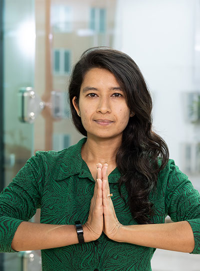
Sugandha (Su) Sharma is a graduate student in MIT’s Department of Brain and Cognitive Sciences (BCS), where she is co-advised by Ila Fiete (McGovern Institute) and Josh Tenenbaum (BCS).
Prior to joining MIT, she studied theoretical neuroscience at the University of Waterloo where she built neural models of context dependent decision making in the prefrontal cortex and spiking neuron models of bayesian inference, based on online learning of priors from life experience.
Today, in the Fiete and Tenenbaum labs, she studies the computational and theoretical principles underlying cognition and intelligence in the human brain. She is currently exploring the coding principles in the hippocampal circuits implicated in spatial navigation, and their role in cognitive computations like structure learning and relational reasoning.
When did you start practicing mindfulness?
When I first learned to meditate, I was challenged to practice it every day for at least 3 months in a row. I took up the challenge, and by the end of it, the results were profound. My whole perspective towards life changed. It made me more empathetic — I could step in other people’s shoes and be mindful of their situations and feelings; my focus shifted from myself to the big picture — it made me realize how insignificant my life was on the grand scale of the universe, and how it was worthless to be caught up in small things that I was usually worrying about. It somehow also brought selflessness to me. This experience hooked me to meditation and mindfulness for life!
What kind of mindfulness do you practice and why?
I practice mindfulness because it brings awareness. It helps me to be aware of myself, my thoughts, my actions, and my surroundings at each moment in my life, thus helping me stay in and enjoy the present moment. Awareness is of utmost importance since an aware mind always does the right thing. Imagine that you are angry, in that moment you have lost awareness of yourself. The moment you become aware of yourself; anger goes away. This is why sometimes counting helps to combat anger. If you start counting, that gives you time to think and become aware of yourself and your actions.
Meditating — sitting with my eyes closed and just observing (being aware of) my thoughts — is a yogic technique that helps me clear the noise in my mind and calm it down making it easier for me to be mindful not only while meditating, but also in general after I am done meditating. Over time, the thoughts vanish, and the mind becomes blank (noiseless). For this reason, practicing meditation regularly makes it easier for me to be mindful all the time.
An added advantage of yoga and meditation is that it helps combat stress by relaxing the mind and body. Many people don’t know what to do when they are stressed, but I am grateful to have this toolkit of yoga and meditation to deal with stressful situations in my life. They help me calm my mind in stressful situations and ensure that instead of reacting to a situation, I instead act mindfully and appropriately to make it right.