Language skills stay strong in older adults, even while other cognitive abilities decline

As people age, many cognitive functions tend to decline. Brain scanning studies have revealed corresponding changes in the function of a brain network that is involved in many of these cognitive functions, including working memory and problem-solving.

When it comes to language skills, however, the picture is different. Unless impaired by a stroke or dementia, most older people retain their language skills and may even improve them as they steadily gain vocabulary throughout their lives.

A new brain imaging study from an MIT-Boston University collaboration now reveals the neural activity underlying this observation. The researchers found that in older adults, activity of the language processing network is nearly identical to that seen in the brains of younger adults during language tasks.

In contrast, the researchers found that activation patterns in the multiple demand network, a brain system involved in executive control tasks such as decision-making, were very different in older and younger adults.

“In the language network, we couldn’t find any differences between older and younger groups. In contrast, the executive system showed decline across almost all of the measures,” says Anne Billot.

“The network synchronization declined in older adults, the extent of activation was reduced, and the magnitude of activation was reduced as well,” adds Billot, one of the lead authors of the new study, who carried out this work while doing her PhD at BU and is now a postdoc at Harvard University.

The findings suggest that parts of the brain that are specialized for specific functions, such as language processing, may be more resilient to aging than the multiple demand network, a more general-purpose network that has greater flexibility in its function, the researchers say.

Former MIT research assistant Niharika Jhingan is also a lead author of the study, which appears today in Nature CommunicationsEvelina Fedorenko, an MIT associate professor of brain and cognitive sciences and member of MIT’s McGovern Institute for Brain Research, and Swathi Kiran, the James and Cecilia Tse Ying Professor in Neurorehabilitation at BU, are the paper’s senior co-authors.

The resilience of language

To study the effects of aging on the brain, the researchers looked at two groups of people, ages 17-39 and 41-80. Based on previous studies, they expected that the multiple demand network, which includes several regions in the frontal and parietal lobes of the brain, would look different in the brains of older people.

“It’s well known that executive functions, such as attention, working memory, and cognitive control, tend to decline with age. And it’s also known that in opposition to that, language skills typically tend to remain quite stable or even improve with age,” Billot says. “These two types of functions really go in opposite directions in healthy aging. In terms of behavior, that’s quite well-established, and we wanted to see if that was also the case in the brain.”

In previous studies of the multiple demand network, scientists have found that as people age network activity becomes less synchronized. Some neuroscientists have hypothesized that this may also happen in the language network, but studies haven’t found definitive evidence for this.

The MIT researchers were able to look at both networks by designing tasks that elicit responses primarily in either the language network or the multiple demand network. This allowed them to identify, for each participant, the brain areas that belong to each network.
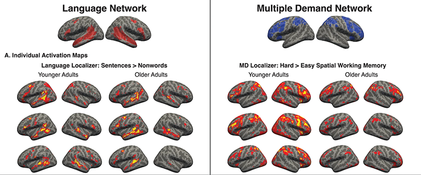
Neural responses in young adults and older adults are compared across four neural measures. Across these measures, the language network shows preserved function with age, whereas the multiple demand network shows consistent decline. Image courtesy of the researchers.

During a spatial memory task — remembering the location of squares in a grid — the researchers confirmed that the multiple demand network showed altered activity in older adults. Compared to the younger subjects, their networks were smaller and less well-synchronized, and the overall activation level was weaker.

To identify the language network, the researchers had participants listen to stories and read sentences. They found that in both groups, brain activity in response to language showed similar levels and spatial distribution across the network.

They also found that younger and older subjects showed similar brain responses when they encountered an unfamiliar word or an unusual grammatical construction.

“We have previously used similar kinds of materials to show that young adults show strong sensitivity to these points of linguistic difficulty: activity in the language areas goes up. Here we found that in older adults, you also see this sensitivity, which suggests that there’s nothing fundamentally different about how they process language,” Fedorenko says.

A language boost

The researchers also showed that in older people, the language network did not show any signs of becoming less synchronized. Additionally, the network did not show signs that it was blurring together with the multiple demand network, as some neuroscientists have hypothesized might happen.

While this study did not evaluate language ability, other studies have shown that not only do language skills not decline with age, for some people, their language processing improves in older age. This might be because vocabulary and reading skill can continually grow over time, the researchers say.

“Vocabulary keeps increasing as long as people have been measuring, which makes sense. People get exposed to more and more language, and older people sometimes start reading more, so they get an extra boost — it’s like a large language model trained on increasingly more data,” Fedorenko says.

Given these findings, one possible generalization is that parts of the brain that are specialized for particular functions such as language may be less susceptible to age-related decline than the multiple demand network. That network is unique in its ability to give the human brain the flexibility to learn new skills and adapt to new situations.

“The multiple demand network is a different system in the sense that it’s not accumulating knowledge over time. It’s more like a flexible resource that you can deploy in all sorts of ways. And somehow that’s the thing that is more vulnerable to aging,” Fedorenko says. “Why it’s so vulnerable — that is a very good question.”

The research was funded by the National Institute on Deafness and Other Communication Disorders, as well as MIT’s McGovern Institute, Simons Center for the Social Brain, Poitras Center for Psychiatric Disorders Research, and Quest for Intelligence.


Tackling rare genetic disorders with patient-focused science

Shannon Knight attributes her interest in neuroscience to an experience she had in high school. She and her sister attended a medical day for students at the nearby University of Illinois Chicago. As they were on their way out of the event, they walked past a room with a person holding a brain.

“We stopped and backpedaled into the room, and I was so fascinated,” says Knight. “I was able to hold the brain of a patient who had passed away of Alzheimer’s. The brain holds so much emotion, decision-making — everything. I realized that this man’s entire memory was in my hands, and something clicked for me. I decided that I really wanted to learn much more about this organ.”

Now in her sixth year of doctoral studies at MIT’s McGovern Institute for Brain Research, Knight is working on developing a novel gene therapy for childhood-onset epilepsy, specifically SYNGAP1 haploinsufficiency. This rare genetic disorder is caused by a mutation in the SYNGAP1 gene, rendering one of the two copies of the gene nonfunctional.

SYNGAP1 is important for brain development and neuronal communication, and the disorder leads to seizures in children starting as young as 4 months old. Other symptoms include intellectual disabilities, challenges with eating and sleeping, and difficulties with movement.

While there are currently methods to address the symptoms of the disorder, such as anti-seizure medications and dietary restrictions, as the child ages, the seizures often become resistant to medications. Knight is working to develop a therapeutic using CRISPR, a biotechnology tool used to edit genes. This therapeutic aims to address the root cause of this medication resistance by focusing on the gene itself.

“The idea of leading science with empathy is something that I feel very deeply,” she says. “I hope my efforts in the lab work toward the benefit of the people affected, rather than just for the benefit of my own science.”

Researching gene therapies

Knight’s interest in the brain flourished as a neuroscience major at Bowdoin College, working with Professor Hadley Horch. While she had originally planned to be pre-med, Knight ultimately decided that it wasn’t the best fit. She enjoyed the research she did as part of her honors thesis, exploring the regeneration of neurons in the auditory system of crickets, and decided that she wanted to pursue more research in molecular neuroscience, as well as genetics.

After graduating, Knight worked at the Perrimon Lab at Harvard University, where she first learned about CRISPR, applying it in a fruit fly model. She worked for two years in the lab, co-authoring a few papers and applying to graduate schools.

She ultimately landed in the lab of MIT Professor Guoping Feng, studying the potential of utilizing CRISPR to develop a gene therapy treatment for Phelan-McDermid Syndrome, a rare genetic disorder caused by a deletion or mutation on the 22nd chromosome.

“Many of our graduate students are passionate about making a positive impact to society through cutting-edge research, and Shannon is a perfect example,” says Feng, the James W. and Patricia T. Poitras Professor and associate director at the McGovern Institute. “She is developing gene therapy technologies that have the potential to help many kids with devastating neurodevelopmental disorders.”

Building off of the gene therapy research around Phelan-McDermid syndrome, which is now in clinical trials in patients, Knight is now in the early phases of testing gene therapy for SYNGAP1 disorder. The goal is to go through the same process for the SYNGAP1 gene therapy as for the Phelan-McDermid gene therapy — eventually obtaining U.S. Food and Drug Administration approval and beginning clinical trials.

The testing of the gene therapy on mice with a version of SYNGAP1 disorder has alleviated seizures and all of the behavioral phenotypes. This promising work is being accelerated by the Rare Brain Disorders Nexus, an MIT initiative that launched in the fall of 2025.

“Something I think about a lot is the idea of who ‘deserves’ the attention of a gene therapy. I feel that, regardless of how rare a genetic disorder might be, it still deserves care,” says Knight. “SYNGAP1 disorder is extremely rare, only impacting one to four out of every 10,000 children. I am very fortunate to be at an institution like MIT that has so many labs and brilliant researchers working on diseases that impact large portions of society, and it was really important to me to spend my PhD years helping a small, often unseen population. Although I don’t actually have a relationship with someone who has SYNGAP1 disorder, I know so many people who feel invisible in systems, and it is really important to me to be able to focus on people who feel unseen and give them hope.”

Inspiring others in the lab

In addition to her passion for neuroscience and genetic research, Knight has also developed a love of teaching. She has been a teaching assistant for class 9.12 (Experimental Molecular Neurobiology), leading the lab portion of the course. She has enjoyed working closely with small classes of students, introducing them to the fundamentals of neuroscience lab research.

“We walked through the process of looking at a specific protein in neurons, and talked about how you can go from cell culture all the way up to a mouse brain — and all the steps in between,” she says. “It was so important to me to be able to teach the students and help them to consider all of the different types of experiments they could do.”

Knight received the Goodwin Medal in 2025 in recognition of her commitment to excellent teaching.

“I’ve talked to many of the students since then,” she says, “and many said it was one of their favorite classes.”

MIT honors employees with 2026 Excellence Awards, Collier Medal, and Staff Award for Distinction in Service

On June 4, colleagues held homemade signs, waved pompoms, and cheered loudly for award recipients in Kresge Auditorium. Twenty individuals and three teams received MIT Excellence Awards — the Institute’s highest honor for staff. Additional honors included the Collier Medal, the Staff Award for Distinction in Service, and the Gordon Y. Billard Award.

The Collier Medal honors the memory of MIT police officer Sean Collier, who gave his life in service to the MIT community. Recipients embody a deep commitment to community and approach their work with compassion for others. The Staff Award for Distinction in Service is presented to an individual who approaches their work with kindness, empathy, and approachability, and serves as a trusted adviser at the Institute. The Gordon Y. Billard Award is given to staff or faculty members, or MIT-affiliated individuals, who make “significant and lasting contributions to the MIT community.”

The 2026 MIT Excellence Award recipients and their categories are:

Bringing Out the Best 

  • Robin Elices
  • Kate McCarthy
  • Jim Mitchell

Embracing Inclusion 

  • Allison Chang
  • Mandana Sassanfar

Innovative Solutions 

  • Kayla S. (KB) Burt
  • Amanda Jarvis
  • Julie Uva
  • Sustainability Team (Yu Cheng, Brian Goldberg, Susy Jones, Steve Lanou, Ellie McLane, and Julie Newman)

Outstanding Contributor 

  • Barry Pugatch
  • Emma Shortall
  • Chao Li
  • Catherine Nunziata
  • Trinidad Carney
  • Gang Liu
  • Laura von Bosau
  • James Daley
  • Craig Rowe
  • MIT Health Housekeeping Team (Michael Batista, Maria Coelho, Mae Evans, Maria Fatima Rosario, Selam Stefanos, Claudia Teixeira, and Claudia Vidal)

Serving Our Community 

  • Olivia Cheo
  • Clayton Hainsworth
  • Atsushi Takahashi
  • MIT Health Ambulatory Safety Net and Population Management Team (Michele M. A. David, Solanlly Mendez, Pamela Mensah, Nicole Napier, Lucus David Sensius, and Stephanie Shaprio)

The 2026 Collier Medal recipient was Michael Grenier, pub manager, dining, Division of Student Life. At the Muddy Charles and the Thirsty Ear pubs, Grenier creates spaces where MIT community members can relax, meet friends, and find unconditional support. His acts of kindness are woven into the atmosphere he creates, and alumni regard him as someone who shaped an important part of their early adult lives.

This year’s winner of the Staff Award for Distinction in Service was Christina Couch, associate director and lecturer, MIT Graduate Program in Science Writing, Comparative Media Studies, School of Humanities, Arts and Social Sciences (SHASS). Couch has been an invaluable member of the SHASS community — as a student, alumna, and staff member. Through her work, she has created opportunities for students to interact and build relationships with professional journalists, and underlying everything she does is her compassion and deep belief in student potential.

Three community members were honored with a 2026 Gordon Y. Billard Award.

  • Cullen R. Buie, professor of mechanical and biological engineering, associate department head of mechanical engineering, and head of house, Maseeh Hall
  • Traci Swartz, assistant director, Community Services Office, Institute Affairs, Office of the President
  • David L. Verrill, executive director, Initiative on the Digital Economy, MIT Sloan School of Management

Presenters included Provost Anantha Chandrakasan; MIT Chief of Police John DiFava and Captain Andrew Turco; Executive Vice President and Treasurer Glen Shor; Associate Provost Maria Yang; Dean of the School of Science Nergis Mavalvala; Lincoln Laboratory Assistant Director Justin Brooke; Vice President for Human Resources Ramona Allen; and Chancellor Melissa Nobles.

Visit the MIT Human Resources website for more information about the award recipients, categories, and to view photos and video of the event.

Powerful shrinking technique could enable devices that compute with light

Using a new technique that can create vacancies at any site across a material and then shrink it to about 1/2,000 of its original volume, MIT researchers have designed nanotechnology devices that could be used for optical computing and other applications involving the manipulation of visible light.

The new fabrication technique, known as “implosion carving,” allows researchers to imprint features throughout a hydrogel using photopatterning. If patterned with a resolution of about 800 nanometers, these features can then be shrunk to less than 100 nanometers.

Because that resolution is smaller than the wavelength of light, the devices can bend light in specific ways that allow them to perform optical computations.

Animation of block resembling three skyscrapers spinning in mid air.
MIT engineers created this photonic device by laser-patterning a hydrogel and then shrinking it to 1/2000 of its original volume.

“In order to enable nanophotonic applications in visible light, we need to make nanostructures with feature sizes with a resolution less than 100 nanometers. Only in that way can we precisely create the structure that can manipulate visible light,” says Quansan Yang, a former MIT postdoc, now an assistant professor at the University of Washington, and one of the lead authors of the new study.

In their paper, the researchers demonstrated a photonic device that can perform a simple digit-classification task, but future versions could be used for high-speed imaging and information processing, they say.

Gaojie Yang, a former MIT postdoc, is the co-lead author of the paper, which appears today in Nature Photonics. The paper’s senior authors are Peter So, director of the MIT Laser Biomedical Research Center (LBCR) and an MIT professor of biological engineering and mechanical engineering, and Edward Boyden, the Y. Eva Tan Professor in Neurotechnology at MIT and a professor of biological engineering, media arts and sciences, and brain and cognitive sciences. Boyden is also a Howard Hughes Medical Institute investigator and a member of MIT’s McGovern Institute for Brain Research, the Yang Tan Collective, and Koch Institute for Integrative Cancer Research.

Nanoscale feature sizes

Photonic devices, which transmit and manipulate light, hold potential for use as optical computer chips that could offer an energy-efficient alternative to semiconductor chips. However, existing techniques for creating 3D photonic devices haven’t yet achieved the 100-nanometer resolution that is needed to channel visible light, which has wavelengths between 380 and 750 nanometers.

Using an additive manufacturing technique called two-photon lithography, researchers can use light to create 3D nanoscale features, but with a resolution larger than 100 nanometers. Another technique, known as electron-beam lithography, can be used to etch smaller-resolution features onto a silicon chip, but it doesn’t generate 3D structures.

To make 3D devices with the necessary feature size, the researchers extended the concept of “implosion fabrication,” which Boyden’s lab developed in 2018, to create a new variant called “implosion carving.” In implosion carving, a laser creates vacancies — tiny voids where the hydrogel material has been removed — at precisely targeted locations. These vacancies exhibit different optical properties than the surrounding hydrogel. The hydrogel is then shrunk to bring the patterned features down to the nanoscale.

The carving process begins with immersing the hydrogel in a photosensitizing dye. Then, the researchers use a laser to excite the photosensitizer at specific places in the gel, which in turn generates reactive oxygen species that cut the bonds holding the hydrogel together. This creates a vacancy in that spot.

Once the desired vacancy pattern has been carved into the hydrogel, the researchers shrink it using a two-step process. First, they soak it in a solution containing ions, which causes it to shrink about tenfold in each dimension. To shrink it a little more, and to remove the watery solution, the hydrogel then undergoes a process called supercritical drying, which can remove liquid from a gel without damaging it.

At the end of the process, the hydrogel has been shrunk more than tenfold in each dimension, leading to a 2,000-fold reduction in volume.

Computing with light

To demonstrate the versatility of this technique, the researchers used it to create several 3D shapes, including a helix and a structure inspired by a butterfly wing. Some of these structures are too thin, and have too high an aspect ratio, to be stably created using conventional two-photon lithography.

The researchers also created a device that could perform a simple calculation known as digit classification, a task that is traditionally used to test the performance of neural networks. During this task, the device was presented with a digit, such as 1 or 5, and had to light up a specific location to indicate which number was detected.

To achieve this, the researchers patterned vacancies throughout the device so that it would act like a neural network. The pattern of vacancies would diffract input light as it passed through many layers of patterned hydrogel, so that the output light was determined by the shape of the digit that was entered into the system.

“This is a purely optical system that effectively performs optical computing,” So says.

“One of the very attractive features of this technology is that you can manipulate the property of the material at every tiny location,” says Dushan Wadduwage, an assistant professor at Old Dominion University and former MIT postdoc, who is also an author of the paper. “You have millions of different locations that you need to decide the property of, and that turns into a really interesting design problem where we can use deep-learning algorithms to find designs over these millions of parameters and come up with parts that go into optical systems in new ways.”

The researchers now plan to use the same principles to build optical devices that could classify cells based on their state as they flow through a microfluidic device. This could help identify rare cells such as circulating tumor cells in a blood sample, they say.

This approach could also enable the creation of high-throughput imaging techniques for applications such as analyzing tissue samples from biopsies or surgical specimens. And, if adapted to work with other materials such as hydrophobic polymers, it could also be used to create channels within 3D nanofluidic devices.

Other authors of the paper include Gaojie Yang, Takahiro Nambara, Hiroyuki Kusaka, Yuichiro Kunai, Alex Matlock, Corban Swain, Brett Pryor, Yannick Salamin, Daniel Oran, Hasindu Kariyawasam, Ramith Hettiarachchi, and Marin Soljacic.

The research was funded, in part, by the MIT-Fujikura Partnership Fund, the U.S. Army Research Office through the Institute for Soldier Nanotechnologies at MIT, Lisa Yang and Y. Eva Tan, John Doerr, the Open Philanthropy Project, the Howard Hughes Medical Institute, and the U.S. National Institutes of Health.

When it comes to language, context matters

In everyday conversation, it’s critical to understand not just the words that are spoken, but the context in which they are said. If it’s pouring rain and someone remarks on the “lovely weather,” you won’t understand their meaning unless you realize that they’re being sarcastic.

Making inferences about what someone really means when it doesn’t match the literal meaning of their words is a skill known as pragmatic language ability. This includes not only interpreting sarcasm but also understanding metaphors and white lies, among many other conversational subtleties.

Portrait of McGovern Investigator Evelina Fedorenko in a black shirt with soft white lights in background. Photo: Alexandra Sokhina
McGovern Investigator Evelina Fedorenko. Photo: Alexandra Sokhina

“Pragmatics is trying to reason about why somebody might say something, and what is the message they’re trying to convey given that they put it in this particular way,” says Evelina Fedorenko, an MIT associate professor of brain and cognitive sciences and a member of MIT’s McGovern Institute for Brain Research.

New research from Fedorenko and her colleagues has revealed that these abilities can be grouped together based on what types of inferences they require. In a study of 800 people, the researchers identified three clusters of pragmatic skills that are based on the same kinds of inferences and may have similar underlying neural processes.

One of these clusters includes inferences that are based on our knowledge of social conventions and rules. Another depends on knowledge of how the physical world works, while the last requires the ability to interpret differences in tone, which can indicate emphasis or emotion.

Fedorenko and Edward Gibson, an MIT professor of brain and cognitive sciences, are the senior authors of the study, which appears today in the Proceedings of the National Academy of Sciences. The paper’s lead authors are Sammy Floyd, a former MIT postdoc who is now an assistant professor of psychology at Sarah Lawrence College, and Olessia Jouravlev, a former MIT postdoc who is now an associate professor of cognitive science at Carleton University.

The importance of context

Much past research on how people understand language has focused on processing the literal meanings of words and how they fit together. To really understand what someone is saying, however, we need to interpret those meanings based on context.

“Language is about getting meanings across, and that often requires taking into account many different kinds of information — such as the social context, the visual context, or the present topic of the conversation,” Fedorenko says.

As one example, the phrase “people are leaving” can mean different things depending on the context, Gibson points out. If it’s late at night and someone asks you how a party is going, you may say “people are leaving,” to convey that the party is ending and everyone’s going home.

“However, if it’s early, and I say ‘people are leaving,’ then the implication is that the party isn’t very good,” Gibson says. “When you say a sentence, there’s a literal meaning to it, but how you interpret that literal meaning depends on the context.”

About 10 years ago, with support from the Simons Center for the Social Brain at MIT, Fedorenko and Gibson decided to explore whether it might be possible to precisely distinguish the types of processing that go into pragmatic language skills.

One way that neuroscientists can approach a question like this is to use functional magnetic resonance imaging (fMRI) to scan the brains of participants as they perform different tasks. This allows them to link brain activity in different locations to different functions. However, the tasks that the researchers designed for this study didn’t easily lend themselves to being performed in a scanner, so they took an alternative approach.

This approach, known as “individual differences,” involves studying a large number of people as they perform a variety of tasks. This technique allows researchers to determine whether the same underlying brain processes may be responsible for performance on different tasks.

To do this, the researchers evaluate whether each participant tends to perform similarly on certain groups of tasks. For example, some people might perform well on tasks that require an understanding of social conventions, such as interpreting indirect requests and irony. The same people might do only so-so on tasks that require understanding how the physical world works, and poorly on tasks that require distinguishing meanings based on changes in intonation — the melody of speech. This would suggest that separate brain processes are being recruited for each set of tasks.

The first phase of the study was led by Jouravlev, who assembled existing tasks that require pragmatic skills and created many more, for a total of 20. These included tasks that require people to understand humor and sarcasm, as well as tasks where changes in intonation can affect the meaning of a sentence. For example, someone who says “I wanted blue and black socks,” with emphasis on the word “black,” is implying that the black socks were forgotten.

“People really find ways to communicate creatively and indirectly and non-literally, and this battery of tasks captures that,” Floyd says.

Components of pragmatic ability

The researchers recruited study participants from an online crowdsourcing platform to perform the tasks, which took about eight hours to complete. From this first set of 400 participants, the researchers found that the tasks formed three clusters, related to social context, general knowledge of the world, and intonation. To test the robustness of the findings, the researchers continued the study with another set of 400 participants, with this second half run by Floyd after Jouravlev had left MIT.

With the second set of participants, the researchers found that tasks clustered into the same three groups. They also confirmed that differences in general intelligence, or in auditory processing ability (which is important for the processing of intonation), did not affect the outcomes that they observed.

In future work, the researchers hope to use brain imaging to explore whether the pragmatic components they identified are correlated with activity in different brain regions. Previous work has found that brain imaging often mirrors the distinctions identified in individual difference studies, but can also help link the relevant abilities to specific neural systems, such as the core language system or the theory of mind system.

This set of tests could also be used to study people with autism, who sometimes have difficulty understanding certain social cues. Such studies could determine more precisely the nature and extent of these difficulties. Another possibility could be studying people who were raised in different cultures, which may have different norms around speaking directly or indirectly.

“In Russian, which happens to be my native language, people are more direct. So perhaps there might be some differences in how native speakers of Russian process indirect requests compared to speakers of English,” Jouravlev says.

The research was funded by the Simons Center for the Social Brain at MIT, the National Institutes of Health, and the National Science Foundation.

A bionic knee integrated into tissue can restore natural movement

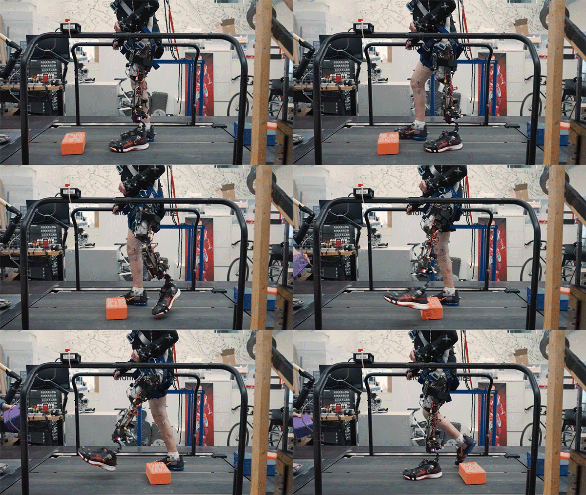
MIT researchers have developed a new bionic knee that can help people with above-the-knee amputations walk faster, climb stairs, and avoid obstacles more easily than they could with a traditional prosthesis.

Unlike prostheses in which the residual limb sits within a socket, the new system is directly integrated with the user’s muscle and bone tissue. This enables greater stability and gives the user much more control over the movement of the prosthesis.

Participants in a small clinical study also reported that the limb felt more like a part of their own body, compared to people who had more traditional above-the-knee amputations.

“A prosthesis that’s tissue-integrated — anchored to the bone and directly controlled by the nervous system — is not merely a lifeless, separate device, but rather a system that is carefully integrated into human physiology, offering a greater level of prosthetic embodiment. It’s not simply a tool that the human employs, but rather an integral part of self,” says Hugh Herr, a professor of media arts and sciences, co-director of the K. Lisa Yang Center for Bionics at MIT, an associate member of MIT’s McGovern Institute for Brain Research, and the senior author of the new study.

Tony Shu PhD ’24 is the lead author of the paper, which appears today in Science.

A subject with the osseointegrated mechanoneural prosthesis overcomes an obstacle placed in their walking path by volitionally flexing and extending their phantom knee joint.

Better control

Over the past several years, Herr’s lab has been working on new prostheses that can extract neural information from muscles left behind after an amputation and use that information to help guide a prosthetic limb.

During a traditional amputation, pairs of muscles that take turns stretching and contracting are usually severed, disrupting the normal agonist-antagonist relationship of the muscles. This disruption makes it very difficult for the nervous system to sense the position of a muscle and how fast it’s contracting.

Using the new surgical approach developed by Herr and his colleagues, known as agonist-antagonist myoneuronal interface (AMI), muscle pairs are reconnected during surgery so that they still dynamically communicate with each other within the residual limb. This sensory feedback helps the wearer of the prosthesis to decide how to move the limb, and also generates electrical signals that can be used to control the prosthetic limb.


In a 2024 study, the researchers showed that people with amputations below the knee who received the AMI surgery were able to walk faster and navigate around obstacles much more naturally than people with traditional below-the-knee amputations.

In the new study, the researchers extended the approach to better serve people with amputations above the knee. They wanted to create a system that could not only read out signals from the muscles using AMI but also be integrated into the bone, offering more stability and better sensory feedback.

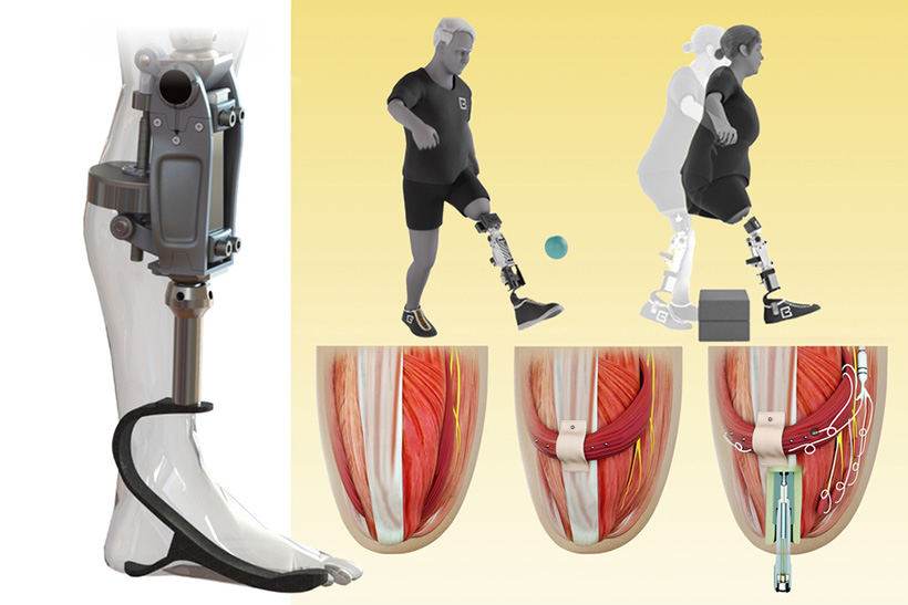
To achieve that, the researchers developed a procedure to insert a titanium rod into the residual femur bone at the amputation site. This implant allows for better mechanical control and load bearing than a traditional prosthesis. Additionally, the implant contains 16 wires that collect information from electrodes located on the AMI muscles inside the body, which enables more accurate transduction of the signals coming from the muscles.

This bone-integrated system, known as e-OPRA, transmits AMI signals to a new robotic controller developed specifically for this study. The controller uses this information to calculate the torque necessary to move the prosthesis the way that the user wants it to move.

The new bionic knee can help people with above-the-knee amputations walk faster, climb stairs, and avoid obstacles more easily than they could with a traditional prosthesis. The new system is directly integrated with the user’s muscle and bone tissue (bottom row right). This enables greater stability and gives the user much more control over the movement of the prosthesis. Image courtesy of the researchers

“All parts work together to better get information into and out of the body and better interface mechanically with the device,” Shu says. “We’re directly loading the skeleton, which is the part of the body that’s supposed to be loaded, as opposed to using sockets, which is uncomfortable and can lead to frequent skin infections.”

In this study, two subjects received the combined AMI and e-OPRA system, known as an osseointegrated mechanoneural prosthesis (OMP). These users were compared with eight who had the AMI surgery but not the e-OPRA implant, and seven users who had neither AMI nor e-OPRA. All subjects took a turn at using an experimental powered knee prosthesis developed by the lab.

The researchers measured the participants’ ability to perform several types of tasks, including bending the knee to a specified angle, climbing stairs, and stepping over obstacles. In most of these tasks, users with the OMP system performed better than the subjects who had the AMI surgery but not the e-OPRA implant, and much better than users of traditional prostheses.

“This paper represents the fulfillment of a vision that the scientific community has had for a long time — the implementation and demonstration of a fully physiologically integrated, volitionally controlled robotic leg,” says Michael Goldfarb, a professor of mechanical engineering and director of the Center for Intelligent Mechatronics at Vanderbilt University, who was not involved in the research. “This is really difficult work, and the authors deserve tremendous credit for their efforts in realizing such a challenging goal.”

A sense of embodiment

In addition to testing gait and other movements, the researchers also asked questions designed to evaluate participants’ sense of embodiment — that is, to what extent their prosthetic limb felt like a part of their own body.

Questions included whether the patients felt as if they had two legs, if they felt as if the prosthesis was part of their body, and if they felt in control of the prosthesis. Each question was designed to evaluate the participants’ feelings of agency, ownership of device, and body representation.

The researchers found that as the study went on, the two participants with the OMP showed much greater increases in their feelings of agency and ownership than the other subjects.

“Another reason this paper is significant is that it looks into these embodiment questions and it shows large improvements in that sensation of embodiment,” Herr says. “No matter how sophisticated you make the AI systems of a robotic prosthesis, it’s still going to feel like a tool to the user, like an external device. But with this tissue-integrated approach, when you ask the human user what is their body, the more it’s integrated, the more they’re going to say the prosthesis is actually part of self.”

The AMI procedure is now done routinely on patients with below-the-knee amputations at Brigham and Women’s Hospital, and Herr expects it will soon become the standard for above-the-knee amputations as well. The combined OMP system will need larger clinical trials to receive FDA approval for commercial use, which Herr expects may take about five years.

The research was funded by the Yang Tan Collective and DARPA.

Researchers present bold ideas for AI at MIT Generative AI Impact Consortium kickoff event

Launched in February of this year, the MIT Generative AI Impact Consortium (MGAIC), a presidential initiative led by MIT’s Office of Innovation and Strategy and administered by the MIT Stephen A. Schwarzman College of Computing, issued a call for proposals, inviting researchers from across MIT to submit ideas for innovative projects studying high-impact uses of generative AI models.

The call received 180 submissions from nearly 250 faculty members, spanning all of MIT’s five schools and the college. The overwhelming response across the Institute exemplifies the growing interest in AI and follows in the wake of MIT’s Generative AI Week and call for impact papers. Fifty-five proposals were selected for MGAIC’s inaugural seed grants, with several more selected to be funded by the consortium’s founding company members.

Over 30 funding recipients presented their proposals to the greater MIT community at a kickoff event on May 13. Anantha P. Chandrakasan, chief innovation and strategy officer and dean of the School of Engineering who is head of the consortium, welcomed the attendees and thanked the consortium’s founding industry members.

“The amazing response to our call for proposals is an incredible testament to the energy and creativity that MGAIC has sparked at MIT. We are especially grateful to our founding members, whose support and vision helped bring this endeavor to life,” adds Chandrakasan. “One of the things that has been most remarkable about MGAIC is that this is a truly cross-Institute initiative. Deans from all five schools and the college collaborated in shaping and implementing it.”

Vivek F. Farias, the Patrick J. McGovern (1959) Professor at the MIT Sloan School of Management and co-faculty director of the consortium with Tim Kraska, associate professor of electrical engineering and computer science in the MIT Computer Science and Artificial Intelligence Laboratory (CSAIL), emceed the afternoon of five-minute lightning presentations.

Presentation highlights include:

“AI-Driven Tutors and Open Datasets for Early Literacy Education,” presented by Ola Ozernov-Palchik, a research scientist at the McGovern Institute for Brain Research, proposed a refinement for AI-tutors for pK-7 students to potentially decrease literacy disparities.

“Developing jam_bots: Real-Time Collaborative Agents for Live Human-AI Musical Improvisation,” presented by Anna Huang, assistant professor of music and assistant professor of electrical engineering and computer science, and Joe Paradiso, the Alexander W. Dreyfoos (1954) Professor in Media Arts and Sciences at the MIT Media Lab, aims to enhance human-AI musical collaboration in real-time for live concert improvisation.

“GENIUS: GENerative Intelligence for Urban Sustainability,” presented by Norhan Bayomi, a postdoc at the MIT Environmental Solutions Initiative and a research assistant in the Urban Metabolism Group, which aims to address the critical gap of a standardized approach in evaluating and benchmarking cities’ climate policies.

Georgia Perakis, the John C Head III Dean (Interim) of the MIT Sloan School of Management and professor of operations management, operations research, and statistics, who serves as co-chair of the GenAI Dean’s oversight group with Dan Huttenlocher, dean of the MIT Schwarzman College of Computing, ended the event with closing remarks that emphasized “the readiness and eagerness of our community to lead in this space.”

“This is only the beginning,” he continued. “We are at the front edge of a historic moment — one where MIT has the opportunity, and the responsibility, to shape the future of generative AI with purpose, with excellence, and with care.”

How the brain solves complicated problems

The human brain is very good at solving complicated problems. One reason for that is that humans can break problems apart into manageable subtasks that are easy to solve one at a time.

This allows us to complete a daily task like going out for coffee by breaking it into steps: getting out of our office building, navigating to the coffee shop, and once there, obtaining the coffee. This strategy helps us to handle obstacles easily. For example, if the elevator is broken, we can revise how we get out of the building without changing the other steps.

While there is a great deal of behavioral evidence demonstrating humans’ skill at these complicated tasks, it has been difficult to devise experimental scenarios that allow precise characterization of the computational strategies we use to solve problems.

In a new study, MIT researchers have successfully modeled how people deploy different decision-making strategies to solve a complicated task — in this case, predicting how a ball will travel through a maze when the ball is hidden from view. The human brain cannot perform this task perfectly because it is impossible to track all of the possible trajectories in parallel, but the researchers found that people can perform reasonably well by flexibly adopting two strategies known as hierarchical reasoning and counterfactual reasoning.

The researchers were also able to determine the circumstances under which people choose each of those strategies.

“What humans are capable of doing is to break down the maze into subsections, and then solve each step using relatively simple algorithms. Effectively, when we don’t have the means to solve a complex problem, we manage by using simpler heuristics that get the job done,” says Mehrdad Jazayeri, a professor of brain and cognitive sciences, a member of MIT’s McGovern Institute for Brain Research, an investigator at the Howard Hughes Medical Institute, and the senior author of the study.

Mahdi Ramadan PhD ’24 and graduate student Cheng Tang are the lead authors of the paper, which appears today in Nature Human Behavior. Nicholas Watters PhD ’25 is also a co-author.

Rational strategies

When humans perform simple tasks that have a clear correct answer, such as categorizing objects, they perform extremely well. When tasks become more complex, such as planning a trip to your favorite cafe, there may no longer be one clearly superior answer. And, at each step, there are many things that could go wrong. In these cases, humans are very good at working out a solution that will get the task done, even though it may not be the optimal solution.

Those solutions often involve problem-solving shortcuts, or heuristics. Two prominent heuristics humans commonly rely on are hierarchical and counterfactual reasoning. Hierarchical reasoning is the process of breaking down a problem into layers, starting from the general and proceeding toward specifics. Counterfactual reasoning involves imagining what would have happened if you had made a different choice. While these strategies are well-known, scientists don’t know much about how the brain decides which one to use in a given situation.

“This is really a big question in cognitive science: How do we problem-solve in a suboptimal way, by coming up with clever heuristics that we chain together in a way that ends up getting us closer and closer until we solve the problem?” Jazayeri says.

To overcome this, Jazayeri and his colleagues devised a task that is just complex enough to require these strategies, yet simple enough that the outcomes and the calculations that go into them can be measured.

The task requires participants to predict the path of a ball as it moves through four possible trajectories in a maze. Once the ball enters the maze, people cannot see which path it travels. At two junctions in the maze, they hear an auditory cue when the ball reaches that point. Predicting the ball’s path is a task that is impossible for humans to solve with perfect accuracy.

“It requires four parallel simulations in your mind, and no human can do that. It’s analogous to having four conversations at a time,” Jazayeri says. “The task allows us to tap into this set of algorithms that the humans use, because you just can’t solve it optimally.”

The researchers recruited about 150 human volunteers to participate in the study. Before each subject began the ball-tracking task, the researchers evaluated how accurately they could estimate timespans of several hundred milliseconds, about the length of time it takes the ball to travel along one arm of the maze.

For each participant, the researchers created computational models that could predict the patterns of errors that would be seen for that participant (based on their timing skill) if they were running parallel simulations, using hierarchical reasoning alone, counterfactual reasoning alone, or combinations of the two reasoning strategies.

The researchers compared the subjects’ performance with the models’ predictions and found that for every subject, their performance was most closely associated with a model that used hierarchical reasoning but sometimes switched to counterfactual reasoning.

That suggests that instead of tracking all the possible paths that the ball could take, people broke up the task. First, they picked the direction (left or right), in which they thought the ball turned at the first junction, and continued to track the ball as it headed for the next turn. If the timing of the next sound they heard wasn’t compatible with the path they had chosen, they would go back and revise their first prediction — but only some of the time.

Switching back to the other side, which represents a shift to counterfactual reasoning, requires people to review their memory of the tones that they heard. However, it turns out that these memories are not always reliable, and the researchers found that people decided whether to go back or not based on how good they believed their memory to be.

“People rely on counterfactuals to the degree that it’s helpful,” Jazayeri says. “People who take a big performance loss when they do counterfactuals avoid doing them. But if you are someone who’s really good at retrieving information from the recent past, you may go back to the other side.”

Human limitations

To further validate their results, the researchers created a machine-learning neural network and trained it to complete the task. A machine-learning model trained on this task will track the ball’s path accurately and make the correct prediction every time, unless the researchers impose limitations on its performance.

When the researchers added cognitive limitations similar to those faced by humans, they found that the model altered its strategies. When they eliminated the model’s ability to follow all possible trajectories, it began to employ hierarchical and counterfactual strategies like humans do. If the researchers reduced the model’s memory recall ability, it began to switch to hierarchical only if it thought its recall would be good enough to get the right answer — just as humans do.

“What we found is that networks mimic human behavior when we impose on them those computational constraints that we found in human behavior,” Jazayeri says. “This is really saying that humans are acting rationally under the constraints that they have to function under.”

By slightly varying the amount of memory impairment programmed into the models, the researchers also saw hints that the switching of strategies appears to happen gradually, rather than at a distinct cut-off point. They are now performing further studies to try to determine what is happening in the brain as these shifts in strategy occur.

The research was funded by a Lisa K. Yang ICoN Fellowship, a Friends of the McGovern Institute Student Fellowship, a National Science Foundation Graduate Research Fellowship, the Simons Foundation, the Howard Hughes Medical Institute, and the McGovern Institute.

How the brain distinguishes between ambiguous hypotheses

When navigating a place that we’re only somewhat familiar with, we often rely on unique landmarks to help make our way. However, if we’re looking for an office in a brick building, and there are many brick buildings along our route, we might use a rule like looking for the second building on a street, rather than relying on distinguishing the building itself.

Man seated on staircase, smiling at camera
McGovern Investigator Mark Harnett. Photo: Adam Glanzman

Until that ambiguity is resolved, we must hold in mind that there are multiple possibilities (or hypotheses) for where we are in relation to our destination. In a study of mice, MIT neuroscientists have now discovered that these hypotheses are explicitly represented in the brain by distinct neural activity patterns.

This is the first time that neural activity patterns that encode simultaneous hypotheses have been seen in the brain. The researchers found that these representations, which were observed in the brain’s retrosplenial cortex (RSC), not only encode hypotheses but also could be used by the animals to choose the correct way to go.

“As far as we know, no one has shown in a complex reasoning task that there’s an area in association cortex that holds two hypotheses in mind and then uses one of those hypotheses, once it gets more information, to actually complete the task,” says Mark Harnett, an associate professor of brain and cognitive sciences, a member of MIT’s McGovern Institute for Brain Research, and the senior author of the study.

Jakob Voigts PhD ’17, a former postdoc in Harnett’s lab and now a group leader at the Howard Hughes Medical Institute Janelia Research Campus, is the lead author of the paper, which appears today in Nature Neuroscience.

Ambiguous landmarks

The RSC receives input from the visual cortex, the hippocampal formation, and the anterior thalamus, which it integrates to help guide navigation.

In a 2020 paper, Harnett’s lab found that the RSC uses both visual and spatial information to encode landmarks used for navigation. In that study, the researchers showed that neurons in the RSC of mice integrate visual information about the surrounding environment with spatial feedback of the mice’s own position along a track, allowing them to learn where to find a reward based on landmarks that they saw.

In their new study, the researchers wanted to delve further into how the RSC uses spatial information and situational context to guide navigational decision-making. To do that, the researchers devised a much more complicated navigational task than typically used in mouse studies. They set up a large, round arena, with 16 small openings, or ports, along the side walls. One of these openings would give the mice a reward when they stuck their nose through it. In the first set of experiments, the researchers trained the mice to go to different reward ports indicated by dots of light on the floor that were only visible when the mice get close to them.

Man in blue shirt wearing glasses building a platform in a lab setting.
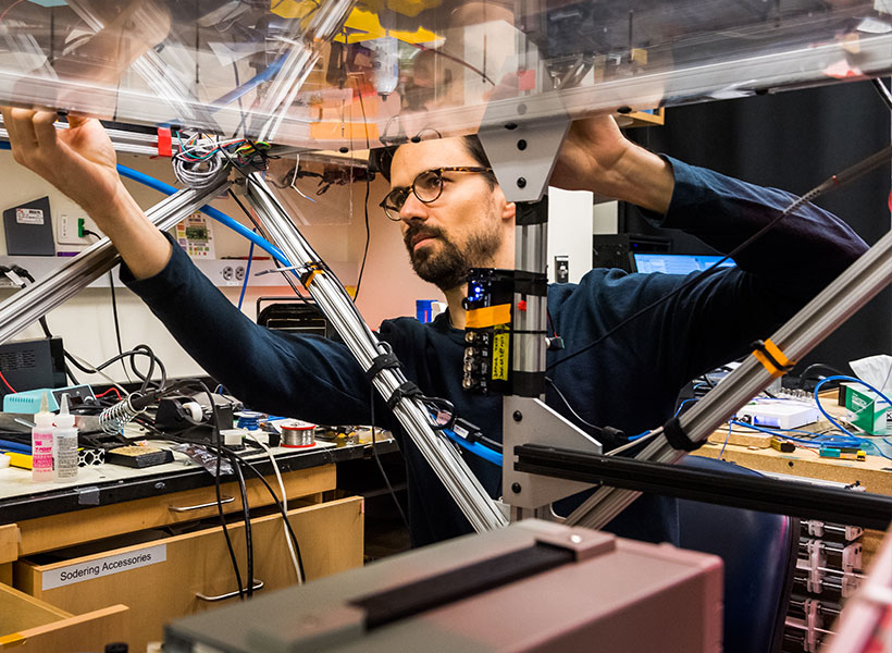
Jakob Voigts PhD ’17, at work in Mark Harnett’s lab. Photo: Justin Knight

Once the mice learned to perform this relatively simple task, the researchers added a second dot. The two dots were always the same distance from each other and from the center of the arena. But now the mice had to go to the port by the counterclockwise dot to get the reward. Because the dots were identical and only became visible at close distances, the mice could never see both dots at once and could not immediately determine which dot was which.

To solve this task, mice therefore had to remember where they expected a dot to show up, integrating their own body position, the direction they were heading, and path they took to figure out which landmark is which. By measuring RSC activity as the mice approached the ambiguous landmarks, the researchers could determine whether the RSC encodes hypotheses about spatial location. The task was carefully designed to require the mice to use the visual landmarks to obtain rewards, instead of other strategies like odor cues or dead reckoning.

“What is important about the behavior in this case is that mice need to remember something and then use that to interpret future input,” says Voigts, who worked on this study while a postdoc in Harnett’s lab.

“It’s not just remembering something, but remembering it in such a way that you can act on it.” – Jakob Voigts

The researchers found that as the mice accumulated information about which dot might be which, populations of RSC neurons displayed distinct activity patterns for incomplete information. Each of these patterns appears to correspond to a hypothesis about where the mouse thought it was with respect to the reward.

When the mice get close enough to figure out which dot was indicating the reward port, these patterns collapsed into the one that represents the correct hypothesis. The findings suggest that these patterns not only passively store hypotheses, they can also be used to compute how to get to the correct location, the researchers say.

“We show that RSC has the required information for using this short-term memory to distinguish the ambiguous landmarks. And we show that this type of hypothesis is encoded and processed in a way that allows the RSC to use it to solve the computation,” Voigts says.

Interconnected neurons

When analyzing their initial results, Harnett and Voigts consulted with MIT Professor Ila Fiete, who had run a study about 10 years ago using an artificial neural network to perform a similar navigation task.

That study, previously published on bioRxiv, showed that the neural network displayed activity patterns that were conceptually similar to those seen in the animal studies run by Harnett’s lab. The neurons of the artificial neural network ended up forming highly interconnected low-dimensional networks, like the neurons of the RSC.

“That interconnectivity seems, in ways that we still don’t understand, to be key to how these dynamics emerge and how they’re controlled. And it’s a key feature of how the RSC holds these two hypotheses in mind at the same time,” Harnett says.

In his lab at Janelia, Voigts now plans to investigate how other brain areas involved in navigation, such as the prefrontal cortex, are engaged as mice explore and forage in a more naturalistic way, without being trained on a specific task.

“We’re looking into whether there are general principles by which tasks are learned,” Voigts says. “We have a lot of knowledge in neuroscience about how brains operate once the animal has learned a task, but in comparison we know extremely little about how mice learn tasks or what they choose to learn when given freedom to behave naturally.”

The research was funded, in part, by the National Institutes of Health, a Simons Center for the Social Brain at MIT postdoctoral fellowship, the National Institute of General Medical Sciences, and the Center for Brains, Minds, and Machines at MIT, funded by the National Science Foundation.

A visual pathway in the brain may do more than recognize objects

When visual information enters the brain, it travels through two pathways that process different aspects of the input. For decades, scientists have hypothesized that one of these pathways, the ventral visual stream, is responsible for recognizing objects, and that it might have been optimized by evolution to do just that.

Consistent with this, in the past decade, MIT scientists have found that when computational models of the anatomy of the ventral stream are optimized to solve the task of object recognition, they are remarkably good predictors of the neural activities in the ventral stream.

However, in a new study, MIT researchers have shown that when they train these types of models on spatial tasks instead, the resulting models are also quite good predictors of the ventral stream’s neural activities. This suggests that the ventral stream may not be exclusively optimized for object recognition.

“This leaves wide open the question about what the ventral stream is being optimized for. I think the dominant perspective a lot of people in our field believe is that the ventral stream is optimized for object recognition, but this study provides a new perspective that the ventral stream could be optimized for spatial tasks as well,” says MIT graduate student Yudi Xie.

Xie is the lead author of the study, which will be presented at the International Conference on Learning Representations. Other authors of the paper include Weichen Huang, a visiting student through MIT’s Research Science Institute program; Esther Alter, a software engineer at the MIT Quest for Intelligence; Jeremy Schwartz, a sponsored research technical staff member; Joshua Tenenbaum, a professor of brain and cognitive sciences; and James DiCarlo, the Peter de Florez Professor of Brain and Cognitive Sciences, director of the Quest for Intelligence, and a member of the McGovern Institute for Brain Research at MIT.

Beyond object recognition

When we look at an object, our visual system can not only identify the object, but also determine other features such as its location, its distance from us, and its orientation in space. Since the early 1980s, neuroscientists have hypothesized that the primate visual system is divided into two pathways: the ventral stream, which performs object-recognition tasks, and the dorsal stream, which processes features related to spatial location.

Over the past decade, researchers have worked to model the ventral stream using a type of deep-learning model known as a convolutional neural network (CNN). Researchers can train these models to perform object-recognition tasks by feeding them datasets containing thousands of images along with category labels describing the images.

The state-of-the-art versions of these CNNs have high success rates at categorizing images. Additionally, researchers have found that the internal activations of the models are very similar to the activities of neurons that process visual information in the ventral stream. Furthermore, the more similar these models are to the ventral stream, the better they perform at object-recognition tasks. This has led many researchers to hypothesize that the dominant function of the ventral stream is recognizing objects.

However, experimental studies, especially a study from the DiCarlo lab in 2016, have found that the ventral stream appears to encode spatial features as well. These features include the object’s size, its orientation (how much it is rotated), and its location within the field of view. Based on these studies, the MIT team aimed to investigate whether the ventral stream might serve additional functions beyond object recognition.

“Our central question in this project was, is it possible that we can think about the ventral stream as being optimized for doing these spatial tasks instead of just categorization tasks?” Xie says.

To test this hypothesis, the researchers set out to train a CNN to identify one or more spatial features of an object, including rotation, location, and distance. To train the models, they created a new dataset of synthetic images. These images show objects such as tea kettles or calculators superimposed on different backgrounds, in locations and orientations that are labeled to help the model learn them.

The researchers found that CNNs that were trained on just one of these spatial tasks showed a high level of “neuro-alignment” with the ventral stream — very similar to the levels seen in CNN models trained on object recognition.

The researchers measure neuro-alignment using a technique that DiCarlo’s lab has developed, which involves asking the models, once trained, to predict the neural activity that a particular image would generate in the brain. The researchers found that the better the models performed on the spatial task they had been trained on, the more neuro-alignment they showed.

“I think we cannot assume that the ventral stream is just doing object categorization, because many of these other functions, such as spatial tasks, also can lead to this strong correlation between models’ neuro-alignment and their performance,” Xie says. “Our conclusion is that you can optimize either through categorization or doing these spatial tasks, and they both give you a ventral-stream-like model, based on our current metrics to evaluate neuro-alignment.”

Comparing models

The researchers then investigated why these two approaches — training for object recognition and training for spatial features — led to similar degrees of neuro-alignment. To do that, they performed an analysis known as centered kernel alignment (CKA), which allows them to measure the degree of similarity between representations in different CNNs. This analysis showed that in the early to middle layers of the models, the representations that the models learn are nearly indistinguishable.

“In these early layers, essentially you cannot tell these models apart by just looking at their representations,” Xie says. “It seems like they learn some very similar or unified representation in the early to middle layers, and in the later stages they diverge to support different tasks.”

The researchers hypothesize that even when models are trained to analyze just one feature, they also take into account “non-target” features — those that they are not trained on. When objects have greater variability in non-target features, the models tend to learn representations more similar to those learned by models trained on other tasks. This suggests that the models are using all of the information available to them, which may result in different models coming up with similar representations, the researchers say.

“More non-target variability actually helps the model learn a better representation, instead of learning a representation that’s ignorant of them,” Xie says. “It’s possible that the models, although they’re trained on one target, are simultaneously learning other things due to the variability of these non-target features.”

In future work, the researchers hope to develop new ways to compare different models, in hopes of learning more about how each one develops internal representations of objects based on differences in training tasks and training data.

“There could be still slight differences between these models, even though our current way of measuring how similar these models are to the brain tells us they’re on a very similar level. That suggests maybe there’s still some work to be done to improve upon how we can compare the model to the brain, so that we can better understand what exactly the ventral stream is optimized for,” Xie says.

The research was funded by the Semiconductor Research Corporation and the U.S. Defense Advanced Research Projects Agency.