A new way to deliver drugs with pinpoint targeting

Most pharmaceuticals must either be ingested or injected into the body to do their work. Either way, it takes some time for them to reach their intended targets, and they also tend to spread out to other areas of the body. Now, researchers at the McGovern Institute at MIT and elsewhere have developed a system to deliver medical treatments that can be released at precise times, minimally-invasively, and that ultimately could also deliver those drugs to specifically targeted areas such as a specific group of neurons in the brain.

The new approach is based on the use of tiny magnetic particles enclosed within a tiny hollow bubble of lipids (fatty molecules) filled with water, known as a liposome. The drug of choice is encapsulated within these bubbles, and can be released by applying a magnetic field to heat up the particles, allowing the drug to escape from the liposome and into the surrounding tissue.

The findings are reported today in the journal Nature Nanotechnology in a paper by MIT postdoc Siyuan Rao, Associate Professor Polina Anikeeva, and 14 others at MIT, Stanford University, Harvard University, and the Swiss Federal Institute of Technology in Zurich.

“We wanted a system that could deliver a drug with temporal precision, and could eventually target a particular location,” Anikeeva explains. “And if we don’t want it to be invasive, we need to find a non-invasive way to trigger the release.”

Magnetic fields, which can easily penetrate through the body — as demonstrated by detailed internal images produced by magnetic resonance imaging, or MRI — were a natural choice. The hard part was finding materials that could be triggered to heat up by using a very weak magnetic field (about one-hundredth the strength of that used for MRI), in order to prevent damage to the drug or surrounding tissues, Rao says.

Rao came up with the idea of taking magnetic nanoparticles, which had already been shown to be capable of being heated by placing them in a magnetic field, and packing them into these spheres called liposomes. These are like little bubbles of lipids, which naturally form a spherical double layer surrounding a water droplet.
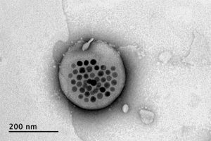
Electron microscope image shows the actual liposome, the white blob at center, with its magnetic particles showing up in black at its center.
Image courtesy of the researchers

When placed inside a high-frequency but low-strength magnetic field, the nanoparticles heat up, warming the lipids and making them undergo a transition from solid to liquid, which makes the layer more porous — just enough to let some of the drug molecules escape into the surrounding areas. When the magnetic field is switched off, the lipids re-solidify, preventing further releases. Over time, this process can be repeated, thus releasing doses of the enclosed drug at precisely controlled intervals.

The drug carriers were engineered to be stable inside the body at the normal body temperature of 37 degrees Celsius, but able to release their payload of drugs at a temperature of 42 degrees. “So we have a magnetic switch for drug delivery,” and that amount of heat is small enough “so that you don’t cause thermal damage to tissues,” says Anikeeva, who also holds appointments in the departments of Materials Science and Engineering and the Brain and Cognitive Sciences.

In principle, this technique could also be used to guide the particles to specific, pinpoint locations in the body, using gradients of magnetic fields to push them along, but that aspect of the work is an ongoing project. For now, the researchers have been injecting the particles directly into the target locations, and using the magnetic fields to control the timing of drug releases. “The technology will allow us to address the spatial aspect,” Anikeeva says, but that has not yet been demonstrated.

This could enable very precise treatments for a wide variety of conditions, she says. “Many brain disorders are characterized by erroneous activity of certain cells. When neurons are too active or not active enough, that manifests as a disorder, such as Parkinson’s, or depression, or epilepsy.” If a medical team wanted to deliver a drug to a specific patch of neurons and at a particular time, such as when an onset of symptoms is detected, without subjecting the rest of the brain to that drug, this system “could give us a very precise way to treat those conditions,” she says.

Rao says that making these nanoparticle-activated liposomes is actually quite a simple process. “We can prepare the liposomes with the particles within minutes in the lab,” she says, and the process should be “very easy to scale up” for manufacturing. And the system is broadly applicable for drug delivery: “we can encapsulate any water-soluble drug,” and with some adaptations, other drugs as well, she says.

One key to developing this system was perfecting and calibrating a way of making liposomes of a highly uniform size and composition. This involves mixing a water base with the fatty acid lipid molecules and magnetic nanoparticles and homogenizing them under precisely controlled conditions. Anikeeva compares it to shaking a bottle of salad dressing to get the oil and vinegar mixed, but controlling the timing, direction and strength of the shaking to ensure a precise mixing.

Anikeeva says that while her team has focused on neurological disorders, as that is their specialty, the drug delivery system is actually quite general and could be applied to almost any part of the body, for example to deliver cancer drugs, or even to deliver painkillers directly to an affected area instead of delivering them systemically and affecting the whole body. “This could deliver it to where it’s needed, and not deliver it continuously,” but only as needed.

Because the magnetic particles themselves are similar to those already in widespread use as contrast agents for MRI scans, the regulatory approval process for their use may be simplified, as their biological compatibility has largely been proven.

The team included researchers in MIT’s departments of Materials Science and Engineering and Brain and Cognitive Sciences, as well as the McGovern Institute for Brain Research, the Simons Center for Social Brain, and the Research Laboratory of Electronics; the Harvard University Department of Chemistry and Chemical Biology and the John A. Paulsen School of Engineering and Applied Sciences; Stanford University; and the Swiss Federal Institute of Technology in Zurich. The work was supported by the Simons Postdoctoral Fellowship, the U.S. Defense Advanced Research Projects Agency, the Bose Research Grant, and the National Institutes of Health.
Brain region linked to altered social interactions in autism model

Although psychiatric disorders can be linked to particular genes, the brain regions and mechanisms underlying particular disorders are not well-understood. Mutations or deletions of the SHANK3 gene are strongly associated with autism spectrum disorder (ASD) and a related rare disorder called Phelan-McDermid syndrome. Mice with SHANK3 mutations also display some of the traits associated with autism, including avoidance of social interactions, but the brain regions responsible for this behavior have not been identified.

A new study by neuroscientists at MIT and colleagues in China provides clues to the neural circuits underlying social deficits associated with ASD. The paper, published in Nature Neuroscience, found that structural and functional impairments in the anterior cingulate cortex (ACC) of SHANK3 mutant mice are linked to altered social interactions.

“Neurobiological mechanisms of social deficits are very complex and involve many brain regions, even in a mouse model,” explains Guoping Feng, the James W. and Patricia T. Poitras Professor at MIT and one of the senior authors of the study. “These findings add another piece of the puzzle to mapping the neural circuits responsible for this social deficit in ASD models.”

The Nature Neuroscience paper is the result of a collaboration between Feng, who is also an investigator at MIT’s McGovern Institute and a senior scientist in the Broad Institute’s Stanley Center for Psychiatric Research, and Wenting Wang and Shengxi Wu at the Fourth Military Medical University, Xi’an, China.

A number of brain regions have been implicated in social interactions, including the prefrontal cortex (PFC) and its projections to brain regions including the nucleus accumbens and habenula, but these studies failed to definitively link the PFC to altered social interactions seen in SHANK3 knockout mice.

In the new study, the authors instead focused on the ACC, a brain region noted for its role in social functions in humans and animal models. The ACC is also known to play a role in fundamental cognitive processes, including cost-benefit calculation, motivation, and decision making.

In mice lacking SHANK3, the researchers found structural and functional disruptions at the synapses, or connections, between excitatory neurons in the ACC. The researchers went on to show that the loss of SHANK3 in excitatory ACC neurons alone was enough to disrupt communication between these neurons and led to unusually reduced activity of these neurons during behavioral tasks reflecting social interaction.

Having implicated these ACC neurons in social preferences and interactions in SHANK3 knockout mice, the authors then tested whether activating these same neurons could rescue these behaviors. Using optogenetics and specfic drugs, the researchers activated the ACC neurons and found improved social behavior in the SHANK3 mutant mice.

“Next, we are planning to explore brain regions downstream of the ACC that modulate social behavior in normal mice and models of autism,” explains Wenting Wang, co-corresponding author on the study. “This will help us to better understand the neural mechanisms of social behavior, as well as social deficits in neurodevelopmental disorders.”

Previous clinical studies reported that anatomical structures in the ACC were altered and/or dysfunctional in people with ASD, an initial indication that the findings from SHANK3 mice may also hold true in these individuals.

The research was funded, in part, by the Natural Science Foundation of China. Guoping Feng was supported by NIMH grant no. MH097104, the  Poitras Center for Psychiatric Disorders Research at the McGovern Institute at MIT, and the Hock E. Tan and K. Lisa Yang Center for Autism Research at the McGovern Institute at MIT.

Four new faces in the School of Science faculty

This fall, the School of Science will welcome four new members joining the faculty in the departments of Biology, Brain and Cognitive Sciences, and Chemistry.

Evelina Fedorenko investigates how our brains process language. She has developed novel analytic approaches for functional magnetic resonance imaging (fMRI) and other brain imaging techniques to help answer the questions of how the language processing network functions and how it relates to other networks in the brain. She works with both neurotypical individuals and individuals with brain disorders. Fedorenko joins the Department of Brain and Cognitive Sciences as an assistant professor. She received her BA from Harvard University in linguistics and psychology and then completed her doctoral studies at MIT in 2007. After graduating from MIT, Fedorenko worked as a postdoc and then as a research scientist at the McGovern Institute for Brain Research. In 2014, she joined the faculty at Massachusetts General Hospital and Harvard Medical School, where she was an associate researcher and an assistant professor, respectively. She is also a member of the McGovern Institute.

Morgan Sheng focuses on the structure, function, and turnover of synapses, the junctions that allow communication between brain cells. His discoveries have improved our understanding of the molecular basis of cognitive function and diseases of the nervous system, such as autism, Alzheimer’s disease, and dementia. Being both a physician and a scientist, he incorporates genetic as well as biological insights to aid the study and treatment of mental illnesses and neurodegenerative diseases. He rejoins the Department of Brain and Cognitive Sciences (BCS), returning as a professor of neuroscience, a position he also held from 2001 to 2008. At that time, he was a member of the Picower Institute for Learning and Memory, a joint appointee in the Department of Biology, and an investigator of the Howard Hughes Medical Institute. Sheng earned his PhD from Harvard University in 1990, completed a postdoc at the University of California at San Francisco in 1994, and finished his medical training with a residency in London in 1986. From 1994 to 2001, he researched molecular and cellular neuroscience at Massachusetts General Hospital and Harvard Medical School. From 2008 to 2019 he was vice president of neuroscience at Genentech, a leading biotech company. In addition to his faculty appointment in BCS, Sheng is core institute member and co-director of the Stanley Center for Psychiatric Research at the Broad Institute of MIT and Harvard, as well as an affiliate member of the McGovern Institute and the Picower Institute.

Seychelle Vos studies genome organization and its effect on gene expression at the intersection of biochemistry and genetics. Vos uses X-ray crystallography, cryo-electron microscopy, and biophysical approaches to understand how transcription is physically coupled to the genome’s organization and structure. She joins the Department of Biology as an assistant professor after completing a postdoc at the Max Plank Institute for Biophysical Chemistry. Vos received her BS in genetics in 2008 from the University of Georgia and her PhD in molecular and cell biology in 2013 from the University of California at Berkeley.

Xiao Wang is a chemist and molecular engineer working to improve our understanding of biology and human health. She focuses on brain function and dysfunction, producing and applying new chemical, biophysical, and genomic tools at the molecular level. Previously, she focused on RNA modifications and how they impact cellular function. Wang is joining MIT as an assistant professor in the Department of Chemistry. She was previously a postdoc of the Life Science Research Foundation at Stanford University. Wang received her BS in chemistry and molecular engineering from Peking University in 2010 and her PhD in chemistry from the University of Chicago in 2015. She is also a core member of the Broad Institute of MIT and Harvard.

How expectation influences perception

For decades, research has shown that our perception of the world is influenced by our expectations. These expectations, also called “prior beliefs,” help us make sense of what we are perceiving in the present, based on similar past experiences. Consider, for instance, how a shadow on a patient’s X-ray image, easily missed by a less experienced intern, jumps out at a seasoned physician. The physician’s prior experience helps her arrive at the most probable interpretation of a weak signal.

The process of combining prior knowledge with uncertain evidence is known as Bayesian integration and is believed to widely impact our perceptions, thoughts, and actions. Now, MIT neuroscientists have discovered distinctive brain signals that encode these prior beliefs. They have also found how the brain uses these signals to make judicious decisions in the face of uncertainty.

“How these beliefs come to influence brain activity and bias our perceptions was the question we wanted to answer,” says Mehrdad Jazayeri, the Robert A. Swanson Career Development Professor of Life Sciences, a member of MIT’s McGovern Institute for Brain Research, and the senior author of the study.

The researchers trained animals to perform a timing task in which they had to reproduce different time intervals. Performing this task is challenging because our sense of time is imperfect and can go too fast or too slow. However, when intervals are consistently within a fixed range, the best strategy is to bias responses toward the middle of the range. This is exactly what animals did. Moreover, recording from neurons in the frontal cortex revealed a simple mechanism for Bayesian integration: Prior experience warped the representation of time in the brain so that patterns of neural activity associated with different intervals were biased toward those that were within the expected range.

MIT postdoc Hansem Sohn, former postdoc Devika Narain, and graduate student Nicolas Meirhaeghe are the lead authors of the study, which appears in the July 15 issue of Neuron.

Ready, set, go

Statisticians have known for centuries that Bayesian integration is the optimal strategy for handling uncertain information. When we are uncertain about something, we automatically rely on our prior experiences to optimize behavior.

“If you can’t quite tell what something is, but from your prior experience you have some expectation of what it ought to be, then you will use that information to guide your judgment,” Jazayeri says. “We do this all the time.”

In this new study, Jazayeri and his team wanted to understand how the brain encodes prior beliefs, and put those beliefs to use in the control of behavior. To that end, the researchers trained animals to reproduce a time interval, using a task called “ready-set-go.” In this task, animals measure the time between two flashes of light (“ready” and “set”) and then generate a “go” signal by making a delayed response after the same amount of time has elapsed.

They trained the animals to perform this task in two contexts. In the “Short” scenario, intervals varied between 480 and 800 milliseconds, and in the “Long” context, intervals were between 800 and 1,200 milliseconds. At the beginning of the task, the animals were given the information about the context (via a visual cue), and therefore knew to expect intervals from either the shorter or longer range.

Jazayeri had previously shown that humans performing this task tend to bias their responses toward the middle of the range. Here, they found that animals do the same. For example, if animals believed the interval would be short, and were given an interval of 800 milliseconds, the interval they produced was a little shorter than 800 milliseconds. Conversely, if they believed it would be longer, and were given the same 800-millisecond interval, they produced an interval a bit longer than 800 milliseconds.

“Trials that were identical in almost every possible way, except the animal’s belief led to different behaviors,” Jazayeri says. “That was compelling experimental evidence that the animal is relying on its own belief.”

Once they had established that the animals relied on their prior beliefs, the researchers set out to find how the brain encodes prior beliefs to guide behavior. They recorded activity from about 1,400 neurons in a region of the frontal cortex, which they have previously shown is involved in timing.

During the “ready-set” epoch, the activity profile of each neuron evolved in its own way, and about 60 percent of the neurons had different activity patterns depending on the context (Short versus Long). To make sense of these signals, the researchers analyzed the evolution of neural activity across the entire population over time, and found that prior beliefs bias behavioral responses by warping the neural representation of time toward the middle of the expected range.

“We have never seen such a concrete example of how the brain uses prior experience to modify the neural dynamics by which it generates sequences of neural activities, to correct for its own imprecision. This is the unique strength of this paper: bringing together perception, neural dynamics, and Bayesian computation into a coherent framework, supported by both theory and measurements of behavior and neural activities,” says Mate Lengyel, a professor of computational neuroscience at Cambridge University, who was not involved in the study.

Embedded knowledge

Researchers believe that prior experiences change the strength of connections between neurons. The strength of these connections, also known as synapses, determines how neurons act upon one another and constrains the patterns of activity that a network of interconnected neurons can generate. The finding that prior experiences warp the patterns of neural activity provides a window onto how experience alters synaptic connections. “The brain seems to embed prior experiences into synaptic connections so that patterns of brain activity are appropriately biased,” Jazayeri says.

As an independent test of these ideas, the researchers developed a computer model consisting of a network of neurons that could perform the same ready-set-go task. Using techniques borrowed from machine learning, they were able to modify the synaptic connections and create a model that behaved like the animals.

These models are extremely valuable as they provide a substrate for the detailed analysis of the underlying mechanisms, a procedure that is known as “reverse-engineering.” Remarkably, reverse-engineering the model revealed that it solved the task the same way the monkeys’ brain did. The model also had a warped representation of time according to prior experience.

The researchers used the computer model to further dissect the underlying mechanisms using perturbation experiments that are currently impossible to do in the brain. Using this approach, they were able to show that unwarping the neural representations removes the bias in the behavior. This important finding validated the critical role of warping in Bayesian integration of prior knowledge.

The researchers now plan to study how the brain builds up and slowly fine-tunes the synaptic connections that encode prior beliefs as an animal is learning to perform the timing task.

The research was funded by the Center for Sensorimotor Neural Engineering, the Netherlands Scientific Organization, the Marie Sklodowska Curie Reintegration Grant, the National Institutes of Health, the Sloan Foundation, the Klingenstein Foundation, the Simons Foundation, the McKnight Foundation, and the McGovern Institute.

Artificial “muscles” achieve powerful pulling force

As a cucumber plant grows, it sprouts tightly coiled tendrils that seek out supports in order to pull the plant upward. This ensures the plant receives as much sunlight exposure as possible. Now, researchers at MIT have found a way to imitate this coiling-and-pulling mechanism to produce contracting fibers that could be used as artificial muscles for robots, prosthetic limbs, or other mechanical and biomedical applications.

While many different approaches have been used for creating artificial muscles, including hydraulic systems, servo motors, shape-memory metals, and polymers that respond to stimuli, they all have limitations, including high weight or slow response times. The new fiber-based system, by contrast, is extremely lightweight and can respond very quickly, the researchers say. The findings are being reported today in the journal Science.

The new fibers were developed by MIT postdoc Mehmet Kanik and MIT graduate student Sirma Örgüç, working with professors Polina Anikeeva, Yoel Fink, Anantha Chandrakasan, and C. Cem Taşan, and five others, using a fiber-drawing technique to combine two dissimilar polymers into a single strand of fiber.

The key to the process is mating together two materials that have very different thermal expansion coefficients — meaning they have different rates of expansion when they are heated. This is the same principle used in many thermostats, for example, using a bimetallic strip as a way of measuring temperature. As the joined material heats up, the side that wants to expand faster is held back by the other material. As a result, the bonded material curls up, bending toward the side that is expanding more slowly.

Credit: Courtesy of the researchers

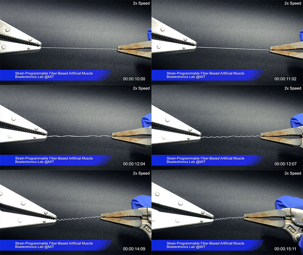
Using two different polymers bonded together, a very stretchable cyclic copolymer elastomer and a much stiffer thermoplastic polyethylene, Kanik, Örgüç and colleagues produced a fiber that, when stretched out to several times its original length, naturally forms itself into a tight coil, very similar to the tendrils that cucumbers produce. But what happened next actually came as a surprise when the researchers first experienced it. “There was a lot of serendipity in this,” Anikeeva recalls.

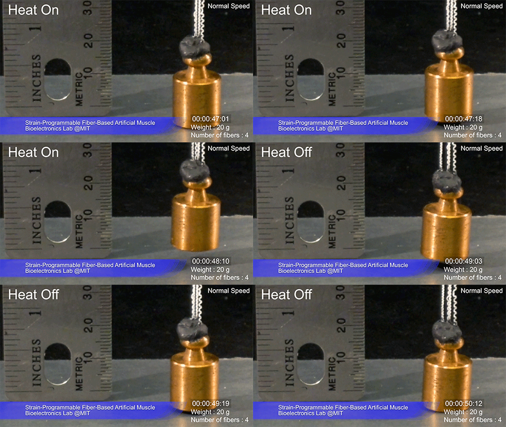
As soon as Kanik picked up the coiled fiber for the first time, the warmth of his hand alone caused the fiber to curl up more tightly. Following up on that observation, he found that even a small increase in temperature could make the coil tighten up, producing a surprisingly strong pulling force. Then, as soon as the temperature went back down, the fiber returned to its original length. In later testing, the team showed that this process of contracting and expanding could be repeated 10,000 times “and it was still going strong,” Anikeeva says.

Credit: Courtesy of the researchers

One of the reasons for that longevity, she says, is that “everything is operating under very moderate conditions,” including low activation temperatures. Just a 1-degree Celsius increase can be enough to start the fiber contraction.

The fibers can span a wide range of sizes, from a few micrometers (millionths of a meter) to a few millimeters (thousandths of a meter) in width, and can easily be manufactured in batches up to hundreds of meters long. Tests have shown that a single fiber is capable of lifting loads of up to 650 times its own weight. For these experiments on individual fibers, Örgüç and Kanik have developed dedicated, miniaturized testing setups.

Credit: Courtesy of the researchers

The degree of tightening that occurs when the fiber is heated can be “programmed” by determining how much of an initial stretch to give the fiber. This allows the material to be tuned to exactly the amount of force needed and the amount of temperature change needed to trigger that force.

The fibers are made using a fiber-drawing system, which makes it possible to incorporate other components into the fiber itself. Fiber drawing is done by creating an oversized version of the material, called a preform, which is then heated to a specific temperature at which the material becomes viscous. It can then be pulled, much like pulling taffy, to create a fiber that retains its internal structure but is a small fraction of the width of the preform.

For testing purposes, the researchers coated the fibers with meshes of conductive nanowires. These meshes can be used as sensors to reveal the exact tension experienced or exerted by the fiber. In the future, these fibers could also include heating elements such as optical fibers or electrodes, providing a way of heating it internally without having to rely on any outside heat source to activate the contraction of the “muscle.”

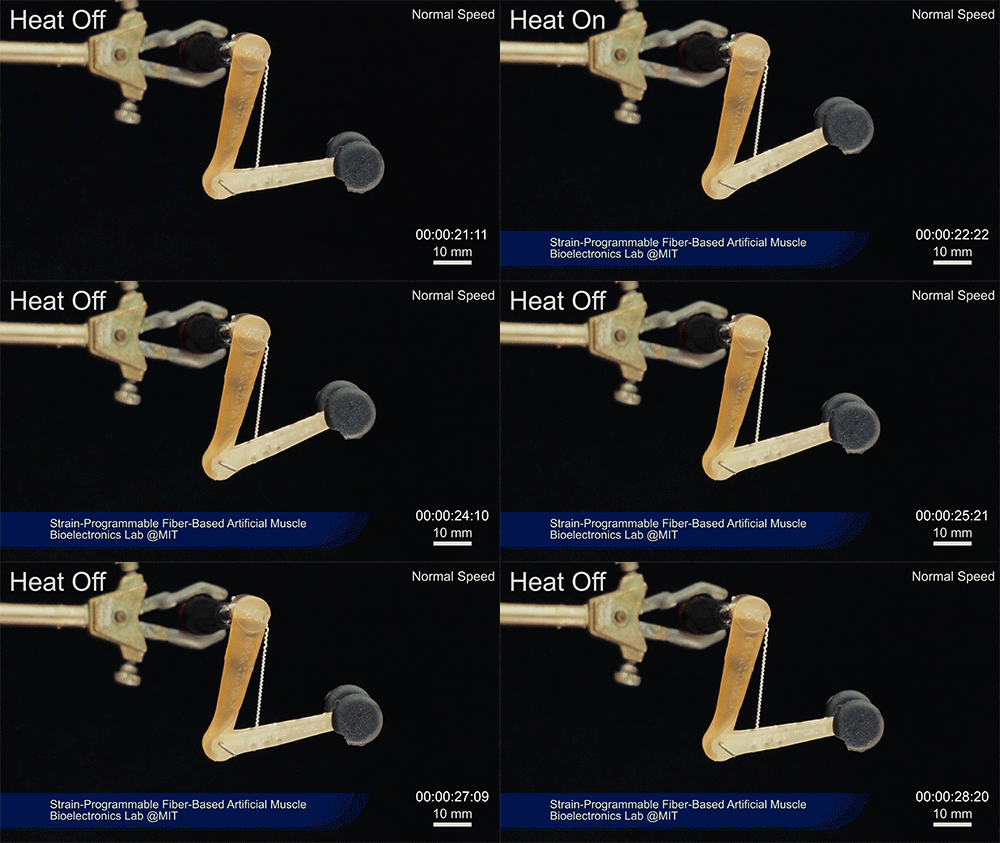
Such fibers could find uses as actuators in robotic arms, legs, or grippers, and in prosthetic limbs, where their slight weight and fast response times could provide a significant advantage.

Some prosthetic limbs today can weigh as much as 30 pounds, with much of the weight coming from actuators, which are often pneumatic or hydraulic; lighter-weight actuators could thus make life much easier for those who use prosthetics. Such fibers might also find uses in tiny biomedical devices, such as a medical robot that works by going into an artery and then being activated,” Anikeeva suggests. “We have activation times on the order of tens of milliseconds to seconds,” depending on the dimensions, she says.

To provide greater strength for lifting heavier loads, the fibers can be bundled together, much as muscle fibers are bundled in the body. The team successfully tested bundles of 100 fibers. Through the fiber drawing process, sensors could also be incorporated in the fibers to provide feedback on conditions they encounter, such as in a prosthetic limb. Örgüç says bundled muscle fibers with a closed-loop feedback mechanism could find applications in robotic systems where automated and precise control are required.

Kanik says that the possibilities for materials of this type are virtually limitless, because almost any combination of two materials with different thermal expansion rates could work, leaving a vast realm of possible combinations to explore. He adds that this new finding was like opening a new window, only to see “a bunch of other windows” waiting to be opened.

“The strength of this work is coming from its simplicity,” he says.

The team also included MIT graduate student Georgios Varnavides, postdoc Jinwoo Kim, and undergraduate students Thomas Benavides, Dani Gonzalez, and Timothy Akintlio. The work was supported by the National Institute of Neurological Disorders and Stroke and the National Science Foundation.

Bridging the gap between research and the classroom

In a moment more reminiscent of a Comic-Con event than a typical MIT symposium, Shawn Robinson, senior research associate at the University of Wisconsin at Madison, helped kick off the first-ever MIT Science of Reading event dressed in full superhero attire as Doctor Dyslexia Dude — the star of a graphic novel series he co-created to engage and encourage young readers, rooted in his own experiences as a student with dyslexia.

The event, co-sponsored by the MIT Integrated Learning Initiative (MITili) and the McGovern Institute for Brain Research at MIT, took place earlier this month and brought together researchers, educators, administrators, parents, and students to explore how scientific research can better inform educational practices and policies — equipping teachers with scientifically-based strategies that may lead to better outcomes for students.

Professor John Gabrieli, MITili director, explained the great need to focus the collective efforts of educators and researchers on literacy.

“Reading is critical to all learning and all areas of knowledge. It is the first great educational experience for all children, and can shape a child’s first sense of self,” he said. “If reading is a challenge or a burden, it affects children’s social and emotional core.”

A great divide

Reading is also a particularly important area to address because so many American students struggle with this fundamental skill. More than six out of every 10 fourth graders in the United States are not proficient readers, and changes in reading scores for fourth and eighth graders have increased only slightly since 1992, according to the National Assessment of Education Progress.

Gabrieli explained that, just as with biomedical research, where there can be a “valley of death” between basic research and clinical application, the same seems to apply to education. Although there is substantial current research aiming to better understand why students might have difficulty reading in the ways they are currently taught, the research often does not necessarily shape the practices of teachers — or how the teachers themselves are trained to teach.

This divide between the research and practical applications in the classroom might stem from a variety of factors. One issue might be the inaccessibility of research publications that are available for free to all — as well as the general need for scientific findings to be communicated in a clear, accessible, engaging way that can lead to actual implementation. Another challenge is the stark difference in pacing between scientific research and classroom teaching. While research can take years to complete and publish, teachers have classrooms full of students — all with different strengths and challenges — who urgently need to learn in real time.

Natalie Wexler, author of “The Knowledge Gap,” described some of the obstacles to getting the findings of cognitive science integrated into the classroom as matters of “head, heart, and habit.” Teacher education programs tend to focus more on some of the outdated psychological models, like Piaget’s theory of cognitive development, and less on recent cognitive science research. Teachers also have to face the emotional realities of working with their students, and might be concerned that a new approach would cause students to feel bored or frustrated. In terms of habit, some new, evidence-based approaches may be, in a practical sense, difficult for teachers to incorporate into the classroom.

“Teaching is an incredibly complex activity,” noted Wexler.

From labs to classrooms

Throughout the day, speakers and panelists highlighted some key insights gained from literacy research, along with some of the implications these might have on education.

Mark Seidenberg, professor of psychology at the University of Wisconsin at Madison and author of “Language at the Speed of Sight,” discussed studies indicating the strong connection between spoken and printed language.

“Reading depends on speech,” said Seidenberg. “Writing systems are codes for expressing spoken language … Spoken language deficits have an enormous impact on children’s reading.”

The integration of speech and reading in the brain increases with reading skill. For skilled readers, the patterns of brain activity (measured using functional magnetic resonance imaging) while comprehending spoken and written language are very similar. Becoming literate affects the neural representation of speech, and knowledge of speech affects the representation of print — thus the two become deeply intertwined.

In addition, researchers have found that the language of books, even for young children, include words and expressions that are rarely encountered in speech to children. Therefore, reading aloud to children exposes them to a broader range of linguistic expressions — including more complex ones that are usually only taught much later. Thus reading to children can be especially important, as research indicates that better knowledge of spoken language facilitates learning to read.

Although behavior and performance on tests are often used as indicators of how well a student can read, neuroscience data can now provide additional information. Neuroimaging of children and young adults identifies brain regions that are critical for integrating speech and print, and can spot differences in the brain activity of a child who might be especially at-risk for reading difficulties. Brain imaging can also show how readers’ brains respond to certain reading and comprehension tasks, and how they adapt to different circumstances and challenges.

“Brain measures can be more sensitive than behavioral measures in identifying true risk,” said Ola Ozernov-Palchik, a postdoc at the McGovern Institute.

Ozernov-Palchik hopes to apply what her team is learning in their current studies to predict reading outcomes for other children, as well as continue to investigate individual differences in dyslexia and dyslexia-risk using behavior and neuroimaging methods.

Identifying certain differences early on can be tremendously helpful in providing much-needed early interventions and tailored solutions. Many speakers noted the problem with the current “wait-to-fail” model of noticing that a child has a difficult time reading in second or third grade, and then intervening. Research suggests that earlier intervention could help the child succeed much more than later intervention.

Speakers and panelists spoke about current efforts, including Reach Every Reader (a collaboration between MITili, the Harvard Graduate School of Education, and the Florida Center for Reading Research), that seek to provide support to students by bringing together education practitioners and scientists.

“We have a lot of information, but we have the challenge of how to enact it in the real world,” said Gabrieli, noting that he is optimistic about the potential for the additional conversations and collaborations that might grow out of the discussions of the Science of Reading event. “We know a lot of things can be better and will require partnerships, but there is a path forward.”

McGovern neuroscientists develop a new model for autism

Using the genome-editing system CRISPR, researchers at MIT and in China have engineered macaque monkeys to express a gene mutation linked to autism and other neurodevelopmental disorders in humans. These monkeys show some behavioral traits and brain connectivity patterns similar to those seen in humans with these conditions.

Mouse studies of autism and other neurodevelopmental disorders have yielded drug candidates that have been tested in clinical trials, but none of them have succeeded. Many pharmaceutical companies have given up on testing such drugs because of the poor track record so far.

The new type of model, however, could help scientists to develop better treatment options for some neurodevelopmental disorders, says Guoping Feng, who is the James W. and Patricia Poitras Professor of Neuroscience, a member of MIT’s McGovern Institute for Brain Research, and one of the senior authors of the study.

“Our goal is to generate a model to help us better understand the neural biological mechanism of autism, and ultimately to discover treatment options that will be much more translatable to humans,” says Feng, who is also an institute member of the Broad Institute of MIT and Harvard and a senior scientist in the Broad’s Stanley Center for Psychiatric Research.

“We urgently need new treatment options for autism spectrum disorder, and treatments developed in mice have so far been disappointing. While the mouse research remains very important, we believe that primate genetic models will help us to develop better medicines and possibly even gene therapies for some severe forms of autism,” says Robert Desimone, the director of MIT’s McGovern Institute for Brain Research, the Doris and Don Berkey Professor of Neuroscience, and an author of the paper.

Huihui Zhou of the Shenzhen Institutes of Advanced Technology, Andy Peng Xiang of Sun Yat-Sen University, and Shihua Yang of South China Agricultural University are also senior authors of the study, which appears in the June 12 online edition of Nature. The paper’s lead authors are former MIT postdoc Yang Zhou, MIT research scientist Jitendra Sharma, Broad Institute group leader Rogier Landman, and Qiong Ke of Sun Yat-Sen University. The research team also includes Mriganka Sur, the Paul and Lilah E. Newton Professor in the Department of Brain and Cognitive Sciences and a member of MIT’s Picower Institute for Learning and Memory.

Gene variants

Scientists have identified hundreds of genetic variants associated with autism spectrum disorder, many of which individually confer only a small degree of risk. In this study, the researchers focused on one gene with a strong association, known as SHANK3. In addition to its link with autism, mutations or deletions of SHANK3 can also cause a related rare disorder called Phelan-McDermid Syndrome, whose most common characteristics include intellectual disability, impaired speech and sleep, and repetitive behaviors. The majority of these individuals are also diagnosed with autism spectrum disorder, as many of the symptoms overlap.

The protein encoded by SHANK3 is found in synapses — the junctions between brain cells that allow them to communicate with each other. It is particularly active in a part of the brain called the striatum, which is involved in motor planning, motivation, and habitual behavior. Feng and his colleagues have previously studied mice with Shank3 mutations and found that they show some of the traits associated with autism, including avoidance of social interaction and obsessive, repetitive behavior.

Although mouse studies can provide a great deal of information on the molecular underpinnings of disease, there are drawbacks to using them to study neurodevelopmental disorders, Feng says. In particular, mice lack the highly developed prefrontal cortex that is the seat of many uniquely primate traits, such as making decisions, sustaining focused attention, and interpreting social cues, which are often affected by brain disorders.

The recent development of the CRISPR genome-editing technique offered a way to engineer gene variants into macaque monkeys, which has previously been very difficult to do. CRISPR consists of a DNA-cutting enzyme called Cas9 and a short RNA sequence that guides the enzyme to a specific area of the genome. It can be used to disrupt genes or to introduce new genetic sequences at a particular location.

Members of the research team based in China, where primate reproductive technology is much more advanced than in the United States, injected the CRISPR components into fertilized macaque eggs, producing embryos that carried the Shank3 mutation.

Researchers at MIT, where much of the data was analyzed, found that the macaques with Shank3 mutations showed behavioral patterns similar to those seen in humans with the mutated gene. They tended to wake up frequently during the night, and they showed repetitive behaviors. They also engaged in fewer social interactions than other macaques.

Magnetic resonance imaging (MRI) scans also revealed similar patterns to humans with autism spectrum disorder. Neurons showed reduced functional connectivity in the striatum as well as the thalamus, which relays sensory and motor signals and is also involved in sleep regulation. Meanwhile, connectivity was strengthened in other regions, including the sensory cortex.

Michael Platt, a professor of neuroscience and psychology at the University of Pennsylvania, says the macaque models should help to overcome some of the limitations of studying neurological disorders in mice, whose behavioral symptoms and underlying neurobiology are often different from those seen in humans.

“Because the macaque model shows a much more complete recapitulation of the human behavioral phenotype, I think we should stand a much greater chance of identifying the degree to which any particular therapy, whether it’s a drug or any other intervention, addresses the core symptoms,” says Platt, who was not involved in the study.

Drug development

Within the next year, the researchers hope to begin testing treatments that may affect autism-related symptoms. They also hope to identify biomarkers, such as the distinctive functional brain connectivity patterns seen in MRI scans, that would help them to evaluate whether drug treatments are having an effect.

A similar approach could also be useful for studying other types of neurological disorders caused by well-characterized genetic mutations, such as Rett Syndrome and Fragile X Syndrome. Fragile X is the most common inherited form of intellectual disability in the world, affecting about 1 in 4,000 males and 1 in 8,000 females. Rett Syndrome, which is more rare and almost exclusively affects girls, produces severe impairments in language and motor skills and can also cause seizures and breathing problems.

“Given the limitations of mouse models, patients really need this kind of advance to bring them hope,” Feng says. “We don’t know whether this will succeed in developing treatments, but we will see in the next few years how this can help us to translate some of the findings from the lab to the clinic.”

The research was funded, in part, by the Shenzhen Overseas Innovation Team Project, the Guangdong Innovative and Entrepreneurial Research Team Program, the National Key R&D Program of China, the External Cooperation Program of the Chinese Academy of Sciences, the Patrick J. McGovern Foundation, the National Natural Science Foundation of China, the Shenzhen Science, Technology Commission, the James and Patricia Poitras Center for Psychiatric Disorders Research at the McGovern Institute at MIT, the Stanley Center for Psychiatric Research at the Broad Institute of MIT and Harvard, and the Hock E. Tan and K. Lisa Yang Center for Autism Research at the McGovern Institute at MIT. The research facilities in China where the primate work was conducted are accredited by AAALAC International, a private, nonprofit organization that promotes the humane treatment of animals in science through voluntary accreditation and assessment programs.

How we tune out distractions

Imagine trying to focus on a friend’s voice at a noisy party, or blocking out the phone conversation of the person sitting next to you on the bus while you try to read. Both of these tasks require your brain to somehow suppress the distracting signal so you can focus on your chosen input.

MIT neuroscientists have now identified a brain circuit that helps us to do just that. The circuit they identified, which is controlled by the prefrontal cortex, filters out unwanted background noise or other distracting sensory stimuli. When this circuit is engaged, the prefrontal cortex selectively suppresses sensory input as it flows into the thalamus, the site where most sensory information enters the brain.

“This is a fundamental operation that cleans up all the signals that come in, in a goal-directed way,” says Michael Halassa, an assistant professor of brain and cognitive sciences, a member of MIT’s McGovern Institute for Brain Research, and the senior author of the study.

The researchers are now exploring whether impairments of this circuit may be involved in the hypersensitivity to noise and other stimuli that is often seen in people with autism.

Miho Nakajima, an MIT postdoc, is the lead author of the paper, which appears in the June 12 issue of Neuron. Research scientist L. Ian Schmitt is also an author of the paper.

Shifting attention

Our brains are constantly bombarded with sensory information, and we are able to tune out much of it automatically, without even realizing it. Other distractions that are more intrusive, such as your seatmate’s phone conversation, require a conscious effort to suppress.

In a 2015 paper, Halassa and his colleagues explored how attention can be consciously shifted between different types of sensory input, by training mice to switch their focus between a visual and auditory cue. They found that during this task, mice suppress the competing sensory input, allowing them to focus on the cue that will earn them a reward.

This process appeared to originate in the prefrontal cortex (PFC), which is critical for complex cognitive behavior such as planning and decision-making. The researchers also found that a part of the thalamus that processes vision was inhibited when the animals were focusing on sound cues. However, there are no direct physical connections from the prefrontal cortex to the sensory thalamus, so it was unclear exactly how the PFC was exerting this control, Halassa says.

In the new study, the researchers again trained mice to switch their attention between visual and auditory stimuli, then mapped the brain connections that were involved. They first examined the outputs of the PFC that were essential for this task, by systematically inhibiting PFC projection terminals in every target. This allowed them to discover that the PFC connection to a brain region known as the striatum is necessary to suppress visual input when the animals are paying attention to the auditory cue.

Further mapping revealed that the striatum then sends input to a region called the globus pallidus, which is part of the basal ganglia. The basal ganglia then suppress activity in the part of the thalamus that processes visual information.

Using a similar experimental setup, the researchers also identified a parallel circuit that suppresses auditory input when animals pay attention to the visual cue. In that case, the circuit travels through parts of the striatum and thalamus that are associated with processing sound, rather than vision.

The findings offer some of the first evidence that the basal ganglia, which are known to be critical for planning movement, also play a role in controlling attention, Halassa says.

“What we realized here is that the connection between PFC and sensory processing at this level is mediated through the basal ganglia, and in that sense, the basal ganglia influence control of sensory processing,” he says. “We now have a very clear idea of how the basal ganglia can be involved in purely attentional processes that have nothing to do with motor preparation.”

Noise sensitivity

The researchers also found that the same circuits are employed not only for switching between different types of sensory input such as visual and auditory stimuli, but also for suppressing distracting input within the same sense — for example, blocking out background noise while focusing on one person’s voice.

The team also showed that when the animals are alerted that the task is going to be noisy, their performance actually improves, as they use this circuit to focus their attention.

“This study uses a dazzling array of techniques for neural circuit dissection to identify a distributed pathway, linking the prefrontal cortex to the basal ganglia to the thalamic reticular nucleus, that allows the mouse brain to enhance relevant sensory features and suppress distractors at opportune moments,” says Daniel Polley, an associate professor of otolaryngology at Harvard Medical School, who was not involved in the research. “By paring down the complexities of the sensory stimulus only to its core relevant features in the thalamus — before it reaches the cortex — our cortex can more efficiently encode just the essential features of the sensory world.”

Halassa’s lab is now doing similar experiments in mice that are genetically engineered to develop symptoms similar to those of people with autism. One common feature of autism spectrum disorder is hypersensitivity to noise, which could be caused by impairments of this brain circuit, Halassa says. He is now studying whether boosting the activity of this circuit might reduce sensitivity to noise.

“Controlling noise is something that patients with autism have trouble with all the time,” he says. “Now there are multiple nodes in the pathway that we can start looking at to try to understand this.”

The research was funded by the National Institutes of Mental Health, the National Institute of Neurological Disorders and Stroke, the Simons Foundation, the Alfred P. Sloan Foundation, the Esther A. and Joseph Klingenstein Fund, and the Human Frontier Science Program.

Antenna-like inputs unexpectedly active in neural computation

Most neurons have many branching extensions called dendrites that receive input from thousands of other neurons. Dendrites aren’t just passive information-carriers, however. According to a new study from MIT, they appear to play a surprisingly large role in neurons’ ability to translate incoming signals into electrical activity.

Neuroscientists had previously suspected that dendrites might be active only rarely, under specific circumstances, but the MIT team found that dendrites are nearly always active when the main cell body of the neuron is active.

“It seems like dendritic spikes are an intrinsic feature of how neurons in our brain can compute information. They’re not a rare event,” says Lou Beaulieu-Laroche, an MIT graduate student and the lead author of the study. “All the neurons that we looked at had these dendritic spikes, and they had dendritic spikes very frequently.”

The findings suggest that the role of dendrites in the brain’s computational ability is much larger than had previously been thought, says Mark Harnett, who is the Fred and Carole Middleton Career Development Assistant Professor of Brain and Cognitive Sciences, a member of the McGovern Institute for Brain Research, and the senior author of the paper.

“It’s really quite different than how the field had been thinking about this,” he says. “This is evidence that dendrites are actively engaged in producing and shaping the outputs of neurons.”

Graduate student Enrique Toloza and technical associate Norma Brown are also authors of the paper, which appears in Neuron on June 6.

“A far-flung antenna”

Dendrites receive input from many other neurons and carry those signals to the cell body, also called the soma. If stimulated enough, a neuron fires an action potential — an electrical impulse that spreads to other neurons. Large networks of these neurons communicate with each other to perform complex cognitive tasks such as producing speech.

Through imaging and electrical recording, neuroscientists have learned a great deal about the anatomical and functional differences between different types of neurons in the brain’s cortex, but little is known about how they incorporate dendritic inputs and decide whether to fire an action potential. Dendrites give neurons their characteristic branching tree shape, and the size of the “dendritic arbor” far exceeds the size of the soma.

“It’s an enormous, far-flung antenna that’s listening to thousands of synaptic inputs distributed in space along that branching structure from all the other neurons in the network,” Harnett says.

Some neuroscientists have hypothesized that dendrites are active only rarely, while others thought it possible that dendrites play a more central role in neurons’ overall activity. Until now, it has been difficult to test which of these ideas is more accurate, Harnett says.

To explore dendrites’ role in neural computation, the MIT team used calcium imaging to simultaneously measure activity in both the soma and dendrites of individual neurons in the visual cortex of the brain. Calcium flows into neurons when they are electrically active, so this measurement allowed the researchers to compare the activity of dendrites and soma of the same neuron. The imaging was done while mice performed simple tasks such as running on a treadmill or watching a movie.

Unexpectedly, the researchers found that activity in the soma was highly correlated with dendrite activity. That is, when the soma of a particular neuron was active, the dendrites of that neuron were also active most of the time. This was particularly surprising because the animals weren’t performing any kind of cognitively demanding task, Harnett says.

“They weren’t engaged in a task where they had to really perform and call upon cognitive processes or memory. This is pretty simple, low-level processing, and already we have evidence for active dendritic processing in almost all the neurons,” he says. “We were really surprised to see that.”

Evolving patterns

The researchers don’t yet know precisely how dendritic input contributes to neurons’ overall activity, or what exactly the neurons they studied are doing.

“We know that some of those neurons respond to some visual stimuli, but we don’t necessarily know what those individual neurons are representing. All we can say is that whatever the neuron is representing, the dendrites are actively participating in that,” Beaulieu-Laroche says.

While more work remains to determine exactly how the activity in the dendrites and the soma are linked, “it is these tour-de-force in vivo measurements that are critical for explicitly testing hypotheses regarding electrical signaling in neurons,” says Marla Feller, a professor of neurobiology at the University of California at Berkeley, who was not involved in the research.

The MIT team now plans to investigate how dendritic activity contributes to overall neuronal function by manipulating dendrite activity and then measuring how it affects the activity of the cell body, Harnett says. They also plan to study whether the activity patterns they observed evolve as animals learn a new task.

“One hypothesis is that dendritic activity will actually sharpen up for representing features of a task you taught the animals, and all the other dendritic activity, and all the other somatic activity, is going to get dampened down in the rest of the cortical cells that are not involved,” Harnett says.

The research was funded by the Natural Sciences and Engineering Research Council of Canada and the U.S. National Institutes of Health.

Putting vision models to the test

MIT neuroscientists have performed the most rigorous testing yet of computational models that mimic the brain’s visual cortex.

Using their current best model of the brain’s visual neural network, the researchers designed a new way to precisely control individual neurons and populations of neurons in the middle of that network. In an animal study, the team then showed that the information gained from the computational model enabled them to create images that strongly activated specific brain neurons of their choosing.

The findings suggest that the current versions of these models are similar enough to the brain that they could be used to control brain states in animals. The study also helps to establish the usefulness of these vision models, which have generated vigorous debate over whether they accurately mimic how the visual cortex works, says James DiCarlo, the head of MIT’s Department of Brain and Cognitive Sciences, an investigator in the McGovern Institute for Brain Research and the Center for Brains, Minds, and Machines, and the senior author of the study.

“People have questioned whether these models provide understanding of the visual system,” he says. “Rather than debate that in an academic sense, we showed that these models are already powerful enough to enable an important new application. Whether you understand how the model works or not, it’s already useful in that sense.”

MIT postdocs Pouya Bashivan and Kohitij Kar are the lead authors of the paper, which appears in the May 2 online edition of Science.

Neural control

Over the past several years, DiCarlo and others have developed models of the visual system based on artificial neural networks. Each network starts out with an arbitrary architecture consisting of model neurons, or nodes, that can be connected to each other with different strengths, also called weights.

The researchers then train the models on a library of more than 1 million images. As the researchers show the model each image, along with a label for the most prominent object in the image, such as an airplane or a chair, the model learns to recognize objects by changing the strengths of its connections.

It’s difficult to determine exactly how the model achieves this kind of recognition, but DiCarlo and his colleagues have previously shown that the “neurons” within these models produce activity patterns very similar to those seen in the animal visual cortex in response to the same images.

In the new study, the researchers wanted to test whether their models could perform some tasks that previously have not been demonstrated. In particular, they wanted to see if the models could be used to control neural activity in the visual cortex of animals.

“So far, what has been done with these models is predicting what the neural responses would be to other stimuli that they have not seen before,” Bashivan says. “The main difference here is that we are going one step further and using the models to drive the neurons into desired states.”

To achieve this, the researchers first created a one-to-one map of neurons in the brain’s visual area V4 to nodes in the computational model. They did this by showing images to animals and to the models, and comparing their responses to the same images. There are millions of neurons in area V4, but for this study, the researchers created maps for subpopulations of five to 40 neurons at a time.

“Once each neuron has an assignment, the model allows you to make predictions about that neuron,” DiCarlo says.

The researchers then set out to see if they could use those predictions to control the activity of individual neurons in the visual cortex. The first type of control, which they called “stretching,” involves showing an image that will drive the activity of a specific neuron far beyond the activity usually elicited by “natural” images similar to those used to train the neural networks.

The researchers found that when they showed animals these “synthetic” images, which are created by the models and do not resemble natural objects, the target neurons did respond as expected. On average, the neurons showed about 40 percent more activity in response to these images than when they were shown natural images like those used to train the model. This kind of control has never been reported before.

“That they succeeded in doing this is really amazing. It’s as if, for that neuron at least, its ideal image suddenly leaped into focus. The neuron was suddenly presented with the stimulus it had always been searching for,” says Aaron Batista, an associate professor of bioengineering at the University of Pittsburgh, who was not involved in the study. “This is a remarkable idea, and to pull it off is quite a feat. It is perhaps the strongest validation so far of the use of artificial neural networks to understand real neural networks.”

In a similar set of experiments, the researchers attempted to generate images that would drive one neuron maximally while also keeping the activity in nearby neurons very low, a more difficult task. For most of the neurons they tested, the researchers were able to enhance the activity of the target neuron with little increase in the surrounding neurons.

“A common trend in neuroscience is that experimental data collection and computational modeling are executed somewhat independently, resulting in very little model validation, and thus no measurable progress. Our efforts bring back to life this ‘closed loop’ approach, engaging model predictions and neural measurements that are critical to the success of building and testing models that will most resemble the brain,” Kar says.

Measuring accuracy

The researchers also showed that they could use the model to predict how neurons of area V4 would respond to synthetic images. Most previous tests of these models have used the same type of naturalistic images that were used to train the model. The MIT team found that the models were about 54 percent accurate at predicting how the brain would respond to the synthetic images, compared to nearly 90 percent accuracy when the natural images are used.

“In a sense, we’re quantifying how accurate these models are at making predictions outside the domain where they were trained,” Bashivan says. “Ideally the model should be able to predict accurately no matter what the input is.”

The researchers now hope to improve the models’ accuracy by allowing them to incorporate the new information they learn from seeing the synthetic images, which was not done in this study.

This kind of control could be useful for neuroscientists who want to study how different neurons interact with each other, and how they might be connected, the researchers say. Farther in the future, this approach could potentially be useful for treating mood disorders such as depression. The researchers are now working on extending their model to the inferotemporal cortex, which feeds into the amygdala, which is involved in processing emotions.

“If we had a good model of the neurons that are engaged in experiencing emotions or causing various kinds of disorders, then we could use that model to drive the neurons in a way that would help to ameliorate those disorders,” Bashivan says.

The research was funded by the Intelligence Advanced Research Projects Agency, the MIT-IBM Watson AI Lab, the National Eye Institute, and the Office of Naval Research.