The National Institutes of Health (NIH) has awarded grants to MIT’s Ariel Furst and Fan Wang, through its High-Risk, High-Reward Research program. The NIH High-Risk, High-Reward Research program awarded 85 new research grants to support exceptionally creative scientists pursuing highly innovative behavioral and biomedical research projects.
Ariel Furst was selected as the recipient of the NIH Director’s New Innovator Award, which has supported unusually innovative research since 2007. Recipients are early-career investigators who are within 10 years of their final degree or clinical residency and have not yet received a research project grant or equivalent NIH grant.
Furst, the Paul M. Cook Career Development Assistant Professor of Chemical Engineering at MIT, invents technologies to improve human and environmental health by increasing equitable access to resources. Her lab develops transformative technologies to solve problems related to health care and sustainability by harnessing the inherent capabilities of biological molecules and cells. She is passionate about STEM outreach and increasing the participation of underrepresented groups in engineering.
After completing her PhD at Caltech, where she developed noninvasive diagnostics for colorectal cancer, Furst became an A. O. Beckman Postdoctoral Fellow at the University of California at Berkeley. There she developed sensors to monitor environmental pollutants. In 2022, Furst was awarded the MIT UROP Outstanding Faculty Mentor Award for her work with undergraduate researchers. She is a now a 2023 Marion Milligan Mason Awardee, a CIFAR Azrieli Global Scholar for Bio-Inspired Solar Energy, and an ARO Early Career Grantee. She is also a co-founder of the regenerative agriculture company, Seia Bio.
Fan Wang received the Pioneer Award, which has been challenging researchers at all career levels to pursue new directions and develop groundbreaking, high impact approaches to a broad area of biomedical and behavioral sciences since 2004.
Wang, a professor in the Department of Brain and Cognitive Sciences and an investigator in the McGovern Institute for Brain Research, is uncovering the neural circuit mechanisms that govern bodily sensations, like touch, pain, and posture, as well as the mechanisms that control sensorimotor behaviors. Researchers in the Wang lab aim to generate an integrated understanding of the sensation-perception-action process, hoping to find better treatments for diseases like chronic pain, addiction, and movement disorders. Wang’s lab uses genetic, viral, in vivo large-scale electrophysiology and imaging techniques to gain traction in these pursuits.
Wang obtained her PhD at Columbia University, working with Professor Richard Axel. She conducted her postdoctoral work at Stanford University with Mark Tessier-Lavigne, and then subsequently joined Duke University as faculty in 2003. Wang was later appointed as the Morris N. Broad Distinguished Professor of Neurobiology at the Duke University School of Medicine. In January 2023, she joined the faculty of the MIT School of Science and the McGovern Institute.
The High-Risk, High-Reward Research program is funded through the NIH Common Fund, which supports a series of exceptionally high-impact programs that cross NIH Institutes and Centers.
“The HRHR program is a pillar for innovation here at NIH, providing support to transformational research, with advances in biomedical and behavioral science,” says Robert W. Eisinger, acting director of the Division of Program Coordination, Planning, and Strategic Initiatives, which oversees the NIH Common Fund. “These awards align with the Common Fund’s mandate to support science expected to have exceptionally high and broadly applicable impact.”
NIH issued eight Pioneer Awards, 58 New Innovator Awards, six Transformative Research Awards, and 13 Early Independence Awards in 2023. Funding for the awards comes from the NIH Common Fund; the National Institute of General Medical Sciences; the National Institute of Mental Health; the National Library of Medicine; the National Institute on Aging; the National Heart, Lung, and Blood Institute; and the Office of Dietary Supplements.
Human sensory systems are very good at recognizing objects that we see or words that we hear, even if the object is upside down or the word is spoken by a voice we’ve never heard.
Computational models known as deep neural networks can be trained to do the same thing, correctly identifying an image of a dog regardless of what color its fur is, or a word regardless of the pitch of the speaker’s voice. However, a new study from MIT neuroscientists has found that these models often also respond the same way to images or words that have no resemblance to the target.
When these neural networks were used to generate an image or a word that they responded to in the same way as a specific natural input, such as a picture of a bear, most of them generated images or sounds that were unrecognizable to human observers. This suggests that these models build up their own idiosyncratic “invariances” — meaning that they respond the same way to stimuli with very different features.
The findings offer a new way for researchers to evaluate how well these models mimic the organization of human sensory perception, says Josh McDermott, an associate professor of brain and cognitive sciences at MIT and a member of MIT’s McGovern Institute for Brain Research and Center for Brains, Minds, and Machines.
“This paper shows that you can use these models to derive unnatural signals that end up being very diagnostic of the representations in the model,” says McDermott, who is the senior author of the study. “This test should become part of a battery of tests that we as a field are using to evaluate models.”
Jenelle Feather PhD ’22, who is now a research fellow at the Flatiron Institute Center for Computational Neuroscience, is the lead author of the open-access paper, which appears today in Nature Neuroscience. Guillaume Leclerc, an MIT graduate student, and Aleksander Mądry, the Cadence Design Systems Professor of Computing at MIT, are also authors of the paper.
Different perceptions
In recent years, researchers have trained deep neural networks that can analyze millions of inputs (sounds or images) and learn common features that allow them to classify a target word or object roughly as accurately as humans do. These models are currently regarded as the leading models of biological sensory systems.
It is believed that when the human sensory system performs this kind of classification, it learns to disregard features that aren’t relevant to an object’s core identity, such as how much light is shining on it or what angle it’s being viewed from. This is known as invariance, meaning that objects are perceived to be the same even if they show differences in those less important features.
“Classically, the way that we have thought about sensory systems is that they build up invariances to all those sources of variation that different examples of the same thing can have,” Feather says. “An organism has to recognize that they’re the same thing even though they show up as very different sensory signals.”
The researchers wondered if deep neural networks that are trained to perform classification tasks might develop similar invariances. To try to answer that question, they used these models to generate stimuli that produce the same kind of response within the model as an example stimulus given to the model by the researchers.
They term these stimuli “model metamers,” reviving an idea from classical perception research whereby stimuli that are indistinguishable to a system can be used to diagnose its invariances. The concept of metamers was originally developed in the study of human perception to describe colors that look identical even though they are made up of different wavelengths of light.
To their surprise, the researchers found that most of the images and sounds produced in this way looked and sounded nothing like the examples that the models were originally given. Most of the images were a jumble of random-looking pixels, and the sounds resembled unintelligible noise. When researchers showed the images to human observers, in most cases the humans did not classify the images synthesized by the models in the same category as the original target example.
“They’re really not recognizable at all by humans. They don’t look or sound natural and they don’t have interpretable features that a person could use to classify an object or word,” Feather says.
The findings suggest that the models have somehow developed their own invariances that are different from those found in human perceptual systems. This causes the models to perceive pairs of stimuli as being the same despite their being wildly different to a human.
Idiosyncratic invariances
The researchers found the same effect across many different vision and auditory models. However, each of these models appeared to develop their own unique invariances. When metamers from one model were shown to another model, the metamers were just as unrecognizable to the second model as they were to human observers.
“The key inference from that is that these models seem to have what we call idiosyncratic invariances,” McDermott says. “They have learned to be invariant to these particular dimensions in the stimulus space, and it’s model-specific, so other models don’t have those same invariances.”
The researchers also found that they could induce a model’s metamers to be more recognizable to humans by using an approach called adversarial training. This approach was originally developed to combat another limitation of object recognition models, which is that introducing tiny, almost imperceptible changes to an image can cause the model to misrecognize it.
The researchers found that adversarial training, which involves including some of these slightly altered images in the training data, yielded models whose metamers were more recognizable to humans, though they were still not as recognizable as the original stimuli. This improvement appears to be independent of the training’s effect on the models’ ability to resist adversarial attacks, the researchers say.
“This particular form of training has a big effect, but we don’t really know why it has that effect,” Feather says. “That’s an area for future research.”
Analyzing the metamers produced by computational models could be a useful tool to help evaluate how closely a computational model mimics the underlying organization of human sensory perception systems, the researchers say.
“This is a behavioral test that you can run on a given model to see whether the invariances are shared between the model and human observers,” Feather says. “It could also be used to evaluate how idiosyncratic the invariances are within a given model, which could help uncover potential ways to improve our models in the future.”
The research was funded by the National Science Foundation, the National Institutes of Health, a Department of Energy Computational Science Graduate Fellowship, and a Friends of the McGovern Institute Fellowship.
Many studies have found that practicing mindfulness — defined as cultivating an open-minded attention to the present moment — has benefits for children. Children who receive mindfulness training at school have demonstrated improvements in attention and behavior, as well as greater mental health.
When the Covid-19 pandemic began in 2020, sending millions of students home from school, a group of MIT researchers wondered if remote, app-based mindfulness practices could offer similar benefits. In a study conducted during 2020 and 2021, they report that children who used a mindfulness app at home for 40 days showed improvements in several aspects of mental health, including reductions in stress and negative emotions such as loneliness and fear.
The findings suggest that remote, app-based mindfulness interventions, which could potentially reach a larger number of children than school-based approaches, could offer mental health benefits, the researchers say.
“There is growing and compelling scientific evidence that mindfulness can support mental well-being and promote mental health in diverse children and adults,” says John Gabrieli, the Grover Hermann Professor of Health Sciences and Technology, a professor of brain and cognitive sciences at MIT, and the senior author of the study, which appears this week in the journal Mindfulness.
Researchers in Gabrieli’s lab also recently reported that children who showed higher levels of mindfulness were more emotionally resilient to the negative impacts of the Covid-19 pandemic.
“To some extent, the impact of Covid is out of your control as an individual, but your ability to respond to it and to interpret it may be something that mindfulness can help with,” says MIT graduate student Isaac Treves, who is the lead author of both studies.
Pandemic resilience
After the pandemic began in early 2020, Gabrieli’s lab decided to investigate the effects of mindfulness on children who had to leave school and isolate from friends. In a study that appeared in the journal PLOS One in July, the researchers explored whether mindfulness could boost children’s resilience to negative emotions that the pandemic generated, such as frustration and loneliness.
Working with students between 8 and 10 years old, the researchers measured the children’s mindfulness using a standardized assessment that captures their tendency to blame themselves, ruminate on negative thoughts, and suppress their feelings.
The researchers also asked the children questions about how much the pandemic had affected different aspects of their lives, as well as questions designed to assess their levels of anxiety, depression, stress, and negative emotions such as worry or fear.
Among children who showed the highest levels of mindfulness, there was no correlation between how much the pandemic impacted them and negative feelings. However, in children with lower levels of mindfulness, there was a strong correlation between Covid-19 impact and negative emotions.
The children in this study did not receive any kind of mindfulness training, so their responses reflect their tendency to be mindful at the time they answered the researchers’ questions. The findings suggest that children with higher levels of mindfulness were less likely to get caught up in negative emotions or blame themselves for the negative things they experienced during the pandemic.
“This paper was our best attempt to look at mindfulness specifically in the context of Covid and to think about what are the factors that may help children adapt to the changing circumstances,” Treves says. “The takeaway is not that we shouldn’t worry about pandemics because we can just help the kids with mindfulness. People are able to be resilient when they’re in systems that support them, and in families that support them.”
Remote interventions
The researchers then built on that study by exploring whether a remote, app-based intervention could effectively increase mindfulness and improve mental health. Researchers in Gabrieli’s lab have previously shown that students who received mindfulness training in middle school showed better academic performance, received fewer suspensions, and reported less stress than those who did not receive the training.
For the new study, reported today in Mindfulness, the researchers worked with the same children they had recruited for the PLOS One study and divided them into three groups of about 80 students each.
One group received mindfulness training through an app created by Inner Explorer, a nonprofit that also develops school-based meditation programs. Those children were instructed to engage in mindfulness training five days a week, including relaxation exercises, breathing exercises, and other forms of meditation.
For comparison purposes, the other two groups were asked to use an app for listening to audiobooks (not related to mindfulness). One group was simply given the audiobook app and encouraged to listen at their own pace, while the other group also had weekly one-on-one virtual meetings with a facilitator.
At the beginning and end of the study, the researchers evaluated each participant’s levels of mindfulness, along with measures of mental health such as anxiety, stress, and depression. They found that in all three groups, mental health improved over the course of the eight-week study, and each group also showed increases in mindfulness and prosociality (engaging in helpful behavior).
Additionally, children in the mindfulness group showed some improvements that the other groups didn’t, including a more significant decrease in stress. They also found that parents in the mindfulness group reported that their children experienced more significant decreases in negative emotions such as anger and sadness. Students who practiced the mindfulness exercises the most days showed the greatest benefits.
The researchers were surprised to see that there were no significant differences in measures of anxiety and depression between the mindfulness group and audiobook groups; they hypothesize that may be because students who interacted with a facilitator in one of the audiobook groups also experienced beneficial effects on their mental health.
Overall, the findings suggest that there is value in remote, app-based mindfulness training, especially if children engage with the exercises consistently and receive encouragement from parents, the researchers say. Apps also offer the ability to reach a larger number of children than school-based programs, which require more training and resources.
“There are a lot of great ways to incorporate mindfulness training into schools, but in general, it’s more resource-intensive than having people download an app. So, in terms of pure scalability and cost-effectiveness, apps are useful,” Treves says. “Another good thing about apps is that the kids can go at their own pace and repeat practices that they like, so there’s more freedom of choice.”
The research was funded by the Chan Zuckerberg Initiative as part of the Reach Every Reader Project, the National Institutes of Health, and the National Science Foundation.
The National Academy of Medicine announced the election of 100 new members to join their esteemed ranks in 2023, among them five MIT faculty members and seven additional affiliates.
MIT professors Daniel Anderson, Regina Barzilay, Guoping Feng, Darrell Irvine, and Morgen Shen were among the new members. Justin Hanes PhD ’96, Said Ibrahim MBA ’16, and Jennifer West ’92, along with three former students in the Harvard-MIT Program in Health Sciences and Technology (HST) — Michael Chiang, Siddhartha Mukherjee, and Robert Vonderheide — were also elected, as was Yi Zhang, an associate member of The Broad Institute of MIT and Harvard.
Election to the academy is considered one of the highest honors in the fields of health and medicine and recognizes individuals who have demonstrated outstanding professional achievement and commitment to service, the academy noted in announcing the election of its new members.
MIT faculty
Daniel G. Anderson, professor in the Department of Chemical Engineering and the Institute for Medical Engineering and Science, was elected “for pioneering the area of non-viral gene therapy and cellular delivery. His work has resulted in fundamental scientific advances; over 500 papers, patents, and patent applications; and the creation of companies, products, and technologies that are now in the clinic.” Anderson is an affiliate of the Broad Institute of MIT and Harvard and of the Ragon Institute at MGH, MIT and Harvard.
Regina Barzilay, the School of Engineering Distinguished Professor for AI and Health within the Department of Electrical Engineering and Computer Science at MIT, was elected “for the development of machine learning tools that have been transformational for breast cancer screening and risk assessment, and for the development of molecular design tools broadly utilized for drug discovery.” Barzilay is the AI faculty lead within the MIT Abdul Latif Jameel Clinic for Machine Learning in Health and an affiliate of the Computer Science and Artificial Intelligence Laboratory and Institute for Medical Engineering and Science.
Guoping Feng, the associate director of the McGovern Institute for Brain Research, James W. (1963) and Patricia T. Professor of Neuroscience in MIT’s Department of Brain and Cognitive Sciences, and an affiliate of the Broad Institute of MIT and Harvard, was elected “for his breakthrough discoveries regarding the pathological mechanisms of neurodevelopmental and psychiatric disorders, providing foundational knowledges and molecular targets for developing effective therapeutics for mental illness such as OCD, ASD, and ADHD.”
Darrell J. Irvine ’00, the Underwood-Prescott Professor of Biological Engineering and Materials Science at MIT and a member of the Koch Institute for Integrative Cancer Research, was elected “for the development of novel methods for delivery of immunotherapies and vaccines for cancer and infectious diseases.”
Morgan Sheng, professor of neuroscience in the Department of Brain and Cognitive Sciences, with affiliations in the McGovern Institute and The Picower Institute for Learning and Memory at MIT, as well as the Broad Institute of MIT and Harvard, was elected “for transforming the understanding of excitatory synapses. He revealed the postsynaptic density as a protein network controlling synaptic signaling and morphology; established the paradigm of signaling complexes organized by PDZ scaffolds; and pioneered the concept of localized regulation of mitochondria, apoptosis, and complement for targeted synapse elimination.”
Additional MIT affiliates
Michael F. Chiang, a former student in the Harvard-MIT Program in Health Sciences and Technology (HST) who is now director of the National Eye Institute of the National Institutes of Health, was honored “for pioneering applications of biomedical informatics to ophthalmology in artificial intelligence, telehealth, pediatric retinal disease, electronic health records, and data science, including methodological and diagnostic advances in AI for pediatric retinopathy of prematurity, and for contributions to developing and implementing the largest ambulatory care registry in the United States.”
Justin Hanes PhD ’96, who earned his PhD from the MIT Department of Chemical Engineering and is now a professor at Johns Hopkins University, was honored “for pioneering discoveries and inventions of innovative drug delivery technologies, especially mucosal, ocular, and central nervous system drug delivery systems; and for international leadership in research and education at the interface of engineering, medicine, and entrepreneurship, leading to clinical translation of drug delivery technologies.”
Said Ibrahim MBA ’16, a graduate of the MIT Sloan School of Management who is now a senior vice president and chair, department of medicine at the Zucker School of Medicine at Hofstra/Northwell, was honored for influential “health services research on racial disparities in elective joint replacement that has provided a national model for advancing health equity research beyond the identification of inequities and toward their remediation, and for his research that has been leveraged to engage diverse and innovative emerging scholars.”
Siddhartha Mukherjee, a former student in HST who is now an associate professor of medicine at Columbia University School of Medicine, was honored “for contributing important research in the immunotherapy of myeloid malignancies, such as acute myeloid leukemia, for establishing international centers for immunotherapy for childhood cancers, and for the discovery of tissue-resident stem cells.”
Robert H. Vonderheide, a former student in HST who is now a professor and vice dean at the Perelman School of Medicine and vice president of cancer programs at the University of Pennsylvania Health System, was honored “for developing immune combination therapies for patients with pancreatic cancer by driving proof-of-concept from lab to clinic, then leading national, randomized clinical trials for therapy, maintenance, and interception; and for improving access of minority individuals to clinical trials while directing an NCI comprehensive cancer center.”
Jennifer West ’92, a graduate of the MIT Department of Chemical Engineering who is now a professor of biomedical engineering and dean of the School of Engineering and Applied Science at the University of Virginia at Charlottesville, was honored “for the invention, development, and translation of novel biomaterials including bioactive, photopolymerizable hydrogels and theranostic nanoparticles.”
Yi Zhang, associate member of the Broad Institute, was honored “for making fundamental contributions to the epigenetics field through systematic identification and characterization of chromatin modifying enzymes, including EZH2, JmjC, and Tet. His proof-of-principle work on EZH2 inhibitors led to the founding of Epizyme and eventual making of tazemetostat, a drug approved for epithelioid sarcoma and follicular lymphoma.”
“It is my honor to welcome this truly exceptional class of new members to the National Academy of Medicine,” said NAM President Victor J. Dzau. “Their contributions to health and medicine are unparalleled, and their leadership and expertise will be essential to helping the NAM tackle today’s urgent health challenges, inform the future of health care, and ensure health equity for the benefit of all around the globe.”
by Maura R. O'Connor | Department of Brain and Cognitive Sciences |
Over a decade ago, the neuroscientist Ev Fedorenko asked 48 English speakers to complete tasks like reading sentences, recalling information, solving math problems, and listening to music. As they did this, she scanned their brains using functional magnetic resonance imaging to see which circuits were activated. If, as linguists have proposed for decades, language is connected to thought in the human brain, then the language processing regions would be activated even during nonlinguistic tasks.
Fedorenko’s experiment, published in 2011 in the Proceedings of the National Academy of Sciences, showed that when it comes to arithmetic, musical processing, general working memory, and other nonlinguistic tasks, language regions of the human brain showed no response. Contrary to what many linguistists have claimed, complex thought and language are separate things. One does not require the other. “We have this highly specialized place in the brain that doesn’t respond to other activities,” says Fedorenko, who is an associate professor at the Department of Brain and Cognitive Sciences (BCS) and the McGovern Institute for Brain Research. “It’s not true that thought critically needs language.”
The design of the experiment, using neuroscience to understand how language works, how it evolved, and its relation to other cognitive functions, is at the heart of Fedorenko’s research. She is part of a unique intellectual triad at MIT’s Department of BCS, along with her colleagues Roger Levy and Ted Gibson. (Gibson and Fedorenko have been married since 2007). Together they have engaged in a years-long collaboration and built a significant body of research focused on some of the biggest questions in linguistics and human cognition. While working in three independent labs — EvLab, TedLab, and the Computational Psycholinguistics Lab — the researchers are motivated by a shared fascination with the human mind and how language works in the brain. “We have a great deal of interaction and collaboration,” says Levy. “It’s a very broadly collaborative, intellectually rich and diverse landscape.”
Using combinations of computational modeling, psycholinguistic experimentation, behavioral data, brain imaging, and large naturalistic language datasets, the researchers also share an answer to a fundamental question: What is the purpose of language? Of all the possible answers to why we have language, perhaps the simplest and most obvious is communication. “Believe it or not,” says Ted Gibson, “that is not the standard answer.”
Gibson first came to MIT in 1993 and joined the faculty of the Linguistics Department in 1997. Recalling the experience today, he describes it as frustrating. The field of linguistics at that time was dominated by the ideas of Noam Chomsky, one of the founders of MIT’s Graduate Program in Linguistics, who has been called the father of modern linguistics. Chomsky’s “nativist” theories of language posited that the purpose of language is the articulation of thought and that language capacity is built-in in advance of any learning. But Gibson, with his training in math and computer science, felt that researchers didn’t satisfyingly test these ideas. He believed that finding the answer to many outstanding questions about language required quantitative research, a departure from standard linguistic methodology. “There’s no reason to rely only on you and your friends, which is how linguistics has worked,” Gibson says. “The data you can get can be much broader if you crowdsource lots of people using experimental methods.” Chomsky’s ascendancy in linguistics presented Gibson with what he saw as a challenge and an opportunity. “I felt like I had to figure it out in detail and see if there was truth in these claims,” he says.
Three decades after he first joined MIT, Gibson believes that the collaborative research at BCS is persuasive and provocative, pointing to new ways of thinking about human culture and cognition. “Now we’re at a stage where it is not just arguments against. We have a lot of positive stuff saying what language is,” he explains. Levy adds: “I would say all three of us are of the view that communication plays a very import role in language learning and processing, but also in the structure of language itself.”
Levy points out that the three researchers completed PhDs in different subjects: Fedorenko in neuroscience, Gibson in computer science, Levy in linguistics. Yet for years before their paths finally converged at MIT, their shared interests in quantitative linguistic research led them to follow each other’s work closely and be influenced by it. The first collaboration between the three was in 2005 and focused on language processing in Russian relative clauses. Around that time, Gibson recalls, Levy was presenting what he describes as “lovely work” that was instrumental in helping him to understand the links between language structure and communication. “Communicative pressures drive the structures,” says Gibson. “Roger was crucial for that. He was the one helping me think about those things a long time ago.”
Levy’s lab is focused on the intersection of artificial intelligence, linguistics, and psychology, using natural language processing tools. “I try to use the tools that are afforded by mathematical and computer science approaches to language to formalize scientific hypotheses about language and the human mind and test those hypotheses,” he says.
Levy points to ongoing research between him and Gibson focused on language comprehension as an example of the benefits of collaboration. “One of the big questions is: When language understanding fails, why does it fail?” Together, the researchers have applied the concept of a “noisy channel,” first developed by the information theorist Claude Shannon in the 1950s, which says that information or messages are corrupted in transmission. “Language understanding unfolds over time, involving an ongoing integration of the past with the present,” says Levy. “Memory itself is an imperfect channel conveying the past from our brain a moment ago to our brain now in order to support successful language understanding.” Indeed, the richness of our linguistic environment, the experience of hundreds of millions of words by adulthood, may create a kind of statistical knowledge guiding our expectations, beliefs, predictions, and interpretations of linguistic meaning. “Statistical knowledge of language actually interacts with the constraints of our memory,” says Levy. “Our experience shapes our memory for language itself.”
All three researchers say they share the belief that by following the evidence, they will eventually discover an even bigger and more complete story about language. “That’s how science goes,” says Fedorenko. “Ted trained me, along with Nancy Kanwisher, and both Ted and Roger are very data-driven. If the data is not giving you the answer you thought, you don’t just keep pushing your story. You think of new hypotheses. Almost everything I have done has been like that.” At times, Fedorenko’s research into parts of the brain’s language system has surprised her and forced her to abandon her hypotheses. “In a certain project I came in with a prior idea that there would be some separation between parts that cared about combinatorics versus words meanings,” she says, “but every little bit of the language system is sensitive to both. At some point, I was like, this is what the data is telling us, and we have to roll with it.”
The researchers’ work pointing to communication as the constitutive purpose of language opens new possibilities for probing and studying non-human language. The standard claim is that human language has a drastically more extensive lexicon than animals, which have no grammar. “But many times, we don’t even know what other species are communicating,” says Gibson. “We say they can’t communicate, but we don’t know. We don’t speak their language.” Fedorenko hopes that more opportunities to make cross-species linguistic comparisons will open up. “Understanding where things are similar and where things diverge would be super useful,” she says.
Meanwhile, the potential applications of language research are far-reaching. One of Levy’s current research projects focuses on how people read and use machine learning algorithms informed by the psychology of eye movements to develop proficiency tests. By tracking the eye movements of people who speak English as a second language while they read texts in English, Levy can predict how good they are at English, an approach that could one day replace the Test of English as a Foreign Language. “It’s an implicit measure of language rather than a much more game-able test,” he says.
The researchers agree that some of the most exciting opportunities in the neuroscience of language lies with large language models that provide new opportunities for asking new questions and making new discoveries. “In the neuroscience of language, the kind of stories that we’ve been able to tell about how the brain does language were limited to verbal, descriptive hypotheses,” says Fedorenko. Computationally implemented models are now amazingly good at language and show some degree of alignment to the brain, she adds. Now, researchers can ask questions such as: what are the actual computations that cells are doing to get meaning from strings of words? “You can now use these models as tools to get insights into how humans might be processing language,” she says. “And you can take the models apart in ways you can’t take apart the brain.”
In 2022, nine MIT faculty were granted tenure in the School of Science:
Gloria Choi examines the interaction of the immune system with the brain and the effects of that interaction on neurodevelopment, behavior, and mood. She also studies how social behaviors are regulated according to sensory stimuli, context, internal state, and physiological status, and how these factors modulate neural circuit function via a combinatorial code of classic neuromodulators and immune-derived cytokines. Choi joined the Department of Brain and Cognitive Sciences after a postdoc at Columbia University. She received her bachelor’s degree from the University of California at Berkeley, and her PhD from Caltech. Choi is also an investigator in The Picower Institute for Learning and Memory.
Nikta Fakhri develops experimental tools and conceptual frameworks to uncover laws governing fluctuations, order, and self-organization in active systems. Such frameworks provide powerful insight into dynamics of nonequilibrium living systems across scales, from the emergence of thermodynamic arrow of time to spatiotemporal organization of signaling protein patterns and discovery of odd elasticity. Fakhri joined the Department of Physics in 2015 following a postdoc at University of Göttingen. She completed her undergraduate degree at Sharif University of Technology and her PhD at Rice University.
Geobiologist Greg Fournier uses a combination of molecular phylogeny insights and geologic records to study major events in planetary history, with the hope of furthering our understanding of the co-evolution of life and environment. Recently, his team developed a new technique to analyze multiple gene evolutionary histories and estimated that photosynthesis evolved between 3.4 and 2.9 billion years ago. Fournier joined the Department of Earth, Atmospheric and Planetary Sciences in 2014 after working as a postdoc at the University of Connecticut and as a NASA Postdoctoral Program Fellow in MIT’s Department of Civil and Environmental Engineering. He earned his BA from Dartmouth College in 2001 and his PhD in genetics and genomics from the University of Connecticut in 2009.
Daniel Harlow researches black holes and cosmology, viewed through the lens of quantum gravity and quantum field theory. His work generates new insights into quantum information, quantum field theory, and gravity. Harlow joined the Department of Physics in 2017 following postdocs at Princeton University and Harvard University. He obtained a BA in physics and mathematics from Columbia University in 2006 and a PhD in physics from Stanford University in 2012. He is also a researcher in the Center for Theoretical Physics.
A biophysicist, Gene-Wei Li studies how bacteria optimize the levels of proteins they produce at both mechanistic and systems levels. His lab focuses on design principles of transcription, translation, and RNA maturation. Li joined the Department of Biology in 2015 after completing a postdoc at the University of California at San Francisco. He earned an BS in physics from National Tsinghua University in 2004 and a PhD in physics from Harvard University in 2010.
Michael McDonald focuses on the evolution of galaxies and clusters of galaxies, and the role that environment plays in dictating this evolution. This research involves the discovery and study of the most distant assemblies of galaxies alongside analyses of the complex interplay between gas, galaxies, and black holes in the closest, most massive systems. McDonald joined the Department of Physics and the Kavli Institute for Astrophysics and Space Research in 2015 after three years as a Hubble Fellow, also at MIT. He obtained his BS and MS degrees in physics at Queen’s University, and his PhD in astronomy at the University of Maryland in College Park.
Gabriela Schlau-Cohen combines tools from chemistry, optics, biology, and microscopy to develop new approaches to probe dynamics. Her group focuses on dynamics in membrane proteins, particularly photosynthetic light-harvesting systems that are of interest for sustainable energy applications. Following a postdoc at Stanford University, Schlau-Cohen joined the Department of Chemistry faculty in 2015. She earned a bachelor’s degree in chemical physics from Brown University in 2003 followed by a PhD in chemistry at the University of California at Berkeley.
Phiala Shanahan’s research interests are focused around theoretical nuclear and particle physics. In particular, she works to understand the structure and interactions of hadrons and nuclei from the fundamental degrees of freedom encoded in the Standard Model of particle physics. After a postdoc at MIT and a joint position as an assistant professor at the College of William and Mary and senior staff scientist at the Thomas Jefferson National Accelerator Facility, Shanahan returned to the Department of Physics as faculty in 2018. She obtained her BS from the University of Adelaide in 2012 and her PhD, also from the University of Adelaide, in 2015.
Omer Yilmaz explores the impact of dietary interventions on stem cells, the immune system, and cancer within the intestine. By better understanding how intestinal stem cells adapt to diverse diets, his group hopes to identify and develop new strategies that prevent and reduce the growth of cancers involving the intestinal tract. Yilmaz joined the Department of Biology in 2014 and is now also a member of Koch Institute for Integrative Cancer Research. After receiving his BS from the University of Michigan in 1999 and his PhD and MD from University of Michigan Medical School in 2008, he was a resident in anatomic pathology at Massachusetts General Hospital and Harvard Medical School until 2013.
In 2023, five MIT faculty were granted tenure in the School of Science:
Physicist Riccardo Comin explores the novel phases of matter that can be found in electronic solids with strong interactions, also known as quantum materials. His group employs a combination of synthesis, scattering, and spectroscopy to obtain a comprehensive picture of these emergent phenomena, including superconductivity, (anti)ferromagnetism, spin-density-waves, charge order, ferroelectricity, and orbital order. Comin joined the Department of Physics in 2016 after postdoctoral work at the University of Toronto. He completed his undergraduate studies at the Universita’ degli Studi di Trieste in Italy, where he also obtained a MS in physics in 2009. Later, he pursued doctoral studies at the University of British Columbia, Canada, earning a PhD in 2013.
Netta Engelhardt researches the dynamics of black holes in quantum gravity and uses holography to study the interplay between gravity and quantum information. Her primary focus is on the black hole information paradox, that black holes seem to be destroying information that, according to quantum physics, cannot be destroyed. Engelhardt was a postdoc at Princeton University and a member of the Princeton Gravity Initiative prior to joining the Department of Physics in 2019. She received her BS in physics and mathematics from Brandeis University and her PhD in physics from the University of California at Santa Barbara. Engelhardt is a researcher in the Center for Theoretical Physics and the Black Hole Initiative at Harvard University.
Mark Harnett studies how the biophysical features of individual neurons endow neural circuits with the ability to process information and perform the complex computations that underlie behavior. As part of this work, his lab was the first to describe the physiological properties of human dendrites. He joined the Department of Brain and Cognitive Sciences and the McGovern Institute for Brain Research in 2015. Prior, he was a postdoc at the Howard Hughes Medical Institute’s Janelia Research Campus. He received his BA in biology from Reed College in Portland, Oregon and his PhD in neuroscience from the University of Texas at Austin.
Or Hen investigates quantum chromodynamic effects in the nuclear medium and the interplay between partonic and nucleonic degrees of freedom in nuclei. Specifically, Hen utilizes high-energy scattering of electron, neutrino, photon, proton and ion off atomic nuclei to study short-range correlations: temporal fluctuations of high-density, high-momentum, nucleon clusters in nuclei with important implications for nuclear, particle, atomic, and astrophysics. Hen was an MIT Pappalardo Fellow in the Department of Physics from 2015 to 2017 before joining the faculty in 2017. He received his undergraduate degree in physics and computer engineering from the Hebrew University and earned his PhD in experimental physics at Tel Aviv University.
Sebastian Lourido is interested in learning about the vulnerabilities of parasites in order to develop treatments for infectious diseases and expand our understanding of eukaryotic diversity. His lab studies many important human pathogens, including Toxoplasma gondii, to model features conserved throughout the phylum. Lourido was a Whitehead Fellow at the Whitehead Institute for Biomedical Research until 2017, when he joined the Department of Biology and became a Whitehead Member. He earned his BS from Tulane University in 2004 and his PhD from Washington University in St. Louis in 2012.
Twenty-four individuals and one team were awarded MIT Excellence Awards — the highest awards for staff at the Institute — at a well-attended and energetic ceremony the afternoon of June 8 in Kresge Auditorium. In addition to the Excellence Awards, two community members were honored with the Collier Medal and Staff Award for Distinction in Service.
The Excellence Awards, Collier Medal, and Staff Award for Distinction in Service recognize the extraordinary dedication of staff and community members who represent all areas of the Institute, both on campus and at the Lincoln Laboratory.
The Collier Medal honors the memory of Officer Sean Collier, who gave his life protecting and serving the MIT community, and celebrates an individual or group whose actions demonstrate the importance of community. The Staff Award for Distinction in Service, now in its second year, is presented to a staff member whose service to the Institute results in a positive lasting impact on the community.
The 2023 MIT Excellence Award recipients and their award categories are:
Sustaining MIT: Erin Genereux; Rachida Kernis; J. Bradley Morrison, and the Tip Box Recycling Team (John R. Collins, Michael A. DeBerio, Normand J. Desrochers III, Mitchell S. Galanek, David M. Pavone, Ryan Samz, Rosario Silvestri, and Lu Zhong);
Innovative Solutions: Abram Barrett, Nicole H. W. Henning
Bringing Out the Best: Patty Eames, Suzy Maholchic Nelson
Serving Our Community: Mahnaz El-Kouedi, Kara Flyg, Timothy J. Meunier, Marie A. Stuppard, Roslyn R. Wesley
Embracing Diversity, Equity, and Inclusion: Farrah A. Belizaire
Outstanding Contributor: Diane Ballestas, Robert J. Bicchieri, Lindsey Megan Charles, Benoit Desbiolles, Dennis C. Hamel, Heather Anne Holland, Gregory L. Long, Linda Mar, Mary Ellen Sinkus, Sarah E. Willis, and Phyl A. Winn
The 2023 Collier Medal recipient was Martin Eric William Nisser, a graduate student fellow in the Department of Electrical Engineering and Computer Science/Computer Science and Artificial Intelligence Laboratory and the School of Engineering/MIT Schwarzman College of Computing.
The 2023 recipient of the Staff Award for Distinction in Service was Kimberly A. Haberlin, chief of staff in the Chancellor’s Office.
Presenters included President Sally Kornbluth; Vice President for Human Resources Ramona Allen; Provost Cynthia Barnhart; School of Engineering Dean Anantha Chandrakasan; MIT Police Chief John DiFava and MIT Police Captain Andrew Turco; Institute Community and Equity Officer John Dozier; Lincoln Laboratory Director Eric Evans; and Chancellor Melissa Nobles. As always, an animated and supportive audience with signs, pompoms, and glow bracelets filled the auditorium with cheers for the honorees.
Visit the MIT Human Resources website for more information about the award categories, selection process, recipients, and to view the archive video of the event.
The brain and the digestive tract are in constant communication, relaying signals that help to control feeding and other behaviors. This extensive communication network also influences our mental state and has been implicated in many neurological disorders.
MIT engineers have designed a new technology for probing those connections. Using fibers embedded with a variety of sensors, as well as light sources for optogenetic stimulation, the researchers have shown that they can control neural circuits connecting the gut and the brain, in mice.
In a new study, the researchers demonstrated that they could induce feelings of fullness or reward-seeking behavior in mice by manipulating cells of the intestine. In future work, they hope to explore some of the correlations that have been observed between digestive health and neurological conditions such as autism and Parkinson’s disease.
“The exciting thing here is that we now have technology that can drive gut function and behaviors such as feeding. More importantly, we have the ability to start accessing the crosstalk between the gut and the brain with the millisecond precision of optogenetics, and we can do it in behaving animals,” says Polina Anikeeva, the Matoula S. Salapatas Professor in Materials Science and Engineering, a professor of brain and cognitive sciences, director of the K. Lisa Yang Brain-Body Center, associate director of MIT’s Research Laboratory of Electronics, and a member of MIT’s McGovern Institute for Brain Research.
McGovern Institute Associate Investigator Polina Anikeeva in her lab. Photo: Steph Stevens
Anikeeva is the senior author of the new study, which appears today in Nature Biotechnology. The paper’s lead authors are MIT graduate student Atharva Sahasrabudhe, Duke University postdoc Laura Rupprecht, MIT postdoc Sirma Orguc, and former MIT postdoc Tural Khudiyev.
The brain-body connection
Last year, the McGovern Institute launched the K. Lisa Yang Brain-Body Center to study the interplay between the brain and other organs of the body. Research at the center focuses on illuminating how these interactions help to shape behavior and overall health, with a goal of developing future therapies for a variety of diseases.
“There’s continuous, bidirectional crosstalk between the body and the brain,” Anikeeva says. “For a long time, we thought the brain is a tyrant that sends output into the organs and controls everything. But now we know there’s a lot of feedback back into the brain, and this feedback potentially controls some of the functions that we have previously attributed exclusively to the central neural control.”
As part of the center’s work, Anikeeva set out to probe the signals that pass between the brain and the nervous system of the gut, also called the enteric nervous system. Sensory cells in the gut influence hunger and satiety via both the neuronal communication and hormone release.
Untangling those hormonal and neural effects has been difficult because there hasn’t been a good way to rapidly measure the neuronal signals, which occur within milliseconds.
“We needed a device that didn’t exist. So, we decided to make it,” says Atharva Sahasrabudhe.
“To be able to perform gut optogenetics and then measure the effects on brain function and behavior, which requires millisecond precision, we needed a device that didn’t exist. So, we decided to make it,” says Sahasrabudhe, who led the development of the gut and brain probes.
The electronic interface that the researchers designed consists of flexible fibers that can carry out a variety of functions and can be inserted into the organs of interest. To create the fibers, Sahasrabudhe used a technique called thermal drawing, which allowed him to create polymer filaments, about as thin as a human hair, that can be embedded with electrodes and temperature sensors.
The filaments also carry microscale light-emitting devices that can be used to optogenetically stimulate cells, and microfluidic channels that can be used to deliver drugs.
The mechanical properties of the fibers can be tailored for use in different parts of the body. For the brain, the researchers created stiffer fibers that could be threaded deep into the brain. For digestive organs such as the intestine, they designed more delicate rubbery fibers that do not damage the lining of the organs but are still sturdy enough to withstand the harsh environment of the digestive tract.
“To study the interaction between the brain and the body, it is necessary to develop technologies that can interface with organs of interest as well as the brain at the same time, while recording physiological signals with high signal-to-noise ratio,” Sahasrabudhe says. “We also need to be able to selectively stimulate different cell types in both organs in mice so that we can test their behaviors and perform causal analyses of these circuits.”
The fibers are also designed so that they can be controlled wirelessly, using an external control circuit that can be temporarily affixed to the animal during an experiment. This wireless control circuit was developed by Orguc, a Schmidt Science Fellow, and Harrison Allen ’20, MEng ’22, who were co-advised between the Anikeeva lab and the lab of Anantha Chandrakasan, dean of MIT’s School of Engineering and the Vannevar Bush Professor of Electrical Engineering and Computer Science.
Driving behavior
Using this interface, the researchers performed a series of experiments to show that they could influence behavior through manipulation of the gut as well as the brain.
First, they used the fibers to deliver optogenetic stimulation to a part of the brain called the ventral tegmental area (VTA), which releases dopamine. They placed mice in a cage with three chambers, and when the mice entered one particular chamber, the researchers activated the dopamine neurons. The resulting dopamine burst made the mice more likely to return to that chamber in search of the dopamine reward.
Then, the researchers tried to see if they could also induce that reward-seeking behavior by influencing the gut. To do that, they used fibers in the gut to release sucrose, which also activated dopamine release in the brain and prompted the animals to seek out the chamber they were in when sucrose was delivered.
Next, working with colleagues from Duke University, the researchers found they could induce the same reward-seeking behavior by skipping the sucrose and optogenetically stimulating nerve endings in the gut that provide input to the vagus nerve, which controls digestion and other bodily functions.
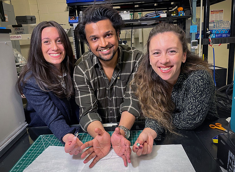
Duke University postdoc Laura Rupprecht, MIT graduate student Atharva Sahasrabudhe, and MIT postdoc Sirma Orguc holding their engineered flexible fiber in Polina Anikeeva’s lab at MIT. Photo: Courtesy of the researchers
“Again, we got this place preference behavior that people have previously seen with stimulation in the brain, but now we are not touching the brain. We are just stimulating the gut, and we are observing control of central function from the periphery,” Anikeeva says.
Sahasrabudhe worked closely with Rupprecht, a postdoc in Professor Diego Bohorquez’ group at Duke, to test the fibers’ ability to control feeding behaviors. They found that the devices could optogenetically stimulate cells that produce cholecystokinin, a hormone that promotes satiety. When this hormone release was activated, the animals’ appetites were suppressed, even though they had been fasting for several hours. The researchers also demonstrated a similar effect when they stimulated cells that produce a peptide called PYY, which normally curbs appetite after very rich foods are consumed.
The researchers now plan to use this interface to study neurological conditions that are believed to have a gut-brain connection. For instance, studies have shown that autistic children are far more likely than their peers to be diagnosed with GI dysfunction, while anxiety and irritable bowel syndrome share genetic risks.
“We can now begin asking, are those coincidences, or is there a connection between the gut and the brain? And maybe there is an opportunity for us to tap into those gut-brain circuits to begin managing some of those conditions by manipulating the peripheral circuits in a way that does not directly ‘touch’ the brain and is less invasive,” Anikeeva says.
The research was funded, in part, by the Hock E. Tan and K. Lisa Yang Center for Autism Research and the K. Lisa Yang Brain-Body Center, the National Institute of Neurological Disorders and Stroke, the National Science Foundation (NSF) Center for Materials Science and Engineering, the NSF Center for Neurotechnology, the National Center for Complementary and Integrative Health, a National Institutes of Health Director’s Pioneer Award, the National Institute of Mental Health, and the National Institute of Diabetes and Digestive and Kidney Diseases.
When interacting with another person, you likely spend part of your time trying to anticipate how they will feel about what you’re saying or doing. This task requires a cognitive skill called theory of mind, which helps us to infer other people’s beliefs, desires, intentions, and emotions.
MIT neuroscientists have now designed a computational model that can predict other people’s emotions — including joy, gratitude, confusion, regret, and embarrassment — approximating human observers’ social intelligence. The model was designed to predict the emotions of people involved in a situation based on the prisoner’s dilemma, a classic game theory scenario in which two people must decide whether to cooperate with their partner or betray them.
To build the model, the researchers incorporated several factors that have been hypothesized to influence people’s emotional reactions, including that person’s desires, their expectations in a particular situation, and whether anyone was watching their actions.
“These are very common, basic intuitions, and what we said is, we can take that very basic grammar and make a model that will learn to predict emotions from those features,” says Rebecca Saxe, the John W. Jarve Professor of Brain and Cognitive Sciences, a member of MIT’s McGovern Institute for Brain Research, and the senior author of the study.
Sean Dae Houlihan PhD ’22, a postdoc at the Neukom Institute for Computational Science at Dartmouth College, is the lead author of the paper, which appears today in Philosophical Transactions A. Other authors include Max Kleiman-Weiner PhD ’18, a postdoc at MIT and Harvard University; Luke Hewitt PhD ’22, a visiting scholar at Stanford University; and Joshua Tenenbaum, a professor of computational cognitive science at MIT and a member of the Center for Brains, Minds, and Machines and MIT’s Computer Science and Artificial Intelligence Laboratory (CSAIL).
Predicting emotions
While a great deal of research has gone into training computer models to infer someone’s emotional state based on their facial expression, that is not the most important aspect of human emotional intelligence, Saxe says. Much more important is the ability to predict someone’s emotional response to events before they occur.
“The most important thing about what it is to understand other people’s emotions is to anticipate what other people will feel before the thing has happened,” she says. “If all of our emotional intelligence was reactive, that would be a catastrophe.”
To try to model how human observers make these predictions, the researchers used scenarios taken from a British game show called “Golden Balls.” On the show, contestants are paired up with a pot of $100,000 at stake. After negotiating with their partner, each contestant decides, secretly, whether to split the pool or try to steal it. If both decide to split, they each receive $50,000. If one splits and one steals, the stealer gets the entire pot. If both try to steal, no one gets anything.
Depending on the outcome, contestants may experience a range of emotions — joy and relief if both contestants split, surprise and fury if one’s opponent steals the pot, and perhaps guilt mingled with excitement if one successfully steals.
To create a computational model that can predict these emotions, the researchers designed three separate modules. The first module is trained to infer a person’s preferences and beliefs based on their action, through a process called inverse planning.
“This is an idea that says if you see just a little bit of somebody’s behavior, you can probabilistically infer things about what they wanted and expected in that situation,” Saxe says.
Using this approach, the first module can predict contestants’ motivations based on their actions in the game. For example, if someone decides to split in an attempt to share the pot, it can be inferred that they also expected the other person to split. If someone decides to steal, they may have expected the other person to steal, and didn’t want to be cheated. Or, they may have expected the other person to split and decided to try to take advantage of them.
The model can also integrate knowledge about specific players, such as the contestant’s occupation, to help it infer the players’ most likely motivation.
The second module compares the outcome of the game with what each player wanted and expected to happen. Then, a third module predicts what emotions the contestants may be feeling, based on the outcome and what was known about their expectations. This third module was trained to predict emotions based on predictions from human observers about how contestants would feel after a particular outcome. The authors emphasize that this is a model of human social intelligence, designed to mimic how observers causally reason about each other’s emotions, not a model of how people actually feel.
“From the data, the model learns that what it means, for example, to feel a lot of joy in this situation, is to get what you wanted, to do it by being fair, and to do it without taking advantage,” Saxe says.
Core intuitions
Once the three modules were up and running, the researchers used them on a new dataset from the game show to determine how the models’ emotion predictions compared with the predictions made by human observers. This model performed much better at that task than any previous model of emotion prediction.
The model’s success stems from its incorporation of key factors that the human brain also uses when predicting how someone else will react to a given situation, Saxe says. Those include computations of how a person will evaluate and emotionally react to a situation, based on their desires and expectations, which relate to not only material gain but also how they are viewed by others.
“Our model has those core intuitions, that the mental states underlying emotion are about what you wanted, what you expected, what happened, and who saw. And what people want is not just stuff. They don’t just want money; they want to be fair, but also not to be the sucker, not to be cheated,” she says.
“The researchers have helped build a deeper understanding of how emotions contribute to determining our actions; and then, by flipping their model around, they explain how we can use people’s actions to infer their underlying emotions. This line of work helps us see emotions not just as ‘feelings’ but as playing a crucial, and subtle, role in human social behavior,” says Nick Chater, a professor of behavioral science at the University of Warwick, who was not involved in the study.
In future work, the researchers hope to adapt the model so that it can perform more general predictions based on situations other than the game-show scenario used in this study. They are also working on creating models that can predict what happened in the game based solely on the expression on the faces of the contestants after the results were announced.
The research was funded by the McGovern Institute; the Paul E. and Lilah Newton Brain Science Award; the Center for Brains, Minds, and Machines; the MIT-IBM Watson AI Lab; and the Multidisciplinary University Research Initiative.
When staff in MIT’s Department of Facilities would visualize energy use and carbon-associated emissions by campus buildings, Building 46 always stood out — attributed to its energy intensity, which accounted for 8 percent of MIT’s total campus energy use. This high energy draw was not surprising, as the building is home of the Brain and Cognitive Sciences Complex and a large amount of lab space, but it also made the building a perfect candidate for an energy performance audit to seek out potential energy saving opportunities.
This audit revealed that several energy efficiency updates to the building mechanical systems infrastructure, including optimization of the room-by-room ventilation rates, could result in an estimated 35 percent reduction of energy use, which would in turn lower MIT’s total greenhouse gas emissions by an estimated 2 percent — driving toward the Institute’s 2026 goal of net-zero and 2050 goal of elimination of direct campus emissions.
Building energy efficiency projects are not new for MIT. Since 2010, MIT has been engaged in a partnership agreement with utility company Eversource establishing the Efficiency Forward program, empowering MIT to invest in more than 300 energy conservation projects to date and lowering energy consumption on campus for a total calculated savings of approximately 70 million kilowatt hours and 4.2 million therms. But at 418,000 gross square feet, Building 46 is the first energy efficiency project of its size on the campus.
“We’ve never tackled a whole building like this — it’s the first capital project that is technically an energy project,” explains Siobhan Carr, energy efficiency program manager, who was part of the team overseeing the energy audit and lab ventilation performance assessment in the building. “That gives you an idea of the magnitude and complexity of this.”
The project started with the full building energy assessment and lab ventilation risk audit. “We had a team go through every corner of the building and look at every possible opportunity to save energy,” explains Jessica Parks, senior project manager for systems performance and turnover in campus construction. “One of the biggest issues we saw was that there’s a lot of dry lab spaces which are basically offices, but they’re all getting the same ventilation as if they were a high-intensity lab.” Higher ventilation and more frequent air exchange rates draw more energy. By optimizing for the required ventilation rates, there was an opportunity to save energy in nearly every space in the building.
In addition to the optimized ventilation, the project team will convert fume hoods from constant volume to variable volume and install equipment to help the building systems run more efficiently. The team also identified opportunities to work with labs to implement programs such as fume hood hibernation and unoccupied setbacks for temperature and ventilation. As different spaces in the building have varying needs, the energy retrofit will touch all 1,254 spaces in the building — one by one — to implement the different energy measures to reach that estimated 35 percent reduction in energy use.
Although time-consuming and complex, this room-by-room approach has a big benefit in that it has allowed research to continue in the space largely uninterrupted. With a few exceptions, the occupants of Building 46, which include the Department of Brain and Cognitive Sciences, The McGovern Institute for Brain Research, and The Picower Institute for Learning and Memory, have remained in place for the duration of the project. Partners in the MIT Environment, Health and Safety Office are instrumental to this balance of renovations and keeping the building operational during the optimization efforts and are one of several teams across MIT contributing to building efficiency efforts.
The completion date of the building efficiency project is set for 2024, but Carr says that some of the impact of this ongoing work may soon be seen. “We should start to see savings as we move through the building, and we expect to fully realize all of our projected savings a year after completion,” she says, noting that the length of time is required for a year-over-year perspective to see the full reduction in energy use.
The impact of the project goes far beyond the footprint of Building 46 as it offers insights and spurred actions for future projects — including buildings 76 and 68, the number two and three top energy users on campus. Both buildings recently underwent their own energy audits and lab ventilation performance assessments. The energy efficiency team is now crafting a plan for full-building approaches, much like Building 46. “To date, 46 has presented many learning opportunities, such as how to touch every space in a building while research continues, as well as how to overcome challenges encountered when working on existing systems,” explains Parks. “The good news is that we have developed solutions for those challenges and the teams have been proactively implementing those lessons in our other projects.”
Communication has proven to be another key for these large projects where occupants see the work happening and often play a role in answering questions about their unique space. “People are really engaged, they ask questions about the work, and we ask them about the space they’re in every day,” says Parks. “The Building 46 occupants have been wonderful partners as we worked in all of their spaces, which is paving the way for a successful project.”
The release of Fast Forward in 2021 has also made communications easier, notes Carr, who says the plan helps to frame these projects as part of the big picture — not just a construction interruption. “Fast Forward has brought a visibility into what we’re doing within [MIT] Facilities on these buildings,” she says. “It brings more eyes and ears, and people understand that these projects are happening throughout campus and not just in their own space — we’re all working to reduce energy and to reduce greenhouse gas across campus.”
The Energy Efficiency team will continue to apply that big-picture approach as ongoing building efficiency projects on campus are assessed to reach toward a 10 to 15 percent reduction in energy use and corresponding emissions over the next several years.