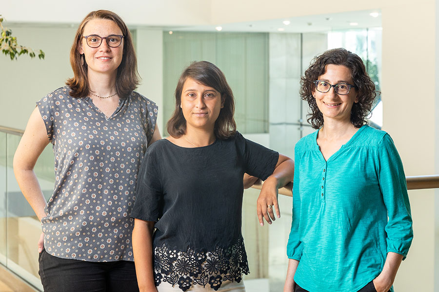
Simons Center’s collaborative approach propels autism research, at MIT and beyond

The secret to the success of MIT’s Simons Center for the Social Brain is in the name. With a founding philosophy of “collaboration and community” that has supported scores of scientists across more than a dozen Boston-area research institutions, the SCSB advances research by being inherently social.

SCSB’s mission is “to understand the neural mechanisms underlying social cognition and behavior and to translate this knowledge into better diagnosis and treatment of autism spectrum disorders.” When Director Mriganka Sur founded the center in 2012 in partnership with the Simons Foundation Autism Research Initiative (SFARI) of Jim and Marilyn Simons, he envisioned a different way to achieve urgently needed research progress than the traditional approach of funding isolated projects in individual labs. Sur wanted SCSB’s contribution to go beyond papers, though it has generated about 350 and counting. He sought the creation of a sustained, engaged autism research community at MIT and beyond.

“When you have a really big problem that spans so many issues  a clinical presentation, a gene, and everything in between  you have to grapple with multiple scales of inquiry,” says Sur, the Newton Professor of Neuroscience in MIT’s Department of Brain and Cognitive Sciences (BCS) and The Picower Institute for Learning and Memory. “This cannot be solved by one person or one lab. We need to span multiple labs and multiple ways of thinking. That was our vision.”

In parallel with a rich calendar of public colloquia, lunches, and special events, SCSB catalyzes multiperspective, multiscale research collaborations in two programmatic ways. Targeted projects fund multidisciplinary teams of scientists with complementary expertise to collectively tackle a pressing scientific question. Meanwhile, the center supports postdoctoral Simons Fellows with not one, but two mentors, ensuring a further cross-pollination of ideas and methods.

Complementary collaboration

In 11 years, SCSB has funded nine targeted projects. Each one, by design, involves a deep and multifaceted exploration of a major question with both fundamental importance and clinical relevance. The first project, back in 2013, for example, marshaled three labs spanning BCS, the Department of Biology, and The Whitehead Institute for Biomedical Research to advance understanding of how mutation of the Shank3 gene leads to the pathophysiology of Phelan-McDermid Syndrome by working across scales ranging from individual neural connections to whole neurons to circuits and behavior.

Other past projects have applied similarly integrated, multiscale approaches to topics ranging from how 16p11.2 gene deletion alters the development of brain circuits and cognition to the critical role of the thalamic reticular nucleus in information flow during sleep and wakefulness. Two others produced deep examinations of cognitive functions: how we go from hearing a string of words to understanding a sentence’s intended meaning, and the neural and behavioral correlates of deficits in making predictions about social and sensory stimuli. Yet another project laid the groundwork for developing a new animal model for autism research.

SFARI is especially excited by SCSB’s team science approach, says Kelsey Martin, executive vice president of autism and neuroscience at the Simons Foundation. “I’m delighted by the collaborative spirit of the SCSB,” Martin says. “It’s wonderful to see and learn about the multidisciplinary team-centered collaborations sponsored by the center.”

New projects

In the last year, SCSB has launched three new targeted projects. One team is investigating why many people with autism experience sensory overload and is testing potential interventions to help. The scientists hypothesize that patients experience a deficit in filtering out the mundane stimuli that neurotypical people predict are safe to ignore. Studies suggest the predictive filter relies on relatively low-frequency “alpha/beta” brain rhythms from deep layers of the cortex moderating the higher frequency “gamma” rhythms in superficial layers that process sensory information.

Together, the labs of Charles Nelson, professor of pediatrics at Boston Children’s Hospital (BCH), and BCS faculty members Bob Desimone, the Doris and Don Berkey Professor of Neuroscience at MIT and director of the McGovern Institute, and Earl K. Miller, the Picower Professor, are testing the hypothesis in two different animal models at MIT and in human volunteers at BCH. In the animals they’ll also try out a new real-time feedback system invented in Miller’s lab that can potentially correct the balance of these rhythms in the brain. And in an animal model engineered with a Shank3 mutation, Desimone’s lab will test a gene therapy, too.

“None of us could do all aspects of this project on our own,” says Miller, an investigator in the Picower Institute. “It could only come about because the three of us are working together, using different approaches.”

Right from the start, Desimone says, close collaboration with Nelson’s group at BCH has been essential. To ensure his and Miller’s measurements in the animals and Nelson’s measurements in the humans are as comparable as possible, they have tightly coordinated their research protocols.

“If we hadn’t had this joint grant we would have chosen a completely different, random set of parameters than Chuck, and the results therefore wouldn’t have been comparable. It would be hard to relate them,” says Desimone, who also directs MIT’s McGovern Institute for Brain Research. “This is a project that could not be accomplished by one lab operating in isolation.”

Another targeted project brings together a coalition of seven labs — six based in BCS (professors Evelina Fedorenko, Edward Gibson, Nancy Kanwisher, Roger Levy, Rebecca Saxe, and Joshua Tenenbaum) and one at Dartmouth College (Caroline Robertson) — for a synergistic study of the cognitive, neural, and computational underpinnings of conversational exchanges. The study will integrate the linguistic and non-linguistic aspects of conversational ability in neurotypical adults and children and those with autism.

Fedorenko said the project builds on advances and collaborations from the earlier language Targeted Project she led with Kanwisher.

“Many directions that we started to pursue continue to be active directions in our labs. But most importantly, it was really fun and allowed the PIs [principal investigators] to interact much more than we normally would and to explore exciting interdisciplinary questions,” Fedorenko says. “When Mriganka approached me a few years after the project’s completion asking about a possible new targeted project, I jumped at the opportunity.”

Gibson and Robertson are studying how people align their dialogue, not only in the content and form of their utterances, but using eye contact. Fedorenko and Kanwisher will employ fMRI to discover key components of a conversation network in the cortex. Saxe will examine the development of conversational ability in toddlers using novel MRI techniques. Levy and Tenenbaum will complement these efforts to improve computational models of language processing and conversation.

The newest Targeted Project posits that the immune system can be harnessed to help treat behavioral symptoms of autism. Four labs — three in BCS and one at Harvard Medical School (HMS) — will study mechanisms by which peripheral immune cells can deliver a potentially therapeutic cytokine to the brain. A study by two of the collaborators, MIT associate professor Gloria Choi and HMS associate professor Jun Huh, showed that when IL-17a reaches excitatory neurons in a region of the mouse cortex, it can calm hyperactivity in circuits associated with social and repetitive behavior symptoms. Huh, an immunologist, will examine how IL-17a can get from the periphery to the brain, while Choi will examine how it has its neurological effects. Sur and MIT associate professor Myriam Heiman will conduct studies of cell types that bridge neural circuits with brain circulatory systems.

“It is quite amazing that we have a core of scientists working on very different things coming together to tackle this one common goal,” Choi says. “I really value that.”

Multiple mentors

While SCSB Targeted Projects unify labs around research, the center’s Simons Fellowships unify labs around young researchers, providing not only funding, but a pair of mentors and free-flowing interactions between their labs. Fellows also gain opportunities to inform and inspire their fundamental research by visiting with patients with autism, Sur says.

“The SCSB postdoctoral program serves a critical role in ensuring that a diversity of outstanding scientists are exposed to autism research during their training, providing a pipeline of new talent and creativity for the field,” adds Martin, of the Simons Foundation.

Simons Fellows praise the extra opportunities afforded by additional mentoring. Postdoc Alex Major was a Simons Fellow in Miller’s lab and that of Nancy Kopell, a mathematics professor at Boston University renowned for her modeling of the brain wave phenomena that the Miller lab studies experimentally.

“The dual mentorship structure is a very useful aspect of the fellowship” Major says. “It is both a chance to network with another PI and provides experience in a different neuroscience sub-field.”

Miller says co-mentoring expands the horizons and capabilities of not only the mentees but also the mentors and their labs. “Collaboration is 21st century neuroscience,” Miller says. “Some our studies of the brain have gotten too big and comprehensive to be encapsulated in just one laboratory. Some of these big questions require multiple approaches and multiple techniques.”

Desimone, who recently co-mentored Seng Bum (Michael Yoo) along with BCS and McGovern colleague Mehrdad Jazayeri in a project studying how animals learn from observing others, agrees.

“We hear from postdocs all the time that they wish they had two mentors, just in general to get another point of view,” Desimone says. “This is a really good thing and it’s a way for faculty members to learn about what other faculty members and their postdocs are doing.”

Indeed, the Simons Center model suggests that research can be very successful when it’s collaborative and social.

The promise of gene therapy

Portrait of Bob Desimone wearing a suit and tie.
McGovern Institute Director Robert Desimone. Photo: Steph Stevens

As we start 2024, I hope you can join me in celebrating a historic recent advance: the FDA approval of Casgevy, a bold new treatment for devastating sickle cell disease and the world’s first approved CRISPR gene therapy.

Developed by Vertex Pharmaceuticals and CRISPR Therapeutics, we are proud to share that this pioneering therapy licenses the CRISPR discoveries of McGovern scientist and Poitras Professor of Neuroscience Feng Zhang.

It is amazing to think that Feng’s breakthrough work adapting CRISPR-Cas9 for genome editing in eukaryotic cells was published only 11 years ago today in Science.

Incredibly, CRISPR-Cas9 rapidly transitioned from proof-of-concept experiments to an approved treatment in just over a decade.

McGovern scientists are determined to maintain the momentum!


Incredibly, CRISPR-Cas9 rapidly transitioned from proof-of-concept experiments to an approved treatment in just over a decade.

Our labs are creating new gene therapies that are already in clinical trials or preparing to enroll patients in trials. For instance, Feng Zhang’s team has developed therapies currently in clinical trials for lymphoblastic leukemia and beta thalassemia, while another McGovern researcher, Guoping Feng, the Poitras Professor of Brain and Cognitive Sciences at MIT, has made advancements that lay the groundwork for a new gene therapy to treat a severe form of autism spectrum disorder. It is expected to enter clinical trials later this year. Moreover, McGovern fellows Omar Abudayyeh and Jonathan Gootenberg created programmable genomic tools that are now licensed for use in monogenic liver diseases and autoimmune disorders.

These exciting innovations stem from your steadfast support of our high-risk, high-reward research. Your generosity is enabling our scientists to pursue basic research in other areas with potential therapeutic applications in the future, such as mechanisms of pain, addiction, the connections between the brain and gut, the workings of memory and attention, and the bi-directional influence of artificial intelligence on brain research. All of this fundamental research is being fueled by major new advances in technology, many of them developed here.

As we enter a new year filled with anticipation following our inaugural gene therapy, I want to express my heartfelt gratitude for your invaluable support in advancing our research programs. Your role in pushing our research to new heights is valued by all faculty, students, and researchers at the McGovern Institute. We can’t wait to share our continued progress with you.

Thank you again for partnering with us to make great scientific achievements possible.

With appreciation and best wishes,

Robert Desimone, PhD
Director, McGovern Institute
Doris and Don Berkey Professor of Neuroscience, MIT

One scientist’s journey from the Middle East to MIT

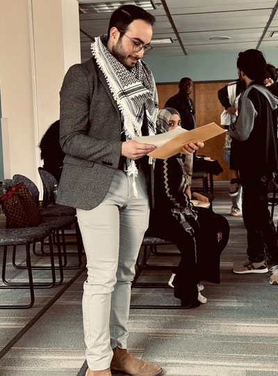
Smiling man holidng paper in a room.
Ubadah Sabbagh, soon after receiving his US citizenship papers, in April 2023. Photo: Ubadah Sabbagh

“I recently exhaled a breath I’ve been holding in for nearly half my life. After applying over a decade ago, I’m finally an American. This means so many things to me. Foremost, it means I can go back to the the Middle East, and see my mama and the family, for the first time in 14 years.” — McGovern Institute Postdoctoral Associate Ubadah Sabbagh, X (formerly Twitter) post, April 27, 2023

The words sit atop a photo of Ubadah Sabbagh, who joined the lab of Guoping Feng, James W. (1963) and Patricia T. Poitras Professor at MIT, as a postdoctoral associate in 2021. Sabbagh, a Syrian national, is dressed in a charcoal grey jacket, a keffiyeh loose around his neck, and holding his US citizenship papers, which he began applying for when he was 19 and an undergraduate at the University of Missouri-Kansas City (UMKC) studying biology and bioinformatics.

In the photo he is 29.

A clarity of vision

Sabbagh’s journey from the Middle East to his research position at MIT has been marked by determination and courage, a multifaceted curiosity, and a role as a scientist-writer/scientist-advocate.  He is particularly committed to the importance of humanity in science.

“For me, a scientist is a person who is not only in the lab but also has a unique perspective to contribute to society,” he says. “The scientific method is an idea, and that can be objective. But the process of doing science is a human endeavor, and like all human endeavors, it is inherently both social and political.”

At just 30 years of age, some of Sabbagh’s ideas have disrupted conventional thinking about how science is done in the United States. He believes nations should do science not primarily to compete, for example, but to be aspirational.

“It is our job to make our work accessible to the public, to educate and inform, and to help ground policy,” he says. “In our technologically advanced society, we need to raise the baseline for public scientific intuition so that people are empowered and better equipped to separate truth from myth.”

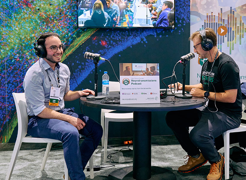
Two men sitting at a booth wearing headphones.
Ubadah Sabbagh is interviewed for Max Planck Forida’s Neurotransmissions podcast at the 2023 Society for Neuroscience conference in San Diego. Photo: Max Planck Florida

His research and advocacy work have won him accolades, including the 2023 Young Arab Pioneers Award from the Arab Youth Center and the 2020 Young Investigator Award from the American Society of Neurochemistry. He was also named to the 2021 Forbes “30 under 30” list, the first Syrian to be selected in the Science category.

A path to knowledge

Sabbagh’s path to that knowledge began when, living on his own at age 16, he attended Longview Community College, in Kansas City, often juggling multiple jobs. It continued at UMKC, where he fell in love with biology and had his first research experience with bioinformatician Gerald Wyckoff at the same time the civil war in Syria escalated, with his family still in the Middle East. “That was a rough time for me,” he says. “I had a lot of survivor’s guilt: I am here, I have all of this stability and security compared to what they have, and while they had suffocation, I had opportunity. I need to make this mean something positive, not just for me, but in as broad a way as possible for other people.”

Child smiles in front of scientific poster.
Ubadah Sabbagh, age 9, presents his first scientific poster. Photo: Ubadah Sabbagh

The war also sparked Sabbagh’s interest in human behavior—“where it originates, what motivates people to do things, but in a biological, not a psychological way,” he says. “What circuitry is engaged? What is the infrastructure of the brain that leads to X, Y, Z?”

His passion for neuroscience blossomed as a graduate student at Virginia Tech, where he earned his PhD in translational biology, medicine, and health. There, he received a six-year NIH F99/K00 Award, and under the mentorship of neuroscientist at the Fralin Biomedical Research Institute he researched the connections between the eye and the brain, specifically, mapping the architecture of the principle neurons in a region of the thalamus essential to visual processing.

“The retina, and the entire visual system, struck me as elegant, with beautiful layers of diverse cells found at every node,” says Sabbagh, his own eyes lighting up.

His research earned him a coveted spot on the Forbes “30 under 30” list, generating enormous visibility, including in the Arab world, adding visitors to his already robust X (formerly Twitter) account, which has more than 9,200 followers. “The increased visibility lets me use my voice to advocate for the things I care about,” he says.

“I need to make this mean something positive, not just for me, but in as broad a way as possible for other people.” — Ubadah Sabbagh

Those causes range from promoting equity and inclusion in science to transforming the American system of doing science for the betterment of science and the scientists themselves. He cofounded the nonprofit Black in Neuro to celebrate and empower Black scholars in neuroscience, and he continues to serve on the board. He is the chair of an advisory committee for the Society for Neuroscience (SfN), recommending ways SfN can better address the needs of its young members, and a member of the Advisory Committee to the National Institutes of Health (NIH) Director working group charged with re-envisioning postdoctoral training. He serves on the advisory board of Community for Rigor, a new NIH initiative that aims to teach scientific rigor at national scale and, in his spare time, he writes articles about the relationship of science and policy for publications including Scientific American and the Washington Post.

Still, there have been obstacles. The same year Sabbagh received the NIH F99/K00 Award, he faced major setbacks in his application to become a citizen. He would not try again until 2021, when he had his PhD in hand and had joined the McGovern Institute.

An MIT postdoc and citizenship

Sabbagh dove into his research in Guoping Feng’s lab with the same vigor and outside-the-box thinking that characterized his previous work. He continues to investigate the thalamus, but in a region that is less involved in processing pure sensory signals, such as light and sound, and more focused on cognitive functions of the brain. He aims to understand how thalamic brain areas orchestrate complex functions we carry out every day, including working memory and cognitive flexibility.

“This is important to understand because when this orchestra goes out of tune it can lead to a range of neurological disorders, including autism spectrum disorder and schizophrenia,” he says. He is also developing new tools for studying the brain using genome editing and viral engineering to expand the toolkit available to neuroscientists.

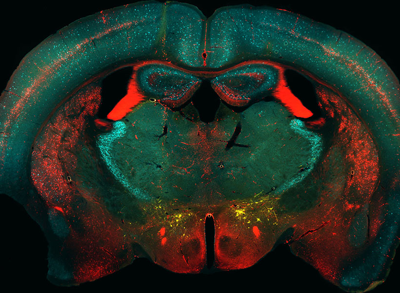
Microscopic image of mouse brain
Neurons in a transgenic mouse brain labeled by Sabbagh using genome editing technology in the Feng lab. Image: Ubadah Sabbagh

The environment at the McGovern Institute is also a source of inspiration for Sabbagh’s research. “The scale and scope of work being done at McGovern is remarkable. It’s an exciting place for me to be as a neuroscientist,” said Sabbagh. “Besides being intellectually enriching, I’ve found great community here – something that’s important to me wherever I work.”

Returning to the Middle East

Profile of scientist Ubadah Sabbagh speaking at a table.
McGovern postdoc Ubadah Sabbagh at the 2023 Young Arab Pioneers Award ceremony in Abu Dhabi. Photo: Arab Youth Center

While at an advisory meeting at the NIH, Sabbagh learned he had been selected as a Young Arab Pioneer by the Arab Youth Center and was flown the next day to Abu Dhabi for a ceremony overseen by Her Excellency Shamma Al Mazrui, Cabinet Member and Minister of Community Development in the United Arab Emirates. The ceremony recognized 20 Arab youth from around the world in sectors ranging from scientific research to entrepreneurship and community development. Sabbagh’s research “presented a unique portrayal of creative Arab youth and an admirable representation of the values of youth beyond the Arab world,” said Sadeq Jarrar, executive director of the center.

“There I was, among other young Arab leaders, learning firsthand about their efforts, aspirations, and their outlook for the future,” says Sabbagh, who was deeply inspired by the experience.

Just a month earlier, his passport finally secured, Sabbagh had reunited with his family in the Middle East after more than a decade in the United States. “I had been away for so long,” he said, describing the experience as a “cultural reawakening.”

Woman hands man an award on stage.
Ubadah Sabbagh receives a Young Arab Pioneer Award by Her Excellency Shamma Al Mazrui, Cabinet Member and Minister of Community Development in the United Arab Emirates. Photo: Arab Youth Center

Sabbagh saw a gaping need he had not been aware of when he left 14 years earlier, as a teen. “The Middle East had such a glorious intellectual past,” he says. “But for years people have been leaving to get their advanced scientific training, and there is no adequate infrastructure to support them if they want to go back.” He wondered: What if there were a scientific renaissance in the region? How would we build infrastructure to cultivate local minds and local talent? What if the next chapter of the Middle East included being a new nexus of global scientific advancements?

“I felt so inspired,” he says. “I have a longing, someday, to meaningfully give back.”

Making invisible therapy targets visible

The lab of Edward Boyden, the Y. Eva Tan Professor in Neurotechnology, has developed a powerful technology called Expansion Revealing (ExR) that makes visible molecular structures that were previously too hidden to be seen with even the most powerful microscopes. It “reveals” the nanoscale alterations in synapses, neural wiring, and other molecular assemblies using ordinary lab microscopes. It does so this way: Inside a cell, proteins and other molecules are often tightly packed together. These dense clusters can be difficult to image because the fluorescent labels used to make them visible can’t wedge themselves between the molecules. ExR “de-crowds” the molecules by expanding the cell using a chemical process, making the molecules accessible to fluorescent tags.

Jinyoung Kang is a J. Douglas Tan Postdoctoral Fellow in the Boyden and Feng labs. Photo: Steph Stevens

“This technology can be used to answer a lot of biological questions about dysfunction in synaptic proteins, which are involved in neurodegenerative diseases,” says Jinyoung Kang, a J. Douglas Tan Postdoctoral Fellow in the labs of Boyden and Guoping Feng, the James W. (1963) and Patricia T. Poitras Professor of Brain and Cognitive Sciences. “Until now, there has been no tool to visualize synapses very well at nanoscale.”

Over the past year, the Boyden team has been using ExR to explore the underlying mechanisms of brain disorders, including autism spectrum disorder (ASD) and Alzheimer’s disease. Since the method can be applied iteratively, Boyden imagines it may one day succeed in creating a 100-fold magnification of molecular structures.

“Using earlier technology, researchers may be missing entire categories of molecular phenomena, both functional and dysfunctional,” says Boyden. “It’s critical to bring these nanostructures into view so that we can identify potential targets for new therapeutics that can restore functional molecular arrangements.”

The team is applying ExR to the study of mutant-animal-model brain slices to expose complex synapse 3D nanoarchitecture and configuration. Among their questions: How do synapses differ when mutations that cause autism and other neurological conditions are present?

Using the new technology, Kang and her collaborator Menglong Zeng characterized the molecular architecture of excitatory synapses on parvalbumin interneurons, cells that drastically influence the downstream effects of neuronal signaling and ultimately change cognitive behaviors. They discovered condensed AMPAR clustering in parvalbumin interneurons is essential for normal brain function. The next step is to explore their role in the function of parvalbumin interneurons, which are vulnerable to stressors and have been implicated in brain disorders including autism and Alzheimer’s disease.

The researchers are now investigating whether ExR can reveal abnormal protein nanostructures in SHANK3 knockout mice and marmosets. Mutations in the SHANK3 gene lead to one of the most severe types of ASD, Phelan-McDermid syndrome, which accounts for about 2 percent of all ASD patients with intellectual disability.

Refining mental health diagnoses

Maedbh King came to MIT to make a difference in mental health. As a postdoctoral fellow in the K. Lisa Yang Integrative Computational Neuroscience (ICoN) Center, she is building computer models aimed at helping clinicians improve diagnosis and treatment, especially for young people with neurodevelopmental and psychiatric disorders.

Tapping two large patient-data sources, King is working to analyze critical biological and behavioral information to better categorize patients’ mental health conditions, including autism spectrum disorder, attention-deficit hyperactivity disorder (ADHD), anxiety, and suicidal thoughts — and to provide more predictive approaches to addressing them. Her strategy reflects the center’s commitment to a holistic understanding of human brain function using theoretical and computa-tional neuroscience.

“Today, treatment decisions for psychiatric disorders are derived entirely from symptoms, which leaves clinicians and patients trying one treatment and, if it doesn’t work, trying another,” says King. “I hope to help change that.”

King grew up in Dublin, Ireland, and studied psychology in college; gained neuroimaging and programming skills while earning a master’s degree from Western University in Canada; and received her doctorate from the University of California, Berkeley, where she built maps and models of the human brain. In fall 2022, King joined the lab of Satrajit Ghosh, a McGovern Institute principal research scientist whose team uses neuroimaging, speech communication, and machine learning to improve assessments and treatments for mental health and neurological disorders.

Big-data insights

King is pursuing several projects using the Healthy Brain Network, a landmark mental health study of children and adolescents in New York City. She and lab colleagues are extracting data from cognitive and other assessments — such as language patterns, favorite school subjects, and family mental illness history — from roughly 4,000 participants to provide more-nuanced understanding of their neurodevelopmental disorders, such as autism or ADHD.

“Computational models are powerful. They can identify patterns that can’t be obtained with the human eye through electronic records,” says King.

With this database, one can develop “very rich clinical profiles of these young people,” including their challenges and adaptive strengths, King explains. “We’re interested in placing these participants within a spectrum of symptoms, rather than just providing a binary label of, ‘has this disorder’ or ‘doesn’t have it.’ It’s an effort to subtype based on these phenotypic assessments.”

In other research, King is developing tools to detect risk factors for suicide among adolescents. Working with psychiatrists at Children’s Hospital of Philadelphia, she is using detailed questionnaires from some 20,000 youths who visited the hospital’s emergency department over several years; about one-tenth had tried to take their own lives. The questionnaires collect information about demographics, lifestyle, relationships, and other aspects of patients’ lives.

“One of the big questions the physicians want to answer is, Are there any risk predictors we can identify that can ultimately prevent, or at least mitigate, future suicide attempts?” King says. “Computational models are powerful. They can identify patterns that can’t be obtained with the human eye through electronic records.”

King is passionate about producing findings to help practitioners, whether they’re clinicians, teachers, parents, or policy makers, and the populations they’re studying. “This applied work,” she says, “should be communicated in a way that can be useful.

How Huntington’s disease affects different neurons

In patients with Huntington’s disease, neurons in a part of the brain called the striatum are among the hardest-hit. Degeneration of these neurons contributes to patients’ loss of motor control, which is one of the major hallmarks of the disease.

Neuroscientists at MIT have now shown that two distinct cell populations in the striatum are affected differently by Huntington’s disease. They believe that neurodegeneration of one of these populations leads to motor impairments, while damage to the other population, located in structures called striosomes, may account for the mood disorders that are often see in the early stages of the disease.

“As many as 10 years ahead of the motor diagnosis, Huntington’s patients can experience mood disorders, and one possibility is that the striosomes might be involved in these,” says Ann Graybiel, an MIT Institute Professor, a member of MIT’s McGovern Institute for Brain Research, and one of the senior authors of the study.

Using single-cell RNA sequencing to analyze the genes expressed in mouse models of Huntington’s disease and postmortem brain samples from Huntington’s patients, the researchers found that cells of the striosomes and another structure, the matrix, begin to lose their distinguishing features as the disease progresses. The researchers hope that their mapping of the striatum and how it is affected by Huntington’s could help lead to new treatments that target specific cells within the brain.

This kind of analysis could also shed light on other brain disorders that affect the striatum, such as Parkinson’s disease and autism spectrum disorder, the researchers say.

Myriam Heiman, an associate professor in MIT’s Department of Brain and Cognitive Sciences and a member of the Picower Institute for Learning and Memory, and Manolis Kellis, a professor of computer science in MIT’s Computer Science and Artificial Intelligence Laboratory (CSAIL) and a member of the Broad Institute of MIT and Harvard, are also senior authors of the study. Ayano Matsushima, a McGovern Institute research scientist, and Sergio Sebastian Pineda, an MIT graduate student, are the lead authors of the paper, which appears in Nature Communications.

Neuron vulnerability

Huntington’s disease leads to degeneration of brain structures called the basal ganglia, which are responsible for control of movement and also play roles in other behaviors, as well as emotions. For many years, Graybiel has been studying the striatum, a part of the basal ganglia that is involved in making decisions that require evaluating the outcomes of a particular action.

Many years ago, Graybiel discovered that the striatum is divided into striosomes, which are clusters of neurons, and the matrix, which surrounds the striosomes. She has also shown that striosomes are necessary for making decisions that require an anxiety-provoking cost-benefit analysis.

In a 2007 study, Richard Faull of the University of Auckland discovered that in postmortem brain tissue from Huntington’s patients, the striosomes showed a great deal of degeneration. Faull also found that while those patients were alive, many of them had shown signs of mood disorders such as depression before their motor symptoms developed.

To further explore the connections between the striatum and the mood and motor effects of Huntington’s, Graybiel teamed up with Kellis and Heiman to study the gene expression patterns of striosomal and matrix cells. To do that, the researchers used single-cell RNA sequencing to analyze human brain samples and brain tissue from two mouse models of Huntington’s disease.

Within the striatum, neurons can be classified as either D1 or D2 neurons. D1 neurons are involved in the “go” pathway, which initiates an action, and D2 neurons are part of the “no-go” pathway, which suppresses an action. D1 and D2 neurons can both be found within either the striosomes and the matrix.

The analysis of RNA expression in each of these types of cells revealed that striosomal neurons are harder hit by Huntington’s than matrix neurons. Furthermore, within the striosomes, D2 neurons are more vulnerable than D1.

The researchers also found that these four major cell types begin to lose their identifying molecular identities and become more difficult to distinguish from one another in Huntington’s disease. “Overall, the distinction between striosomes and matrix becomes really blurry,” Graybiel says.

Striosomal disorders

The findings suggest that damage to the striosomes, which are known to be involved in regulating mood, may be responsible for the mood disorders that strike Huntington’s patients in the early stages of the disease. Later on, degeneration of the matrix neurons likely contributes to the decline of motor function, the researchers say.

In future work, the researchers hope to explore how degeneration or abnormal gene expression in the striosomes may contribute to other brain disorders.

Previous research has shown that overactivity of striosomes can lead to the development of repetitive behaviors such as those seen in autism, obsessive compulsive disorder, and Tourette’s syndrome. In this study, at least one of the genes that the researchers discovered was overexpressed in the striosomes of Huntington’s brains is also linked to autism.

Additionally, many striosome neurons project to the part of the brain that is most affected by Parkinson’s disease (the substantia nigra, which produces most of the brain’s dopamine).

“There are many, many disorders that probably involve the striatum, and now, partly through transcriptomics, we’re working to understand how all of this could fit together,” Graybiel says.

The research was funded by the Saks Kavanaugh Foundation, the CHDI Foundation, the National Institutes of Health, the Nancy Lurie Marks Family Foundation, the Simons Foundation, the JPB Foundation, the Kristin R. Pressman and Jessica J. Pourian ’13 Fund, and Robert Buxton.

Personal pursuits

This story originally appeared in the Fall 2022 issue of BrainScan.

***

Many neuroscientists were drawn to their careers out of curiosity and wonder. Their deep desire to understand how the brain works drew them into the lab and keeps them coming back, digging deeper and exploring more each day. But for some, the work is more personal.

Several McGovern faculty say they entered their field because someone in their lives was dealing with a brain disorder that they wanted to better understand. They are committed to unraveling the basic biology of those conditions, knowing that knowledge is essential to guide the development of better treatments.

The distance from basic research to clinical progress is shortening, and many young neuroscientists hope not just to deepen scientific understanding of the brain, but to have direct impact on the lives of patients. Some want to know why people they love are suffering from neurological disorders or mental illness; others seek to understand the ways in which their own brains work differently than others. But above all, they want better treatments for people affected by such disorders.

Seeking answers

That’s true for Kian Caplan, a graduate student in MIT’s Department of Brain and Cognitive Sciences who was diagnosed with Tourette syndrome around age 13. At the time, learning that the repetitive, uncontrollable movements and vocal tics he had been making for most of his life were caused by a neurological disorder was something of a relief. But it didn’t take long for Caplan to realize his diagnosis came with few answers.

Graduate student Kian Caplan studies the brain circuits associated with Tourette syndrome and obsessive-compulsive disorder in Guoping Feng and Fan Wang’s labs at the McGovern Institute. Photo: Steph Stevens

Tourette syndrome has been estimated to occur in about six of every 1,000 children, but its neurobiology remains poorly understood.

“The doctors couldn’t really explain why I can’t control the movements and sounds I make,” he says. “They couldn’t really explain why my symptoms wax and wane, or why the tics I have aren’t always the same.”

That lack of understanding is not just frustrating for curious kids like Caplan. It means that researchers have been unable to develop treatments that target the root cause of Tourette syndrome. Drugs that dampen signaling in parts of the brain that control movement can help suppress tics, but not without significant side effects. Caplan has tried those drugs. For him, he says, “they’re not worth the suppression.”

Advised by Fan Wang and McGovern Associate Director Guoping Feng, Caplan is looking for answers. A mouse model of obsessive-compulsive disorder developed in Feng’s lab was recently found to exhibit repetitive movements similar to those of people with Tourette syndrome, and Caplan is working to characterize those tic-like movements. He will use the mouse model to examine the brain circuits underlying the two conditions, which often co-occur in people. Broadly, researchers think Tourette syndrome arises due to dysregulation of cortico-striatal-thalamo-cortical circuits, which connect distant parts of the brain to control movement. Caplan and Wang suspect that the brainstem — a structure found where the brain connects to the spinal cord, known for organizing motor movement into different modules — is probably involved, too.

Wang’s research group studies the brainstem’s role in movement, but she says that like most researchers, she hadn’t considered its role in Tourette syndrome until Caplan joined her lab. That’s one reason Caplan, who has long been a mentor and advocate for students with neurodevelopmental disorders, thinks neuroscience needs more neurodiversity.

“I think we need more representation in basic science research by the people who actually live with those conditions,” he says. Their experiences can lead to insights that may be inaccessible to others, he says, but significant barriers in academia often prevent this kind of representation. Caplan wants to see institutions make systemic changes to ensure that neurodiverse and otherwise minority individuals are able to thrive in academia. “I’m not an exception,” he says, “there should be more people like me here, but the present system makes that incredibly difficult.”

Overcoming adversity

Like Caplan, Lace Riggs faced significant challenges in her pursuit to study the brain. She grew up in Southern California’s Inland Empire, where issues of social disparity, chronic stress, drug addiction, and mental illness were a part of everyday life.

Postdoctoral fellow Lace Riggs studies the origins of neurodevelopmental conditions in Guoping Feng’s lab at the McGovern Institute. Photo: Lace Riggs

“Living in severe poverty and relying on government assistance without access to adequate education and resources led everyone I know and love to suffer tremendously, myself included,” says Riggs, a postdoctoral fellow in the Feng lab.

“There are not a lot of people like me who make it to this stage,” says Riggs, who has lost friends and family members to addiction, mental illness, and suicide. “There’s a reason for that,” she adds. “It’s really, really difficult to get through the educational system and to overcome socioeconomic barriers.”

Today, Riggs is investigating the origins of neurodevelopmental conditions, hoping to pave the way to better treatments for brain disorders by uncovering the molecular changes that alter the structure and function of neural circuits.

Riggs says that the adversities she faced early in life offered valuable insights in the pursuit of these goals. She first became interested in the brain because she wanted to understand how our experiences have a lasting impact on who we are — including in ways that leave people vulnerable to psychiatric problems.

“While the need for more effective treatments led me to become interested in psychiatry, my fascination with the brain’s unique ability to adapt is what led me to neuroscience,” says Riggs.

After finishing high school, Riggs attended California State University in San Bernardino and became the only member of her family to attend university or attempt a four-year degree. Today, she spends her days working with mice that carry mutations linked to autism or ADHD in humans, studying the animals’ behavior and monitoring their neural activity. She expects that aberrant neural circuit activity in these conditions may also contribute to mood disorders, whose origins are harder to tease apart because they often arise when genetic and environmental factors intersect. Ultimately, Riggs says, she wants to understand how our genes dictate whether an experience will alter neural signaling and impact mental health in a long-lasting way.

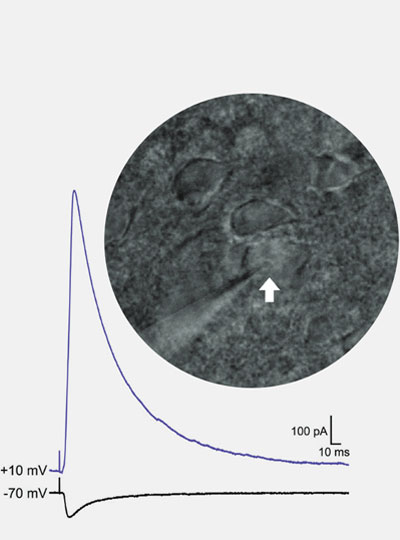
Riggs uses patch clamp electrophysiology to record the strength of inhibitory and excitatory synaptic input onto individual neurons (white arrow) in an animal model of autism. Image: Lace Riggs

“If we understand how these long-lasting synaptic changes come about, then we might be able to leverage these mechanisms to develop new and more effective treatments.”

While the turmoil of her childhood is in the past, Riggs says it is not forgotten — in part, because of its lasting effects on her own mental health.  She talks openly about her ongoing struggle with social anxiety and complex post-traumatic stress disorder because she is passionate about dismantling the stigma surrounding these conditions. “It’s something I have to deal with every day,” Riggs says. That means coping with symptoms like difficulty concentrating, hypervigilance, and heightened sensitivity to stress. “It’s like a constant hum in the background of my life, it never stops,” she says.

“I urge all of us to strive, not only to make scientific discoveries to move the field forward,” says Riggs, “but to improve the accessibility of this career to those whose lived experiences are required to truly accomplish that goal.”

Studies of autism tend to exclude women, researchers find

In recent years, researchers who study autism have made an effort to include more women and girls in their studies. However, despite these efforts, most studies of autism consistently enroll small numbers of female subjects or exclude them altogether, according to a new study from MIT.

The researchers found that a screening test commonly used to determine eligibility for studies of autism consistently winnows out a much higher percentage of women than men, creating a “leaky pipeline” that results in severe underrepresentation of women in studies of autism.

This lack of representation makes it more difficult to develop useful interventions or provide accurate diagnoses for girls and women, the researchers say.

“I think the findings favor having a more inclusive approach and widening the lens to end up being less biased in terms of who participates in research,” says John Gabrieli, the Grover Hermann Professor of Health Sciences and Technology and a professor of brain and cognitive sciences at MIT. “The more we understand autism in men and women and nonbinary individuals, the better services and more accurate diagnoses we can provide.”

Gabrieli, who is also a member of MIT’s McGovern Institute for Brain Research, is the senior author of the study, which appears in the journal Autism Research. Anila D’Mello, a former MIT postdoc who is now an assistant professor at the University of Texas Southwestern, is the lead author of the paper. MIT Technical Associate Isabelle Frosch, Research Coordinator Cindy Li, and Research Specialist Annie Cardinaux are also authors of the paper.

Gabrieli lab researchers Annie Cardinaux (left), Anila D’Mello (center), Cindy Li (right), and Isabelle Frosch (not pictured) have
uncovered sex biases in ASD research. Photo: Steph Stevens

Screening out females

Autism spectrum disorders are diagnosed based on observation of traits such as repetitive behaviors and difficulty with language and social interaction. Doctors may use a variety of screening tests to help them make a diagnosis, but these screens are not required.

For research studies of autism, it is routine to use a screening test called the Autism Diagnostic Observation Schedule (ADOS) to determine eligibility for the study. This test, which assesses social interaction, communication, play, and repetitive behaviors, provides a quantitative score in each category, and only participants who reach certain scores qualify for inclusion in studies.

While doing a study exploring how quickly the brains of autistic adults adapt to novel events in the environment, scientists in Gabrieli’s lab began to notice that the ADOS appeared to have unequal effects on male and female participation in research. As the study progressed, D’Mello noticed some significant brain differences between the male and female subjects in the study.

To investigate these differences further, D’Mello tried to find more female participants using an MIT database of autistic adults who have expressed interest in participating in research studies. However, when she sorted through the subjects, she found that only about half of the women in the database had met the ADOS cutoff scores typically required for inclusion in autism studies, compared to 80 percent of the males.

“We realized then that there’s a discrepancy and that the ADOS is essentially screening out who eventually participated in research,” D’Mello says. “We were really surprised at how many males we retained and how many females we lost to the ADOS.”

To see if this phenomenon was more widespread, the researchers looked at six publicly available datasets, which include more than 40,000 adults who have been diagnosed as autistic. For some of these datasets, participants were screened with ADOS to determine their eligibility to participate in studies, while for others, a “community diagnosis” — diagnosis from a doctor or other health care provider — was sufficient.

The researchers found that in datasets that required ADOS screening for eligibility, the ratio of male to female participants ended up being around 8:1, while in those that required only a community diagnosis the ratios ranged from about 2:1 to 1:1.

Previous studies have found differences between behavioral patterns in autistic men and women, but the ADOS test was originally developed using a largely male sample, which may explain why it often excludes women from research studies, D’Mello says.

“There were few females in the sample that was used to create this assessment, so it might be that it’s not great at picking up the female phenotype, which may differ in certain ways — primarily in domains like social communication,” she says.

Effects of exclusion

Failure to include more women and girls in studies of autism may contribute to shortcomings in the definitions of the disorder, the researchers say.

“The way we think about it is that the field evolved perhaps an implicit bias in how autism is defined, and it was driven disproportionately by analysis of males, and recruitment of males, and so on,” Gabrieli says. “So, the definition doesn’t fit as well, on average, with the different expression of autism that seems to be more common in females.”

This implicit bias has led to documented difficulties in receiving a diagnosis for girls and women, even when their symptoms are the same as those presented by autistic boys and men.

“Many females might be missed altogether in terms of diagnoses, and then our study shows that in the research setting, what is already a small pool gets whittled down at a much larger rate than that of males,” D’Mello says.

Excluding girls and women from this kind of research study can lead to treatments that don’t work as well for them, and it contributes to the perception that autism doesn’t affect women as much as men.

“The goal is that research should directly inform treatment, therapies, and public perception,” D’Mello says. “If the research is saying that there aren’t females with autism, or that the brain basis of autism only looks like the patterns established in males, then you’re not really helping females as much as you could be, and you’re not really getting at the truth of what the disorder might be.”

The researchers now plan to further explore some of the gender and sex-based differences that appear in autism, and how they arise. They also plan to expand the gender categories that they include. In the current study, the surveys that each participant filled out asked them to choose male or female, but the researchers have updated their questionnaire to include nonbinary and transgender options.

The research was funded by the Hock E. Tan and K. Lisa Yang Center for Autism Research, the Simons Center for the Social Brain at MIT, and the National Institutes of Mental Health.

Artificial neural networks model face processing in autism

Many of us easily recognize emotions expressed in others’ faces. A smile may mean happiness, while a frown may indicate anger. Autistic people often have a more difficult time with this task. It’s unclear why. But new research, published today in The Journal of Neuroscience, sheds light on the inner workings of the brain to suggest an answer. And it does so using a tool that opens new pathways to modeling the computation in our heads: artificial intelligence.

Researchers have primarily suggested two brain areas where the differences might lie. A region on the side of the primate (including human) brain called the inferior temporal (IT) cortex contributes to facial recognition. Meanwhile, a deeper region called the amygdala receives input from the IT cortex and other sources and helps process emotions.

Kohitij Kar, a research scientist in the lab of MIT Professor James DiCarlo, hoped to zero in on the answer. (DiCarlo, the Peter de Florez Professor in the Department of Brain and Cognitive Sciences, is a member of the McGovern Institute for Brain Research and director of MIT’s Quest for Intelligence.)

Kar began by looking at data provided by two other researchers: Shuo Wang, at Washington University in St. Louis, and Ralph Adolphs, at the California Institute of Technology. In one experiment, they showed images of faces to autistic adults and to neurotypical controls. The images had been generated by software to vary on a spectrum from fearful to happy, and the participants judged, quickly, whether the faces depicted happiness. Compared with controls, autistic adults required higher levels of happiness in the faces to report them as happy.

Modeling the brain

Kar, who is also a member of the Center for Brains, Minds and Machines, trained an artificial neural network, a complex mathematical function inspired by the brain’s architecture, to perform the same task. The network contained layers of units that roughly resemble biological neurons that process visual information. These layers process information as it passes from an input image to a final judgment indicating the probability that the face is happy. Kar found that the network’s behavior more closely matched the neurotypical controls than it did the autistic adults.

The network also served two more interesting functions. First, Kar could dissect it. He stripped off layers and retested its performance, measuring the difference between how well it matched controls and how well it matched autistic adults. This difference was greatest when the output was based on the last network layer. Previous work has shown that this layer in some ways mimics the IT cortex, which sits near the end of the primate brain’s ventral visual processing pipeline. Kar’s results implicate the IT cortex in differentiating neurotypical controls from autistic adults.

The other function is that the network can be used to select images that might be more efficient in autism diagnoses. If the difference between how closely the network matches neurotypical controls versus autistic adults is greater when judging one set of images versus another set of images, the first set could be used in the clinic to detect autistic behavioral traits. “These are promising results,” Kar says. Better models of the brain will come along, “but oftentimes in the clinic, we don’t need to wait for the absolute best product.”

Next, Kar evaluated the role of the amygdala. Again, he used data from Wang and colleagues. They had used electrodes to record the activity of neurons in the amygdala of people undergoing surgery for epilepsy as they performed the face task. The team found that they could predict a person’s judgment based on these neurons’ activity. Kar re-analyzed the data, this time controlling for the ability of the IT-cortex-like network layer to predict whether a face truly was happy. Now, the amygdala provided very little information of its own. Kar concludes that the IT cortex is the driving force behind the amygdala’s role in judging facial emotion.

Noisy networks

Finally, Kar trained separate neural networks to match the judgments of neurotypical controls and autistic adults. He looked at the strengths or “weights” of the connections between the final layers and the decision nodes. The weights in the network matching autistic adults, both the positive or “excitatory” and negative or “inhibitory” weights, were weaker than in the network matching neurotypical controls. This suggests that sensory neural connections in autistic adults might be noisy or inefficient.

To further test the noise hypothesis, which is popular in the field, Kar added various levels of fluctuation to the activity of the final layer in the network modeling autistic adults. Within a certain range, added noise greatly increased the similarity between its performance and that of the autistic adults. Adding noise to the control network did much less to improve its similarity to the control participants. This further suggest that sensory perception in autistic people may be the result of a so-called “noisy” brain.

Computational power

Looking forward, Kar sees several uses for computational models of visual processing. They can be further prodded, providing hypotheses that researchers might test in animal models. “I think facial emotion recognition is just the tip of the iceberg,” Kar says. They can also be used to select or even generate diagnostic content. Artificial intelligence could be used to generate content like movies and educational materials that optimally engages autistic children and adults. One might even tweak facial and other relevant pixels in what autistic people see in augmented reality goggles, work that Kar plans to pursue in the future.

Ultimately, Kar says, the work helps to validate the usefulness of computational models, especially image-processing neural networks. They formalize hypotheses and make them testable. Does one model or another better match behavioral data? “Even if these models are very far off from brains, they are falsifiable, rather than people just making up stories,” he says. “To me, that’s a more powerful version of science.”

Circuit that focuses attention brings in wide array of inputs

In a new brain-wide circuit tracing study, scientists at MIT’s Picower Institute for Learning and Memory focused selective attention on a circuit that governs, fittingly enough, selective attention. The comprehensive maps they produced illustrate how broadly the mammalian brain incorporates and integrates information to focus its sensory resources on its goals.

Working in mice, the team traced thousands of inputs into the circuit, a communication loop between the anterior cingulate cortex (ACC) and the lateral posterior (LP) thalamus. In primates the LP is called the pulvinar. Studies in humans and nonhuman primates have indicated that the byplay of these two regions is critical for brain functions like being able to focus on an object of interest in a crowded scene, says study co-lead author Yi Ning Leow, a graduate student in the lab of senior author Mriganka Sur, the Newton Professor in MIT’s Department of Brain and Cognitive Sciences. Research has implicated dysfunction in the circuit in attention-affecting disorders such as autism and attention deficit/hyperactivity disorder.

The new study in the Journal of Comparative Neurology extends what’s known about the circuit by detailing it in mice, Leow says, importantly showing that the mouse circuit is closely analogous to the primate version even if the LP is proportionately smaller and less evolved than the pulvinar.

“In these rodent models we were able to find very similar circuits,” Leow says. “So we can possibly study these higher-order functions in mice as well. We have a lot more genetic tools in mice so we are better able to look at this circuit.”

The study, also co-led by former MIT undergraduate Blake Zhou, therefore provides a detailed roadmap in the experimentally accessible mouse model for understanding how the ACC and LP cooperate to produce selective attention. For instance, now that Leow and Zhou have located all the inputs that are wired into the circuit, Leow is tapping into those feeds to eavesdrop on the information they are carrying. Meanwhile, she is correlating that information flow with behavior.

“This study lays the groundwork for understanding one of the most important, yet most elusive, components of brain function, namely our ability to selectively attend to one thing out of several, as well as switch attention,” Sur says.

Using virally mediated circuit-tracing techniques pioneered by co-author Ian Wickersham, principal research scientist in brain and cognitive sciences and the McGovern Institute for Brain Research at MIT, the team found distinct sources of input for the ACC and the LP. Generally speaking, the detailed study finds that the majority of inputs to the ACC were from frontal cortex areas that typically govern goal-directed planning, and from higher visual areas. The bulk of inputs to the LP, meanwhile, were from deeper regions capable of providing context such as the mouse’s needs, location and spatial cues, information about movement, and general information from a mix of senses.

So even though focusing attention might seem like a matter of controlling the senses, Leow says, the circuit pulls in a lot of other information as well.

“We’re seeing that it’s not just sensory — there are so many inputs that are coming from non-sensory areas as well, both sub-cortically and cortically,” she says. “It seems to be integrating a lot of different aspects that might relate to the behavioral state of the animal at a given time. It provides a way to provide a lot of internal and special context for that sensory information.”

Given the distinct sets of inputs to each region, the ACC may be tasked with focusing attention on a desired object, while the LP is modulating how the ACC goes about making those computations, accounting for what’s going on both inside and outside the animal. Decoding just what that incoming contextual information is, and what the LP tells the ACC, are the key next steps, Leow says. Another clear set of questions the study raises are what are the circuit’s outputs. In other words, after it integrates all this information, what does it do with it?

The paper’s other authors are Heather Sullivan and Alexandria Barlowe.

A National Science Scholarship, the National Institutes of Health, and the JPB Foundation provided support for the study.