Five MIT faculty elected 2021 AAAS Fellows

Five MIT faculty members have been elected as fellows of the American Association for the Advancement of Science (AAAS).

The 2021 class of AAAS Fellows includes 564 scientists, engineers, and innovators spanning 24 scientific disciplines who are being recognized for their scientifically and socially distinguished achievements.

Mircea Dincă is the W. M. Keck Professor of Energy in the Department of Chemistry. His group’s research focuses on addressing challenges related to the storage and consumption of energy, and global environmental concerns. Central to these efforts are the synthesis of novel organic-inorganic hybrid materials and the manipulation of their electrochemical and photophysical properties, with a current emphasis on porous materials and extended one-dimensional van der Waals materials.

Guoping Feng is the James W. and Patricia T. Poitras Professor of Neuroscience in the Department of Brain and Cognitive Sciences, associate director of MIT’s McGovern Institute for Brain Research, director of Model Systems and Neurobiology at the Stanley Center for Psychiatric Research, and an institute member of the Broad Institute of MIT and Harvard. His research is devoted to understanding the development and function of synapses in the brain and how synaptic dysfunction may contribute to neurodevelopmental and psychiatric disorders. By understanding the molecular, cellular, and circuitry mechanisms of these disorders, Feng hopes his work will eventually lead to the development of new and effective treatments for the millions of people suffering from these devastating diseases.

David Shoemaker is a senior research scientist with the MIT Kavli Institute for Astrophysics and Space Research. His work is focused on gravitational-wave observation and includes developing technologies for the detectors (LIGO, LISA), developing proposals for new instruments (Cosmic Explorer), managing the teams to build them and the consortia which exploit the data (LIGO Scientific Collaboration, LISA Consortium), and supporting the overall growth of the field (Gravitational-Wave International Committee).

Ian Hunter is the Hatsopoulos Professor of Mechanical Engineering and runs the Bioinstrumentation Lab at MIT. His main areas of research are instrumentation, microrobotics, medical devices, and biomimetic materials. Over the years he and his students have developed many instruments and devices including: confocal laser microscopes, scanning tunneling electron microscopes, miniature mass spectrometers, new forms of Raman spectroscopy, needle-free drug delivery technologies, nano- and micro-robots, microsurgical robots, robotic endoscopes, high-performance Lorentz force motors, and microarray technologies for massively parallel chemical and biological assays.

Evelyn N. Wang is the Ford Professor of Engineering and head of the Department of Mechanical Engineering. Her research program combines fundamental studies of micro/nanoscale heat and mass transport processes with the development of novel engineered structures to create innovative solutions in thermal management, energy, and water harvesting systems. Her work in thermophotovoltaics was named to Technology Review’s lists of Biggest Clean Energy Advances, in 2016, and Ten Breakthrough Technologies, in 2017, and to the Department of Energy Frontiers Research Center’s Ten of Ten awards. Her work extracting water from air has won her the title of 2017 Foreign Policy’s Global ReThinker and the 2018 Eighth Prince Sultan bin Abdulaziz International Prize for Water.

Single gene linked to repetitive behaviors, drug addiction

Making and breaking habits is a prime function of the striatum, a large forebrain region that underlies the cerebral cortex. McGovern researchers have identified a particular gene that controls striatal function as well as repetitive behaviors that are linked to drug addiction vulnerability.

To identify genes involved specifically in striatal functions, MIT Institute Professor Ann Graybiel previously identified genes that are preferentially expressed in striatal neurons. One identified gene encodes CalDAG-GEFI (CDGI), a signaling molecule that effects changes inside of cells in response to extracellular signals that are received by receptors on the cell surface. In a paper to be published in the October issue of Neurobiology of Disease and now available online, Graybiel, along with former Research Scientist Jill Crittenden and collaborators James Surmeier and Shenyu Zhai at the Feinman School of Medicine at Northwestern University, show that CDGI is key for controlling behavioral responses to drugs of abuse and underlying neuronal plasticity (cellular changes induced by experience) in the striatum.

“This paper represents years of intensive research, which paid off in the end by identifying a specific cellular signaling cascade for controlling repetitive behaviors and neuronal plasticity,” says Graybiel, who is also an investigator at the McGovern Institute and a professor of brain and cognitive sciences at MIT.

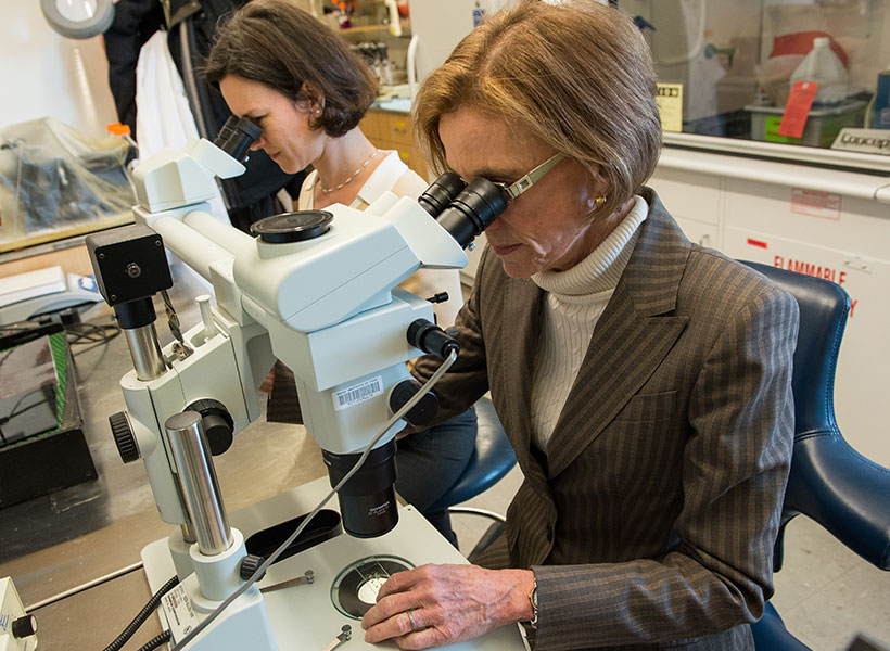
McGovern Investigator Ann Graybiel (right) with former Research Scientist Jill Crittenden. Photo: Justin Knight

Surprise discovery

To understand the essential roles of CDGI, Crittenden first engineered “knockout” mice that lack the gene encoding CDGI. Then the Graybiel team began looking for abnormalities in the CDGI knockout mice that could be tied to the loss of CDGI’s function.

Initially, they noticed that the rodent ear-tag IDs often fell off in the knockout mice, an observation that ultimately led to the surprise discovery by the Graybiel team and others that CDGI is expressed in blood platelets and is responsible for a bleeding disorder in humans, dogs, and other animals. The CDGI knockout mice were otherwise healthy and seemed just like their “wildtype” brothers and sisters, which did not carry the gene mutation. To figure out the role of CDGI in the brain, the Graybiel team would have to scrutinize the mice more closely.

Challenging the striatum

Both the CDGI knockout and wildtype mice were given an extensive set of behavioral and neurological tests and the CDGI mice showed deficits in two tests designed to challenge the striatum.

In one test, mice must find their way through a maze by relying on egocentric (i.e. self-referential) cues, such as their turning right or turning left, and not competing allocentric (i.e. external) cues, such as going toward a bright poster on the wall. Egocentric cues are thought to be processed by the striatum whereas allocentric cues are thought to rely on the hippocampus.

In a second test of striatal function, mice learned various gait patterns to match different patterns of rungs on their running wheel, a task designed to test the mouse’s ability to learn and remember a motor sequence.

The CDGI mice learned both of these striatal tasks more slowly than their wildtype siblings, suggesting that the CDGI mice might perform normally in general tests of behavior because they are able to compensate for striatal deficits by using other brain regions such as the hippocampus to solve standard tasks.

The team then decided to give the mice a completely different type of test that relies on the striatum. Because the striatum is strongly activated by drugs of abuse, which elevate dopamine and drive motor habits, Crittenden and collaborator Morgane Thomsen (now at the University of Copenhagen) looked to see whether the CDGI knockout mice respond normally to amphetamine and cocaine.

Psychomotor stimulants like cocaine and amphetamine normally induce a mixture of hyperactive behaviors such as pacing and focused repetitive behaviors like skin-picking (also called stereotypy or punding in humans). The researchers found however, that the drug-induced behaviors in the CDGI knockout mice were less varied than the normal mice and consisted of abnormally prolonged stereotypy, as though the mice were unable to switch between behaviors. The researchers were able to map the abnormal behavior to CDGI function in the striatum by showing that the same vulnerability to drug-induced stereotypy was observed in mice that were engineered to delete CDGI in the striatum after birth (“conditional knockouts”), but to otherwise have normal CDGI throughout the body.

Controlling cravings

In addition to exhibiting prolonged, repetitive behaviors, the CDGI knockout mice had a vulnerability to self-administer drugs. Although previous research had shown that treatments that activate the M1 acetylcholine receptor can block cocaine self-administration, the team found that this therapy was ineffective in CDGI knockout mice. Knockouts continued to self-administer cocaine (suggesting increased craving for the drug) at the same rate before and after M1 receptor activation treatment, even though the treatment succeeded with their sibling control mice. The researchers concluded that CDGI is critically important for controlling repetitive behaviors and the ability to stop self-administration of addictive stimulants.

mouse brain images
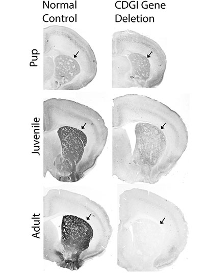
Brain sections from control mice (left) and mice engineered for deletion of the CDGI gene after birth. The expression of CDGI in the striatum (arrows) grows stronger as mice grow from pups to adulthood in control mice, but is gradually lost in the CDGI engineered mice (“conditional knockouts”). Image courtesy of the researchers

To better understand how CDGI is linked to the M1 receptor at the cellular level, the team turned to slice physiologists, scientists who record the electrical activity of neurons in brain slices. Their recordings showed that striatal neurons from CDGI knockouts fail to undergo the normal, expected electrophysiological changes after receiving treatments that target the M1 receptor. In particular, the neurons of the striatum that function broadly to stop ongoing behaviors, did not integrate cellular signals properly and failed to undergo “long-term potentiation,” a type of neuronal plasticity thought to underlie learning.

The new findings suggest that excessive repetitive movements are controlled by M1 receptor signaling through CDGI in indirect pathway neurons of the striatum, a neuronal subtype that degenerates in Huntington’s disease and is affected by dopamine loss and l-DOPA replacement therapy in Parkinson’s disease.

“The M1 acetylcholine receptor is a target for therapeutic drug development in treating cognitive and behavioral problems in multiple disorders, but progress has been severely hampered by off-target side-effects related to the wide-spread expression of the M1 receptor,” Graybiel explains. “Our findings suggest that CDGI offers the possibility for forebrain-specific targeting of M1 receptor signaling cascades that are of interest for blocking pathologically repetitive and unwanted behaviors that are common to numerous brain disorders including Huntington’s disease, drug addiction, autism, and schizophrenia as well as drug-induced dyskinesias. We hope that this work can help therapeutic development for these major health problems.”

This work was funded by the James W. (1963) and Patricia T. Poitras Fund, the William N. & Bernice E. Bumpus Foundation, the Saks Kavanaugh Foundation, the Simons Foundation, and the National Institute of Health.

Some brain disorders exhibit similar circuit malfunctions

Many neurodevelopmental disorders share similar symptoms, such as learning disabilities or attention deficits. A new study from MIT has uncovered a common neural mechanism for a type of cognitive impairment seen in some people with autism and schizophrenia, even though the genetic variations that produce the impairments are different for each condition.

In a study of mice, the researchers found that certain genes that are mutated or missing in some people with those disorders cause similar dysfunctions in a neural circuit in the thalamus. If scientists could develop drugs that target this circuit, they could be used to treat people who have different disorders with common behavioral symptoms, the researchers say.

“This study reveals a new circuit mechanism for cognitive impairment and points to a future direction for developing new therapeutics, by dividing patients into specific groups not by their behavioral profile, but by the underlying neurobiological mechanisms,” says Guoping Feng, the James W. and Patricia T. Poitras Professor in Brain and Cognitive Sciences at MIT, a member of the Broad Institute of Harvard and MIT, the associate director of the McGovern Institute for Brain Research at MIT, and the senior author of the new study.

Dheeraj Roy, a Warren Alpert Distinguished Scholar and a McGovern Fellow at the Broad Institute, and Ying Zhang, a postdoc at the McGovern Institute, are the lead authors of the paper, which appears today in Neuron.

Thalamic connections

The thalamus plays a key role in cognitive tasks such as memory formation and learning. Previous studies have shown that many of the gene variants linked to brain disorders such as autism and schizophrenia are highly expressed in the thalamus, suggesting that it may play a role in those disorders.

One such gene is called Ptchd1, which Feng has studied extensively. In boys, loss of this gene, which is carried on the X chromosome, can lead to attention deficits, hyperactivity, aggression, intellectual disability, and autism spectrum disorders.

In a study published in 2016, Feng and his colleagues showed that Ptchd1 exerts many of its effects in a part of the thalamus called the thalamic reticular nucleus (TRN). When the gene is knocked out in the TRN of mice, the mice show attention deficits and hyperactivity. However, that study did not find any role for the TRN in the learning disabilities also seen in people with mutations in Ptchd1.

In the new study, the researchers decided to look elsewhere in the thalamus to try to figure out how Ptchd1 loss might affect learning and memory. Another area they identified that highly expresses Ptchd1 is called the anterodorsal (AD) thalamus, a tiny region that is involved in spatial learning and communicates closely with the hippocampus.

Using novel techniques that allowed them to trace the connections between the AD thalamus and another brain region called the retrosplenial cortex (RSC), the researchers determined a key function of this circuit. They found that in mice, the AD-to-RSC circuit is essential for encoding fearful memories of a chamber in which they received a mild foot shock. It is also necessary for working memory, such as creating mental maps of physical spaces to help in decision-making.

The researchers found that a nearby part of the thalamus called the anteroventral (AV) thalamus also plays a role in this memory formation process: AV-to-RSC communication regulates the specificity of the encoded memory, which helps us distinguish this memory from others of similar nature.

“These experiments showed that two neighboring subdivisions in the thalamus contribute differentially to memory formation, which is not what we expected,” Roy says.

Circuit malfunction

Once the researchers discovered the roles of the AV and AD thalamic regions in memory formation, they began to investigate how this circuit is affected by loss of Ptchd1. When they knocked down expression of Ptchd1 in neurons of the AD thalamus, they found a striking deficit in memory encoding, for both fearful memories and working memory.

The researchers then did the same experiments with a series of four other genes — one that is linked with autism and three linked with schizophrenia. In all of these mice, they found that knocking down gene expression produced the same memory impairments. They also found that each of these knockdowns produced hyperexcitability in neurons of the AD thalamus.

These results are consistent with existing theories that learning occurs through the strengthening of synapses that occurs as a memory is formed, the researchers say.

“The dominant theory in the field is that when an animal is learning, these neurons have to fire more, and that increase correlates with how well you learn,” Zhang says. “Our simple idea was if a neuron fires too high at baseline, you may lack a learning-induced increase.”

The researchers demonstrated that each of the genes they studied affects different ion channels that influence neurons’ firing rates. The overall effect of each mutation is an increase in neuron excitability, which leads to the same circuit-level dysfunction and behavioral symptoms.

The researchers also showed that they could restore normal cognitive function in mice with these genetic mutations by artificially turning down hyperactivity in neurons of the AD thalamus. The approach they used, chemogenetics, is not yet approved for use in humans. However, it may be possible to target this circuit in other ways, the researchers say.

The findings lend support to the idea that grouping diseases by the circuit malfunctions that underlie them may help to identify potential drug targets that could help many patients, Feng says.

“There are so many genetic factors and environmental factors that can contribute to a particular disease, but in the end, it has to cause some type of neuronal change that affects a circuit or a few circuits involved in this behavior,” he says. “From a therapeutic point of view, in such cases you may not want to go after individual molecules because they may be unique to a very small percentage of patients, but at a higher level, at the cellular or circuit level, patients may have more commonalities.”

The research was funded by the Stanley Center at the Broad Institute, the Hock E. Tan and K. Lisa Yang Center for Autism Research at MIT, the James and Patricia Poitras Center for Psychiatric Disorders Research at MIT, and the National Institutes of Health BRAIN Initiative.

New technique corrects disease-causing mutations

Gene editing, or purposefully changing a gene’s DNA sequence, is a powerful tool for studying how mutations cause disease, and for making changes in an individual’s DNA for therapeutic purposes. A novel method of gene editing that can be used for both purposes has now been developed by a team led by Guoping Feng, the James W. (1963) and Patricia T. Poitras Professor in Brain and Cognitive Sciences at MIT.

“This technical advance can accelerate the production of disease models in animals and, critically, opens up a brand-new methodology for correcting disease-causing mutations,” says Feng, who is also a member of the Broad Institute of Harvard and MIT and the associate director of the McGovern Institute for Brain Research at MIT. The new findings publish online May 26 and in print June 10 in the journal Cell.

Genetic models of disease

A major goal of the Feng lab is to precisely define what goes wrong in neurodevelopmental and neuropsychiatric disorders by engineering animal models that carry the gene mutations that cause these disorders in humans. New models can be generated by injecting embryos with gene editing tools, along with a piece of DNA carrying the desired mutation.

In one such method, the gene editing tool CRISPR is programmed to cut a targeted gene, thereby activating natural DNA mechanisms that “repair” the broken gene with the injected template DNA. The engineered cells are then used to generate offspring capable of passing the genetic change on to further generations, creating a stable genetic line in which the disease, and therapies, are tested.

Although CRISPR has accelerated the process of generating such disease models, the process can still take months or years. Reasons for the inefficiency are that many treated cells do not undergo the desired DNA sequence change at all, and the change only occurs on one of the two gene copies (for most genes, each cell contains two versions, one from the father and one from the mother).

In an effort to increase the efficiency of the gene editing process, the Feng lab team initially hypothesized that adding a DNA repair protein called RAD51 to a standard mixture of CRISPR gene editing tools would increase the chances that a cell (in this case a fertilized mouse egg, or one-cell embryo) would undergo the desired genetic change.

As a test case, they measured the rate at which they were able to insert (“knock-in”) a mutation in the gene Chd2 that is associated with autism.  The overall proportion of embryos that were correctly edited remained unchanged, but to their surprise, a significantly higher percentage carried the desired gene edit on both chromosomes. Tests with a different gene yielded the same unexpected outcome.

“Editing of both chromosomes simultaneously is normally very uncommon,” explains postdoctoral fellow Jonathan Wilde.  “The high rate of editing seen with RAD51 was really striking and what started as a simple attempt to make mutant Chd2 mice quickly turned into a much bigger project focused on RAD51 and its applications in genome editing,” said Wilde, who co-authored the Cell paper with research scientist Tomomi Aida.

A molecular copy machine

The Feng lab team next set out to understand the mechanism by which RAD51 enhances gene editing. They hypothesized that RAD51 engages a process called interhomolog repair (IHR), whereby a DNA break on one chromosome is repaired using the second copy of the chromosome (from the other parent) as the template.

To test this, they injected mouse embryos with RAD51 and CRISPR but left out the template DNA. They programmed CRISPR to cut only the gene sequence on one of the chromosomes, and then tested whether it was repaired to match the sequence on the uncut chromosome. For this experiment, they had to use mice in which the sequences on the maternal and paternal chromosomes were different.

They found that control embryos injected with CRISPR alone rarely showed IHR repair. However, addition of RAD51 significantly increased the number of embryos in which the CRISPR-targeted gene was edited to match the uncut chromosome.

“Previous studies of IHR found that it is incredibly inefficient in most cells,” says Wilde. “Our finding that it occurs much more readily in embryonic cells and can be enhanced by RAD51 suggest that a deeper understanding of what makes the embryo permissive to this type of DNA repair could help us design safer and more efficient gene therapies.”

A new way to correct disease-causing mutations          

Standard gene therapy strategies that rely on injecting a corrective piece of DNA to serve as a template for repairing the mutation engage a process called homology-directed repair (HDR).

“HDR-based strategies still suffer from low efficiency and carry the risk of unwanted integration of donor DNA throughout the genome,” explains Feng. “IHR has the potential to overcome these problems because it relies upon natural cellular pathways and the patient’s own normal chromosome for correction of the deleterious mutation.”

Feng’s team went on to identify additional DNA repair-associated proteins that can stimulate IHR, including several that not only promote high levels of IHR, but also repress errors in the DNA repair process. Additional experiments that allowed the team to examine the genomic features of IHR events gave deeper insight into the mechanism of IHR and suggested ways that the technique can be used to make gene therapies safer.

“While there is still a great deal to learn about this new application of IHR, our findings are the foundation for a new gene therapy approach that could help solve some of the big problems with current approaches,” says Aida.

This study was supported by the Hock E. Tan and K. Lisa Yang Center for Autism Research at MIT, the Poitras Center for Psychiatric Disorders Research at MIT, NIH/NIMH Conte Center Grant (P50 MH094271) and NIH Office of the Director (U24 OD026638).

Individual neurons responsible for complex social reasoning in humans identified

This story is adapted from a January 27, 2021 press release from Massachusetts General Hospital.

The ability to understand others’ hidden thoughts and beliefs is an essential component of human social behavior. Now, neuroscientists have for the first time identified specific neurons critical for social reasoning, a cognitive process that requires individuals to acknowledge and predict others’ hidden beliefs and thoughts.

The findings, published in Nature, open new avenues of study into disorders that affect social behavior, according to the authors.

In the study, a team of Harvard Medical School investigators based at Massachusetts General Hospital and colleagues from MIT took a rare look at how individual neurons represent the beliefs of others. They did so by recording neuron activity in patients undergoing neurosurgery to alleviate symptoms of motor disorders such as Parkinson’s disease.

Theory of mind

The researcher team, which included McGovern scientists Ev Fedorenko and Rebecca Saxe, focused on a complex social cognitive process called “theory of mind.” To illustrate this, let’s say a friend appears to be sad on her birthday. One may infer she is sad because she didn’t get a present or she is upset at growing older.

“When we interact, we must be able to form predictions about another person’s unstated intentions and thoughts,” said senior author Ziv Williams, HMS associate professor of neurosurgery at Mass General. “This ability requires us to paint a mental picture of someone’s beliefs, which involves acknowledging that those beliefs may be different from our own and assessing whether they are true or false.”

This social reasoning process develops during early childhood and is fundamental to successful social behavior. Individuals with autism, schizophrenia, bipolar affective disorder, and traumatic brain injuries are believed to have a deficit of theory-of-mind ability.

For the study, 15 patients agreed to perform brief behavioral tasks before undergoing neurosurgery for placement of deep-brain stimulation for motor disorders. Microelectrodes inserted into the dorsomedial prefrontal cortex recorded the behavior of individual neurons as patients listened to short narratives and answered questions about them.

For example, participants were presented with the following scenario to evaluate how they considered another’s belief of reality: “You and Tom see a jar on the table. After Tom leaves, you move the jar to a cabinet. Where does Tom believe the jar to be?”

Social computation

The participants had to make inferences about another’s beliefs after hearing each story. The experiment did not change the planned surgical approach or alter clinical care.

“Our study provides evidence to support theory of mind by individual neurons,” said study first author Mohsen Jamali, HMS instructor in neurosurgery at Mass General. “Until now, it wasn’t clear whether or how neurons were able to perform these social cognitive computations.”

The investigators found that some neurons are specialized and respond only when assessing another’s belief as false, for example. Other neurons encode information to distinguish one person’s beliefs from another’s. Still other neurons create a representation of a specific item, such as a cup or food item, mentioned in the story. Some neurons may multitask and aren’t dedicated solely to social reasoning.

“Each neuron is encoding different bits of information,” Jamali said. “By combining the computations of all the neurons, you get a very detailed representation of the contents of another’s beliefs and an accurate prediction of whether they are true or false.”

Now that scientists understand the basic cellular mechanism that underlies human theory of mind, they have an operational framework to begin investigating disorders in which social behavior is affected, according to Williams.

“Understanding social reasoning is also important to many different fields, such as child development, economics, and sociology, and could help in the development of more effective treatments for conditions such as autism spectrum disorder,” Williams said.

Previous research on the cognitive processes that underlie theory of mind has involved functional MRI studies, where scientists watch which parts of the brain are active as volunteers perform cognitive tasks.

But the imaging studies capture the activity of many thousands of neurons all at once. In contrast, Williams and colleagues recorded the computations of individual neurons. This provided a detailed picture of how neurons encode social information.

“Individual neurons, even within a small area of the brain, are doing very different things, not all of which are involved in social reasoning,” Williams said. “Without delving into the computations of single cells, it’s very hard to build an understanding of the complex cognitive processes underlying human social behavior and how they go awry in mental disorders.”

Adapted from a Mass General news release.

A large-scale tool to investigate the function of autism spectrum disorder genes

Scientists at Harvard University, the Broad Institute of MIT and Harvard, and MIT have developed a technology to investigate the function of many different genes in many different cell types at once, in a living organism. They applied the large-scale method to study dozens of genes that are associated with autism spectrum disorder, identifying how specific cell types in the developing mouse brain are impacted by mutations.

The “Perturb-Seq” method, published in the journal Science, is an efficient way to identify potential biological mechanisms underlying autism spectrum disorder, which is an important first step toward developing treatments for the complex disease. The method is also broadly applicable to other organs, enabling scientists to better understand a wide range of disease and normal processes.

“For many years, genetic studies have identified a multitude of risk genes that are associated with the development of autism spectrum disorder. The challenge in the field has been to make the connection between knowing what the genes are, to understanding how the genes actually affect cells and ultimately behavior,” said co-senior author Paola Arlotta, the Golub Family Professor of Stem Cell and Regenerative Biology at Harvard. “We applied the Perturb-Seq technology to an intact developing organism for the first time, showing the potential of measuring gene function at scale to better understand a complex disorder.”

The study was also led by co-senior authors Aviv Regev, who was a core member of the Broad Institute during the study and is currently Executive Vice President of Genentech Research and Early Development, and Feng Zhang, a core member of the Broad Institute and an investigator at MIT’s McGovern Institute.

To investigate gene function at a large scale, the researchers combined two powerful genomic technologies. They used CRISPR-Cas9 genome editing to make precise changes, or perturbations, in 35 different genes linked to autism spectrum disorder risk. Then, they analyzed changes in the developing mouse brain using single-cell RNA sequencing, which allowed them to see how gene expression changed in over 40,000 individual cells.

By looking at the level of individual cells, the researchers could compare how the risk genes affected different cell types in the cortex — the part of the brain responsible for complex functions including cognition and sensation. They analyzed networks of risk genes together to find common effects.

“We found that both neurons and glia — the non-neuronal cells in the brain — are directly affected by different sets of these risk genes,” said Xin Jin, lead author of the study and a Junior Fellow of the Harvard Society of Fellows. “Genes and molecules don’t generate cognition per se — they need to impact specific cell types in the brain to do so. We are interested in understanding how these different cell types can contribute to the disorder.”

To get a sense of the model’s potential relevance to the disorder in humans, the researchers compared their results to data from post-mortem human brains. In general, they found that in the post-mortem human brains with autism spectrum disorder, some of the key genes with altered expression were also affected in the Perturb-seq data.

“We now have a really rich dataset that allows us to draw insights, and we’re still learning a lot about it every day,” Jin said. “As we move forward with studying disease mechanisms in more depth, we can focus on the cell types that may be really important.”

“The field has been limited by the sheer time and effort that it takes to make one model at a time to test the function of single genes. Now, we have shown the potential of studying gene function in a developing organism in a scalable way, which is an exciting first step to understanding the mechanisms that lead to autism spectrum disorder and other complex psychiatric conditions, and to eventually develop treatments for these devastating conditions,” said Arlotta, who is also an institute member of the Broad Institute and part of the Broad’s Stanley Center for Psychiatric Research. “Our work also paves the way for Perturb-Seq to be applied to organs beyond the brain, to enable scientists to better understand the development or function of different tissue types, as well as pathological conditions.”

“Through genome sequencing efforts, a very large number of genes have been identified that, when mutated, are associated with human diseases. Traditionally, understanding the role of these genes would involve in-depth studies of each gene individually. By developing Perturb-seq for in vivo applications, we can start to screen all of these genes in animal models in a much more efficient manner, enabling us to understand mechanistically how mutations in these genes can lead to disease,” said Zhang, who is also the James and Patricia Poitras Professor of Neuroscience at MIT and a professor of brain and cognitive sciences and biological engineering at MIT.

This study was funded by the Stanley Center for Psychiatric Research at the Broad Institute, the National Institutes of Health, the Brain and Behavior Research Foundation’s NARSAD Young Investigator Grant, Harvard University’s William F. Milton Fund, the Klarman Cell Observatory, the Howard Hughes Medical Institute, a Center for Cell Circuits grant from the National Human Genome Research Institute’s Centers of Excellence in Genomic Science, the New York Stem Cell Foundation, the Mathers Foundation, the Poitras Center for Psychiatric Disorders Research at MIT, the Hock E. Tan and K. Lisa Yang Center for Autism Research at MIT, and J. and P. Poitras.

New neuron type discovered only in primate brains

Neuropsychiatric illnesses like schizophrenia and autism are a complex interplay of brain chemicals, environment, and genetics that requires careful study to understand the root causes. Scientists have traditionally relied on samples taken from mice and non-human primates to study how these diseases develop. But the question has lingered: are the brains of these subjects similar enough to humans to yield useful insights?

Now work from the Broad Institute of MIT and Harvard and the McGovern Institute for Brain Research is pointing towards an answer. In a study published in Nature, researchers from the Broad’s Stanley Center for Psychiatric Research report several key differences in the brains of ferrets, mice, nonhuman primates, and humans, all focused on a type of neuron called interneurons. Most surprisingly, the team found a new type of interneuron only in primates, located in a part of the brain called the striatum, which is associated with Huntington’s disease and potentially schizophrenia.

The findings could help accelerate research into causes of and treatments for neuropsychiatric illnesses, by helping scientists choose the lab model that best mimics features of the human brain that may be involved in these diseases.

“The data from this work will inform the study of human brain disorders because it helps us think about which features of the human brain can be studied in mice, which features require higher organisms such as marmosets, and why mouse models often don’t reflect the effects of the corresponding mutations in human,” said Steven McCarroll, senior author of the study, director of genetics at the Stanley Center, and a professor of genetics at Harvard Medical School.

“Dysfunctions of interneurons have been strongly linked to several brain disorders including autism spectrum disorder and schizophrenia,” said Guoping Feng, co-author of the study, director of model systems and neurobiology at the Stanley Center, and professor of neuroscience at MIT’s McGovern Institute for Brain Research. “These data further demonstrate the unique importance of non-human primate models in understanding neurobiological mechanisms of brain disorders and in developing and testing therapeutic approaches.”

Enter the interneuron

Interneurons form key nodes within neural circuitry in the brain, and help regulate neuronal activity by releasing the neurotransmitter GABA, which inhibits the firing of other neurons.

Fenna Krienen, a postdoctoral fellow in the McCarroll Lab and first author on the Nature paper, and her colleagues wanted to track the natural history of interneurons.

“We wanted to gain an understanding of the evolutionary trajectory of the cell types that make up the brain,” said Krienen. “And then we went about acquiring samples from species that could inform this understanding of evolutionary divergence between humans and the models that so often stand in for humans in neuroscience studies.”

One of the tools the researchers used was Drop-seq, a high-throughput single nucleus RNA sequencing technique developed by McCarroll’s lab, to classify the roles and locations of more than 184,000 telencephalic interneurons in the brains of ferrets, humans, macaques, marmosets, and mice. Using tissue from frozen samples, the team isolated the nuclei of interneurons from the cortex, the hippocampus, and the striatum, and profiled the RNA from the cells.

The researchers thought that because interneurons are found in all vertebrates, the cells would be relatively static from species to species.

“But with these sensitive measurements and a lot of data from the various species, we got a different picture about how lively interneurons are, in terms of the ways that evolution has tweaked their programs or their populations from one species to the next,” said Krienen.

She and her collaborators identified four main differences in interneurons between the species they studied: the cells change their proportions across brain regions, alter the programs they use to link up with other neurons, and can migrate to different regions of the brain.

But most strikingly, the scientists discovered that primates have a novel interneuron not found in other species. The interneuron is located in the striatum—the brain structure responsible for cognition, reward, and coordinated movements that has existed as far back on the evolutionary tree as ancient primitive fish. The researchers were amazed to find the new neuron type made up a third of all interneurons in the striatum.

“Although we expected the big innovations in human and primate brains to be in the cerebral cortex, which we tend to associate with human intelligence, it was in fact in the venerable striatum that Fenna uncovered the most dramatic cellular innovation in the primate brain,” said McCarroll. “This cell type had never been discovered before, because mice have nothing like it.”

“The question of what provides the “human advantage” in cognitive abilities is one of the fundamental issues neurobiologists have endeavored to answer,” said Gordon Fishell, group leader at the Stanley Center, a professor of neurobiology at Harvard Medical School, and a collaborator on the study. “These findings turn on end the question of ‘how do we build better brains?’. It seems at least part of the answer stems from creating a new list of parts.”

A better understanding of how these inhibitory neurons vary between humans and lab models will provide researchers with new tools for investigating various brain disorders. Next, the researchers will build on this work to determine the specific functions of each type of interneuron.

“In studying neurodevelopmental disorders, you would like to be convinced that your model is an appropriate one for really complex social behaviors,” Krienen said. “And the major overarching theme of the study was that primates in general seem to be very similar to one another in all of those interneuron innovations.”

Support for this work was provided in part by the Broad Institute’s Stanley Center for Psychiatric Research and the NIH Brain Initiative, the Dean’s Innovation Award (Harvard Medical School), the Hock E. Tan and K. Lisa Yang Center for Autism Research at MIT, the Poitras Center for Psychiatric Disorders Research at MIT, the McGovern Institute for Brain Research at MIT, and the National Institute of Neurological Disorders and Stroke.

New molecular therapeutics center established at MIT’s McGovern Institute

More than one million Americans are diagnosed with a chronic brain disorder each year, yet effective treatments for most complex brain disorders are inadequate or even nonexistent.

A major new research effort at MIT’s McGovern Institute aims to change how we treat brain disorders by developing innovative molecular tools that precisely target dysfunctional genetic, molecular, and circuit pathways.

The K. Lisa Yang and Hock E. Tan Center for Molecular Therapeutics in Neuroscience was established at MIT through a $28 million gift from philanthropist Lisa Yang and MIT alumnus Hock Tan ’75. Yang is a former investment banker who has devoted much of her time to advocacy for individuals with disabilities and autism spectrum disorders. Tan is President and CEO of Broadcom, a global technology infrastructure company. This latest gift brings Yang and Tan’s total philanthropy to MIT to more than $72 million.

Lisa Yang (center) and MIT alumnus Hock Tan ’75 with their daughter Eva (far left) pictured at the opening of the Hock E. Tan and K. Lisa Yang Center for Autism Research in 2017. Photo: Justin Knight

“In the best MIT spirit, Lisa and Hock have always focused their generosity on insights that lead to real impact,” says MIT President L. Rafael Reif. “Scientifically, we stand at a moment when the tools and insights to make progress against major brain disorders are finally within reach. By accelerating the development of promising treatments, the new center opens the door to a hopeful new future for all those who suffer from these disorders and those who love them. I am deeply grateful to Lisa and Hock for making MIT the home of this pivotal research.”

Engineering with precision

Research at the K. Lisa Yang and Hock E. Tan Center for Molecular Therapeutics in Neuroscience will initially focus on three major lines of investigation: genetic engineering using CRISPR tools, delivery of genetic and molecular cargo across the blood-brain barrier, and the translation of basic research into the clinical setting. The center will serve as a hub for researchers with backgrounds ranging from biological engineering and genetics to computer science and medicine.

“Developing the next generation of molecular therapeutics demands collaboration among researchers with diverse backgrounds,” says Robert Desimone, McGovern Institute Director and Doris and Don Berkey Professor of Neuroscience at MIT. “I am confident that the multidisciplinary expertise convened by this center will revolutionize how we improve our health and fight disease in the coming decade. Although our initial focus will be on the brain and its relationship to the body, many of the new therapies could have other health applications.”

There are an estimated 19,000 to 22,000 genes in the human genome and a third of those genes are active in the brain–the highest proportion of genes expressed in any part of the body.

Variations in genetic code have been linked to many complex brain disorders, including depression and Parkinson’s. Emerging genetic technologies, such as the CRISPR gene editing platform pioneered by McGovern Investigator Feng Zhang, hold great potential in both targeting and fixing these errant genes. But the safe and effective delivery of this genetic cargo to the brain remains a challenge.

Researchers within the new Yang-Tan Center will improve and fine-tune CRISPR gene therapies and develop innovative ways of delivering gene therapy cargo into the brain and other organs. In addition, the center will leverage newly developed single cell analysis technologies that are revealing cellular targets for modulating brain functions with unprecedented precision, opening the door for noninvasive neuromodulation as well as the development of medicines. The center will also focus on developing novel engineering approaches to delivering small molecules and proteins from the bloodstream into the brain. Desimone will direct the center and some of the initial research initiatives will be led by Associate Professor of Materials Science and Engineering Polina Anikeeva; Ed Boyden, the Y. Eva Tan Professor in Neurotechnology at MIT; Guoping Feng, the James W. (1963) and Patricia T. Poitras Professor of Brain and Cognitive Sciences at MIT; and Feng Zhang, James and Patricia Poitras Professor of Neuroscience at MIT.

Building a research hub

“My goal in creating this center is to cement the Cambridge and Boston region as the global epicenter of next-generation therapeutics research. The novel ideas I have seen undertaken at MIT’s McGovern Institute and Broad Institute of MIT and Harvard leave no doubt in my mind that major therapeutic breakthroughs for mental illness, neurodegenerative disease, autism and epilepsy are just around the corner,” says Yang.

Center funding will also be earmarked to create the Y. Eva Tan Fellows program, named for Tan and Yang’s daughter Eva, which will support fellowships for young neuroscientists and engineers eager to design revolutionary treatments for human diseases.

“We want to build a strong pipeline for tomorrow’s scientists and neuroengineers,” explains Hock Tan. “We depend on the next generation of bright young minds to help improve the lives of people suffering from chronic illnesses, and I can think of no better place to provide the very best education and training than MIT.”

The molecular therapeutics center is the second research center established by Yang and Tan at MIT. In 2017, they launched the Hock E. Tan and K. Lisa Yang Center for Autism Research, and, two years later, they created a sister center at Harvard Medical School, with the unique strengths of each institution converging toward a shared goal: understanding the basic biology of autism and how genetic and environmental influences converge to give rise to the condition, then translating those insights into novel treatment approaches.

All tools developed at the molecular therapeutics center will be shared globally with academic and clinical researchers with the goal of bringing one or more novel molecular tools to human clinical trials by 2025.

“We are hopeful that our centers, located in the heart of the Cambridge-Boston biotech ecosystem, will spur further innovation and fuel critical new insights to our understanding of health and disease,” says Yang.


Fan Wang joins the McGovern Institute

The McGovern Institute is pleased to announce that Fan Wang, currently a Professor at Duke University, will be joining its team of investigators in 2021. Wang is well-known for her work on sensory perception, pain, and behavior. She takes a broad, and very practical approach to these questions, knowing that sensory perception has broad implications for biomedicine when it comes to pain management, addiction, anesthesia, and hypersensitivity.

“McGovern is a dream place for doing innovative and transformative neuroscience.” – Fan Wang

“I am so thrilled that Fan is coming to the McGovern Institute,” says Robert Desimone, director of the institute and the Doris and Don Berkey Professor of Neuroscience at MIT. “I’ve followed her work for a number of years, and she is making inroads into questions that are relevant to a number of societal problems, such as how we can turn off the perception of chronic pain.”

Wang brings with her a range of techniques developed in her lab, including CANE, which precisely highlights neurons that become activated in response to a stimulus. CANE is highlighting new neuronal subtypes in long-studied brain regions such as the amygdala, and recently elucidated previously undefined neurons in the lateral parabrachial nucleus involved in pain processing.

“I am so excited to join the McGovern Institute,” says Wang. “It is a dream place for doing innovative and transformative neuroscience. McGovern researchers are known for using the most cutting-edge, multi-disciplinary technologies to understand how the brain works. I can’t wait to join the team.”

Wang earned her PhD in 1998 with Richard Axel at Columbia University, subsequently conducting postdoctoral research at Stanford University with Mark Tessier-Lavigne. Wang joined Duke University as a Professor in the Department of Neurobiology in 2003, and was later appointed the Morris N. Broad Distinguished Professor of Neurobiology at Duke University School of Medicine. Wang will join the McGovern Institute as an investigator in January 2021.

Fan Wang

Sensing the World

The Wang lab studies the neural circuit basis of sensory perception. Wang is specifically interested in uncovering the neural circuits underlying: (1) Active touch sensation including the tactile processing stream and motor control of touch sensors on the face, (2) pain sensation including both sensory-discriminative and affective aspects of pain and (3) general anesthesia including the process of active pain-suppression. Wang uses a range of techniques to gain traction on these questions, including genetic, viral, electrophysiology, and in vivo imaging.

Virtual Tour of Wang Lab