“Overall, a big portion of my job has been to support our fantastic researchers during the rampdown period, so the transition has been tough. We supported the wind-down period and ensured those who did scan before the shutdown, were taking every precaution to keep all researchers and study participants safe.
I was out of the office during the first week of rampdown with an oscillating fever I kept wondering, do I have the coronavirus? I also played the “is it allergies or coronavirus” game. I struggled with my mood and motivation. My son is a nurse at the Montreal Children’s Hospital emergency room so I have also been deeply concerned about his well-being.
“I am one of the few people permitted to enter Building 46 to check on our imaging center equipment – and the experience has been surreal.”
Knowing that the McGovern Institute and MIT is doing so much to assist us with our mental well-being is comforting and very much appreciated.
Now, I am just trying to keep to a regular routine. I am one of the few people permitted to enter Building 46 to do equipment checks. Recently, our original magnet (MRI scanner) had a spontaneous quench, or loss of liquid helium, so I am working with engineers to get current flowing back to the magnet.
I have entered the building three times in two weeks, and each time there has been zero traffic. The parking garage is almost empty and there is parking available on the street – which never happens in Cambridge! When I see someone on the street, we look at each other in disbelief and shock. Our building is clearly in lockdown; all the doors are locked and I rarely see another person.
When this crisis is over, I most look forward to seeing people smile again — or maybe I just look forward to seeing people!
Steve Shannon has been working at the McGovern Institute since 2006, serving as operations manager of the Martinos Imaging Center for more than fourteen years.
“It’s been really heartening to see the compassion that’s emerged during this situation. People are looking out for each other, and thinking about each other, and checking in with each other.
Usually our social interactions are just built into the day, and now we need to be more deliberate.
The need for human connection has become so apparent these last few weeks as we’ve all been physically distancing. Usually our social interactions are just built into the day, and now we need to be more deliberate.
I’ve started writing a letter to a different person every day – something that I never took the time to do before! Especially as scientists, communication and collaboration are central to what we do. I’ve been amazed at how quickly we’re adapting to this situation and finding ways to keep connecting with each other – whether it’s virtual conferences or Zoom lab meetings or Slack channels. Plus seeing other people’s pets has been a bonus!
Overall I’ve just been really grateful and awed to see people come together, and support each other, and keep things moving forward during a tough time.”
Halie Olson, a graduate student in the labs of John Gabrieli and Rebecca Saxe, studies how early life experiences and environments impact brain development.
“Two weeks ago I joined the Greater Boston Pandemic Fabrication group (PanFab) which is coordinated by the Harvard MIT Center for Regulatory Science and has close connections with Brigham and Women’s Hospital.
My motivation for joining the PanFab group stemmed from my growing frustration with not being able to help with the current pandemic.
While following the various volunteers’ initiatives that aim to address the shortage of personal protective equipment (PPE), I felt that my training in medical engineering and medical physics in the Harvard-MIT Program in Health Sciences and Technology would be useful for interfacing clinicians at the hospital and engineers and hobbyists designing replacement solutions.
PanFab was established to meet urgent demands for medical supplies and equipment arising from the COVID19 pandemic. We have several initiatives ongoing such as 3d-printed nasopharyngeal swabs, face-shield for healthcare workers, and investigation of multiple PPE sterilization methods.
Personally, I focus on the face-shield project and am leading its production scale-up, and dissemination to local hospitals.
If anyone would like to volunteer their skills with us, send an email to panfabteam@gmail.com, we are always looking for new volunteers!”
“I was never good at working out. Every time I was about to go to the gym, I would always come up with an excuse to postpone the workout. Last winter break, however, my sister introduced me to some YouTube fitness classes, and I actually had fun doing them with her. I realized that, to me, working out in my living room was much more enjoyable that dragging my feet to the gym.
Just like in the lab, [my advisor] encourages us to do our very best but is always respectful of our limits.
When COVID hit, I knew I had to do something to keep me in shape, now that I was spending all my days on the couch. I signed up for Wellbeats, an online class platform that MIT offers [as part of its virtual fitness offerings]. Soon, I was doing their online workouts almost every day. Some of the time, I am joined by my roommates. The workouts provide a great way for us to bond, take a break from work, and relieve some of the stress that tends to build up so quickly these days.
More recently, my advisor Ev Fedorenko has started to lead her own workouts for the lab over Zoom. She carefully walks us through every exercise, showing how to do it correctly. Just like in the lab, she encourages us to do our very best but is always respectful of our limits. So, not only am I the most fit I’ve ever been in my life, but I’ve also been able to connect with my lab in a new and meaningful way.”
Anna Ivanova is a graduate student who studies how the brain processes language in the labs of Evelina Fedorenko and Nancy Kanwisher. She is also an editor and regular contributor to the MIT Grad Blog.
One key to stopping the spread of COVID-19 is knowing who has it. A delay in reliable tests and COVID-19 diagnostics in the US has unfortunately painted an unreliable picture of just how many people are infected and how the epidemic is evolving. But new testing options are now becoming available and the information from these diagnostics will help guide decisions and actions important for public health.
To find out more about the current state of COVID-19 testing, we contacted McGovern Institute Fellows, Omar Abuddayeh and Jonathan Gootenberg, who have been developing CRISPR technologies to rapidly diagnose COVID-19 and other infectious diseases.
Q: How do COVID-19 tests work?
A. There are three main types of tests:
1) Detection of nucleic acid. These tests directly test for the RNA genome of the virus in a variety of sample types, such as nasopharyngeal swabs or sputum. These tests are most commonly performed using polymerase chain reaction (PCR), which can amplify a small part of the virus RNA sequence billions of fold higher to allow detection with a fluorescence measuring instrument. These types of tests are highly sensitive, allowing for early detection of the virus days after infection. PCR tests require complex instrumentation and are usually performed by skilled personnel in an advanced laboratory setting. An alternative method is SHERLOCK, a nucleic acid based test that does not need complex instrumentation and can be read out using a paper strip akin to a pregnancy test, without any loss of sensitivity or specificity. The test is also low cost and can be performed in less than an hour. Because of these features, we are hoping to gain FDA approval that allows deployment at the point of care or at home testing with our COVID-19 SHERLOCK test kit.
2) Detection of viral proteins. Some tests use a paper strip that have antibodies against COVID-19 proteins. These allow for easy detection of the virus in less than an hour but are at least a million-fold less sensitive than nucleic acid based tests because there is no amplification step. This makes them less ideal for screening purposes as many patients will not have enough viral load in sputum or swabs and will receive false negative results.
3) Serology tests detecting antibodies against the virus. These tests can also be used as a paper strip with antibodies that detect other antibodies that develop in someone’s blood in response to COVID-19 infection. Antibodies do not show up in blood until 1-2 weeks after symptoms present, so these tests are not great for catching infection at early stages. Serology tests are more useful for determining if someone has had the infection, recovered, and developed immunity. They may serve a purpose for finding immune people and deciding whether they can go back to work, or for developing antibody-based therapies.
Q. Why aren’t there more COVID-19 tests available?
A. The difficulties in getting nucleic acid detection tests stem from a confluence of multiple factors, including limited supplies of tests, limited supplies of other consumables needed for testing (such as nasal swabs or RNA purification kits), insufficient testing bandwidth at sites that can perform tests (often due to bottlenecks in labor or instruments), and complications behind the logistics of assigning tests or reporting back results. Therefore, just producing more testing material would not solve the issue outright, and either more instrumentation and labor is required, or newer, more rapid tests need to be developed that can be performed in a more distributed manner with reduced dependence on equipment, centralized labs, or RNA purification kits.
Q. What kind of COVID-19 test are you developing now?
A. We are working on a nucleic acid-based test that does not require complex instrumentation, rapidly returns results (with a goal of under one hour), and can be performed at a point-of-care location without trained professionals. We hope to accomplish this using a combination of techniques. First we are incorporating isothermal amplification technologies, which, unlike current PCR-based tests, do not require intricate heating and cooling to operate. We are combining this with our CRISPR-based diagnostics, allowing for sensitive detection and readout in a simple visual format, akin to a pregnancy test. We hope that this test will significantly lower the barrier for accurate diagnosis and provide another approach for COVID-19 surveillance.
Dear members and friends of the McGovern Institute,
I am writing to you under unprecedented circumstances. Rather than walking through the vast atrium of our building, stopping to talk with researchers about their work, I am at home, as are many of you. The last couple of weeks have been a whirlwind as we downsized personnel within the institute from 100% to 10% capacity. Thank you tremendously to everybody that helped this huge transition to go smoothly.
As the dust settles, what is striking is how we are all still finding ways to connect. Faculty meetings have resumed, and have included vibrant discussions. Grants are still being written, and processed by the excellent finance team, and papers are being published. In addition, some of our researchers have turned their attention to COVID-19. To name a few, Feng Zhang is not only continuing to develop SHERLOCK, his CRISPR-based diagnostic, to rapidly detect the novel coronavirus. He also just released the How We Feel app with Ben Silbermann, CEO of Pinterest, and a team of global researchers. This app will allow symptom tracking and researchers to ask pressing questions about the symptoms and progression of the virus. McGovern Fellows, Omar Abudayyeh and Jonathan Gootenberg, are also working on rapid COVID-19 diagnostics.
Other researchers are mobilizing to bring their knowledge and skills to mitigate some of the unexpected shortages. Jill Crittenden, a research scientist in the Graybiel lab, has been working with a consortium to gather and curate information about the three main approaches for decontaminating N95 face masks. Shortages of these masks are causing health workers to resort to reusing these masks. The consortium has put together a website and a document that help hospitals and other frontline organizations to quickly, easily examine the effectiveness of, and use, different decontamination protocols. Michael Wells, a former graduate student in Guoping Feng‘s lab has been collaborating to set up a database where researchers that want to volunteer to help can offer up their skills.
Labs are also look at the effects of the response to COVID-19. Rebecca Saxe is working to understand some of the effects of social isolation. Her lab recently posted their findings indicating that loneliness in social isolation leads to neural craving responses similar to hunger. Also from the Saxe lab, Heather Kosakowski and Michelle Hung are also examining the effects of social isolation.
We also have a new page on our website that features stories from members of the McGovern community who have risen to the challenge during this pandemic. I have been so heartened to read about the ways in which our members are supporting one another during this unprecedented time.
But those not working directly on COVID-19 have also greatly impressed me. The diligent, efficient, and calm way in which everybody responded to help to wind down research will help us to ramp up quickly when the time comes, and it will come. In the meantime, please be assured that my team and I are here to help however is needed. If you are a researcher, we are still here to support your communications, grant submissions, and resolve logistical issues that may come up.
If you are interested in following our research, continue to stay tuned as excellent research continues to emerge. And if you are one of the Friends and donors that has come forward to support our research, thank you. Indeed, thank you to all readers for everything that you do to support the research missions of the McGovern Institute. Wishing all the best to you and your families at this difficult time,
SHERLOCK is a relatively new tool from the Zhang lab that uses unique properties of CRISPR enzymes to turn them into easily reprogrammable diagnostics. The technology really shines in this particular situation because it contains the plug-and-play features that makes all CRISPR technologies so transformative while also being amenable to low-resource settings. This allowed Feng to develop a test in a matter of days and send it out for testing by collaborators across the globe. We’ve already seen promising results from these collaborations that demonstrates the test is effective and we are excited to see how it may be adopted in countries that do not have the resources to expand PCR-based testing.
Our dream is to see someone who has never used a pipette before perform a SHERLOCK test in the comfort of their own kitchen.
In the US, appropriate testing has remained a significant barrier to proper control of this pandemic, regardless of the available resources. The bulk of the remaining work for this technology is aimed at tackling that problem. We want to turn SHERLOCK into an at-home test, allowing for widespread and scalable testing while maintaining the sensitivity of the gold-standard PCR test.
Our dream is to see someone who has never used a pipette before perform a SHERLOCK test in the comfort of their own kitchen. Thanks to all of the amazing support we have received, this dream has the very real opportunity to become a reality.”
Alim Ladha is a graduate student in Feng Zhang‘s lab and the 2019-2020 Tan-Yang Center for Autism Research Fellow. In the Zhang lab, Alim tinkers with CRISPR gene-editing tools to make them work efficiently in cells.
A major challenge with containing the spread of COVID-19 in many countries, has been an ability to quickly detect infection. Feng Zhang, along with Pinterest CEO Ben Silberman, and collaborators across scientific and medical disciplines, are coming together to launch an app called How We Feel, that will allow citizen scientists to self-report symptoms.
“It is so important to find a way to connect scientists to fight this pandemic,” explained Zhang. We wanted to find a fast and agile way to ultimately build a dynamic picture of symptoms associated with the virus.”
Designed to help scientists track and stop the spread of the novel coronavirus by creating an exchange of information between the citizens and scientists at scale, the new How We Feel app does just this. The app lets people self-report symptoms in 30 seconds or less and see how others in their area are feeling. To protect user privacy, the app explicitly does not require an account sign in, and doesn’t ask for identifying information such as the user’s name, phone number, or email address before they donate their data. Reporting symptoms only takes about 30 seconds, but the data shared by users has the potential to reveal and even predict outbreak hotspots, potentially providing insight into the spread and progression of COVID-19. To further contribute to the fight against COVID-19, Ben and Divya Silbermann will donate a meal to Feeding America for every download of the How We Feel app—up to 10 million meals.
The app was created by the How We Feel Project, a nonprofit collaboration between Silbermann, doctors, and an interdisciplinary group of researchers including Feng Zhang, investigator at the McGovern Institute for Brain Research, Broad Institute, and the James and Patricia Poitras Professor of Neuroscience at MIT. Other institutions currently involved include Harvard University T.H. Chan School of Public Health and Faculty of Arts and Sciences, University of Pennsylvania, Stanford University, University of Maryland School of Medicine, and the Weizmann Institute of Science.
Silbermann partnered closely with Feng Zhang, best known for his work on CRISPR, a pioneering gene-editing technique designed to treat diseases. Zhang and Silbermann first met in high school in Iowa. As the outbreak grew in the US, they called each other to figure out how the fields of biochemistry and technology could come together to find a solution for the lack of reliable health data from testing.
“Since high school, my friend Feng Zhang and I have been talking about the potential of the internet to connect regular people and scientists for the public good,” said Ben Silbermann, co-founder and CEO of, Pinterest. “When we saw how quickly COVID-19 was spreading, it felt like a critical moment to finally build that bridge between citizens and scientists that we’ve always wanted. I believe we’ve done that with How We Feel.”
Silbermann and Zhang formed the new HWF nonprofit because they believed a fully independent organization with a keen understanding of the needs of doctors and researchers should develop and manage the app. Now, they’re looking for opportunities to collaborate globally. Zhang is working to organize an international consortium of researchers from 11 countries that have developed similar health status surveys. The consortium is called the Coronavirus Census Collective (CCC).
The How We Feel app is available for download today in the US on iOS and Android, and via the web at http://www.howwefeel.org.
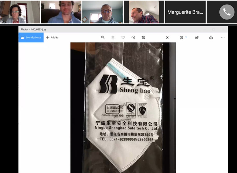
“When the COVID-19 crisis hit the US this March, my biggest concern was the shortage of face masks, which are a key weapon for healthcare providers, frontline service workers, and the public to protect against respiratory transmission of COVID-19. In mid-March I kicked off a gofundme campaign for simple masks to protect frontline service workers but, when it was first announced that frontline healthcare providers were short, I completed the campaign and joined groups of scientists and physicians working on N95 mask reuse in Boston (MGB Center for COVID Innovation) and nation-wide (N95DECON). The N95DECON team and used zoom to connect volunteer scientists, engineers, clinicians and students from across the US to address this problem.
I am deeply committed to helping conserve and decontaminate the N95 masks that are essential for our healthcare workers to most safely treat COVID-19 patients.
I personally love zoom meetings from home for many reasons. For one thing, you can meet people instantaneously from all over the world, no need to travel at all. Also, it is less hierarchical than a typical conference because people all have the same place at the table, rather than some people being relegated to ‘the back of the room.’
McGovern research scientist Jill Crittenden (top left) in a zoom meeting with the Boston-based COVID-19 Innovation Center N95 Reuse team. Photo: Jill Crittenden
For two weeks, we met online daily and exchanged information, suggestions and ideas in a free, open, and transparent way. We reviewed a large body of the information on N95 decontamination and deliberated different methods based on evidence from scientific literature and available data. Our discussions followed the same principles I use in my own work in the Graybiel lab; exploring whether data is convincing, definitive, complete, and reproducible. I am so proud of our resulting report, which provides a summary of this critical information.
I am deeply committed to helping conserve and decontaminate the N95 masks that are essential for our healthcare workers to most safely treat COVID-19 patients. I know physicians personally who are very grateful that teams of scientists are doing the in-depth data analysis so that they can feel confident in what is best for their own health.”
Jill Crittenden is a research scientist in Ann Graybiel‘s lab at the McGovern Institute. She studies neural microcircuits in the basal ganglia that are relevant to Huntington’s and Parkinson’s diseases, dystonia, drug addiction, and repetitive movement disorders such as autism and obsessive-compulsive disorder. Read more about her N95DECON project on our news site.
Jill has also developed a set of helpful guidelines for face masks (either purchased or DIY). She discussed these guidelines, among other COVID-19 related topics on the podcast Dear Discreet Guide.
When the COVID-19 crisis hit the United States this March, McGovern scientist Jill Crittenden wanted to help. One of her greatest concerns was the shortage of face masks, which are a key weapon for healthcare providers, frontline service workers, and the public to protect against respiratory transmission of COVID-19. For those caring for COVID-19 patients, face masks that provide a near 100% seal are essential. These critical pieces of equipment, called N95 masks, are now scarce, and healthcare workers are now faced with reusing potentially contaminated masks.
To address this, Crittenden joined a team of 60 scientists and engineers, students and clinicians, drawn from universities and the private sector to synthesize the scientific literature about mask decontamination and create a set of best practices for bad times. Today the group unveiled its website, N95decon.org, which provides a summary of this critical information.
McGovern research scientist Jill Crittenden helped the N95DECON consortium assess face mask decontamination protocols so healthcare workers can easily access them for COVID-19 protection. Photo: Caitlin Cunningham
“I first heard about the group from Larissa Little, a Harvard graduate student with John Doyle,” explains Crittenden, who is a research scientist in Ann Graybiel‘s lab at the McGovern Institute. “The three of us began communicating because we are all also members of the Boston-based MGB COVID-19 Innovation Center and we agreed that helping to assess the flood of information on N95 decontamination would be an important contribution.”
The team members who came together over several weeks scoured hundreds of peer-reviewed publications, and held continuous online meetings to review studies of decontamination methods that had been used to inactivate previous viral and bacterial pathogens, and to then assess the potential for these methods to neutralize the novel SARS-CoV-2 virus that causes COVID-19.
“This group is absolutely amazing,” says Crittenden. “The zoom meetings are very productive because it is all data and solutions driven. Everyone throws out ideas, what they know and what the literature source is, with the only goal being to get to a data-based consensus efficiently.”
Reliable resource
The goal of the consortium was to provide overwhelmed health officials who don’t have the time to study the literature for themselves, reliable, pre-digested scientific information about the pros and cons of three decontamination methods that offer the best options should local shortages force a choice between decontamination and reuse, or going unmasked.
The three methods involve (1) heat and humidity (2) a specific wavelength of light called ultraviolet C (UVC) and (3) treatment with hydrogen peroxide vapors (HPV). The scientists did not endorse any one method but instead sought to describe the circumstances under which each could inactivate the virus provided rigorous procedures were followed. Devices that rely on heat, for instance, could be used under specific temperature, humidity, and time parameters. With UVC devices – which emit a particular wavelength and energy level of light – considerations involve making sure masks are properly oriented to the light so the entire surface is bathed in sufficient energy. The HPV method has the potential advantage of decontaminating masks in volume, as the U.S. Food and Drug Administration, acting in this emergency, has certified certain vendors to offer hydrogen peroxide vapor treatments on a large scale. In addition to giving health officials the scientific information to assess the methods best suited to their circumstances, N95decon.org points decision makers to sources of reliable and detailed how-to information provided by other organizations, institutions, and commercial services.
“While there is no perfect method for decontamination of N95 masks, it is crucial that decision-makers and users have as much information as possible about the strengths and weaknesses of various approaches,” said Manu Prakash, an associate professor of bioengineering at Stanford who helped coordinate this ad hoc, volunteer undertaking. “Manufacturers currently do not recommend N95 mask reuse. We aim to provide information and evidence in this critical time to help those on the front lines of this crisis make risk-management decisions given the specific conditions and limitations they face.”
The researchers stressed that decontamination does not solve the N95 shortage, and expressed the hope that new masks should be made available in large numbers as soon as possible so that health care workers and first providers could be issued fresh protective gear whenever needed as specified by the non-emergency guidelines set by the U.S. the Centers for Disease Control.
Forward thinking
Meanwhile, these ad hoc volunteers have pledged to continue working together to update N95decon.org website as new information becomes available, and to coordinate their efforts to do research to plug the gaps in current knowledge to avoid duplication of effort.
“We are, at heart, a group of people that want to help better equip hospitals and healthcare personnel in this time of crisis,” says Brian Fleischer, a surgeon at the University of Chicago Medical Center and a member of the N95DECON consortium. “As a healthcare provider, many of my colleagues across the country have expressed concern with a lack of quality information in this ever-evolving landscape. I have learned a great deal from this team and I look forward to our continued collaboration to positively affect change.”
Crittenden is hopeful that the new website will help healthcare workers make informed decisions about the safest methods available for decontamination and reuse of N95 masks. “I know physicians personally who are very grateful that teams of scientists are doing the in-depth data analysis so that they can feel confident in what is best for their own health,” she says.
The members of the N95decon.org team come from institutions including UC Berkeley, the University of Chicago, Stanford, Georgetown University, Harvard University, Seattle University, University of Utah, the McGovern Institute for Brain Research at MIT, the University of Michigan, and from Consolidated Sterilizers and X, the Moonshot Factory.