More than 300 million people worldwide are living with rare disorders — many of which have a genetic cause and affect the brain and nervous system — yet the vast majority of these conditions lack an approved therapy. Because each rare disorder affects fewer than 65 out of every 100,000 people, studying these disorders and creating new treatments for them is especially challenging.
Thanks to a generous philanthropic gift from Ana Méndez ’91 and Rajeev Jayavant ’86, EE ’88, SM ’88, MIT is now poised to fill the gaps in this research landscape. By establishing the Rare Brain Disorders Nexus — or RareNet — at MIT’s McGovern Institute, the alumni aim to convene leaders in neuroscience research, clinical medicine, patient advocacy, and industry to streamline the lab-to-clinic pipeline for rare brain disorder treatments.
“Ana and Rajeev’s commitment to MIT will form crucial partnerships to propel the translation of scientific discoveries into promising therapeutics and expand the Institute’s impact on the rare brain disorders community,” says MIT President Sally Kornbluth. “We are deeply grateful for their pivotal role in advancing such critical science and bringing attention to conditions that have long been overlooked.”
Building new coalitions
Several hurdles have slowed the lab-to-clinic pipeline for rare brain disorder research. It is difficult to secure a sufficient number of patients per study, and current research efforts are fragmented since each study typically focuses on a single disorder (there are more than 7,000 known rare disorders, according to the World Health Organization). Pharmaceutical companies are often reluctant to invest in emerging treatments due to a limited market size and the high costs associated with preparing drugs for commercialization.
Méndez and Jayavant envision that RareNet will finally break down these barriers. “Our hope is that RareNet will allow leaders in the field to come together under a shared framework and ignite scientific breakthroughs across multiple conditions. A discovery for one rare brain disorder could unlock new insights that are relevant to another,” says Jayavant. “By congregating the best minds in the field, we are confident that MIT will create the right scientific climate to produce drug candidates that may benefit a spectrum of uncommon conditions.”
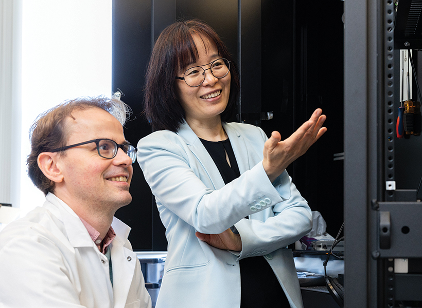
Guoping Feng, the James W. (1963) and Patricia T. Poitras Professor in Neuroscience and associate director of the McGovern Institute for Brain Research at MIT, will serve as RareNet’s inaugural faculty director. Feng holds a strong record of advancing studies on therapies for neurodevelopmental disorders, including autism spectrum disorders, Williams syndrome, and uncommon forms of epilepsy. His team’s gene therapy for Phelan-McDermid syndrome, a rare and profound autism spectrum disorder, has been licensed to Jaguar Gene Therapy and is currently undergoing clinical trials. “RareNet pioneers a unique model for biomedical research — one that is reimagining the role academia can play in developing therapeutics,” says Feng.
RareNet plans to deploy two major initiatives: a global consortium and a therapeutic pipeline accelerator. The consortium will form an international network of researchers, clinicians, and patient groups from the outset. It seeks to connect siloed research efforts, secure more patient samples, promote data sharing, and drive a strong sense of trust and goal alignment across the RareNet community. Partnerships within the consortium will support the aim of the therapeutic pipeline accelerator: to de-risk early lab discoveries and expedite their translation to clinic. By fostering more targeted collaborations — especially between academia and industry — the accelerator will prepare potential treatments for clinical use as efficiently as possible.
MIT labs are focusing on four uncommon conditions in the first wave of RareNet projects: Rett syndrome, prion disease, disorders linked to SYNGAP1 mutations, and Sturge-Weber syndrome. The teams are working to develop novel therapies that can slow, halt, or reverse dysfunctions in the brain and nervous system.
These efforts will build new bridges to connect key stakeholders across the rare brain disorders community and disrupt conventional research approaches. “Rajeev and I are motivated to seed powerful collaborations between MIT researchers, clinicians, patients, and industry,” says Méndez. “Guoping Feng clearly understands our goal to create an environment where foundational studies can thrive and seamlessly move toward clinical impact.”
“Patient and caregiver experiences, and our foreseeable impact on their lives, will guide us and remain at the forefront of our work,” Feng adds. “For far too long the rare brain disorders community has been deprived of life-changing treatments — and, importantly, hope. RareNet gives us the opportunity to transform how we study these conditions and to do so at a moment when it’s needed more than ever.”