And the winner of this year’s costume contest (by popular vote) goes to Ruth Rosenholtz for her depiction of presidential candidate Donald Trump! See below for a full gallery of images from our annual Halloween party.


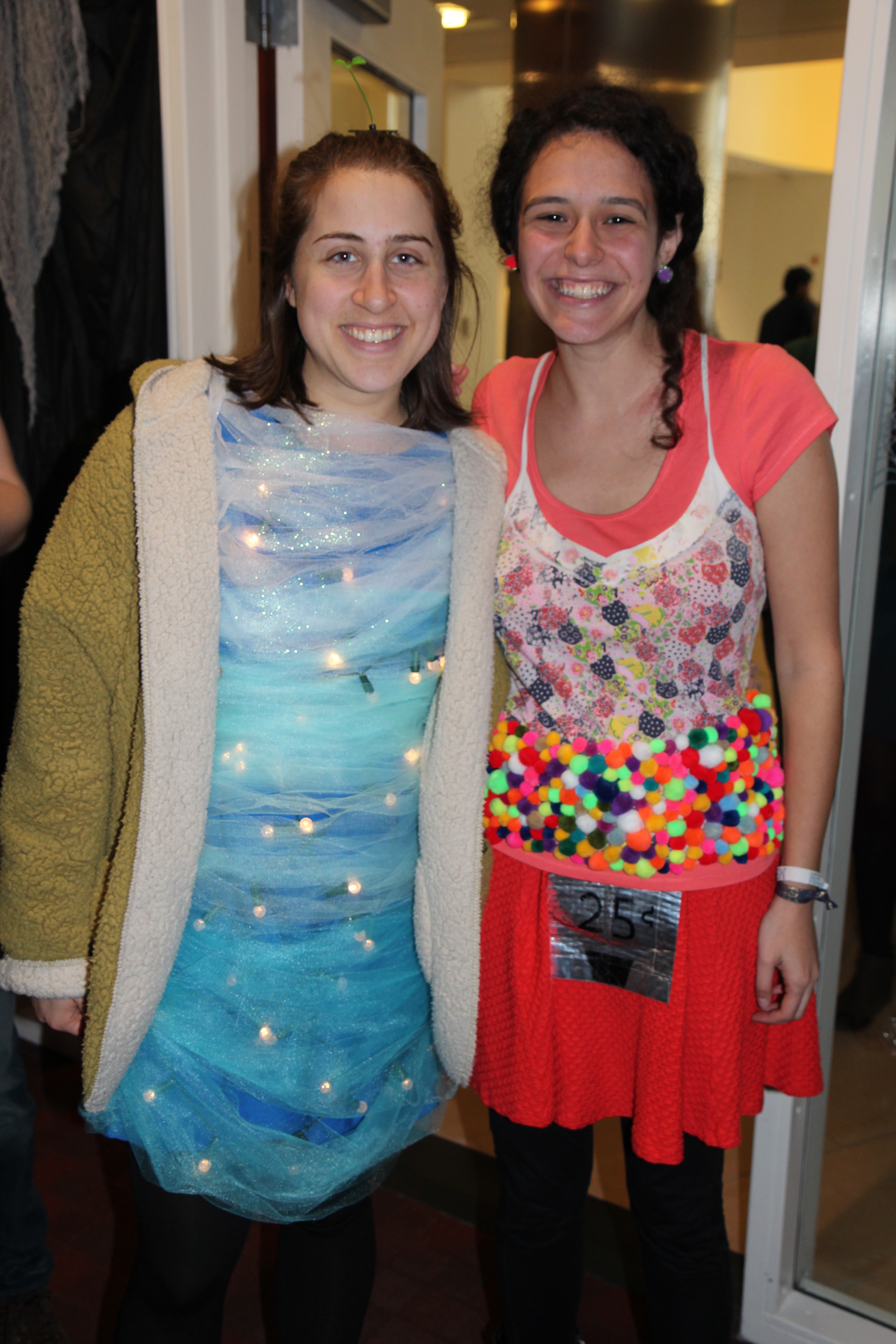


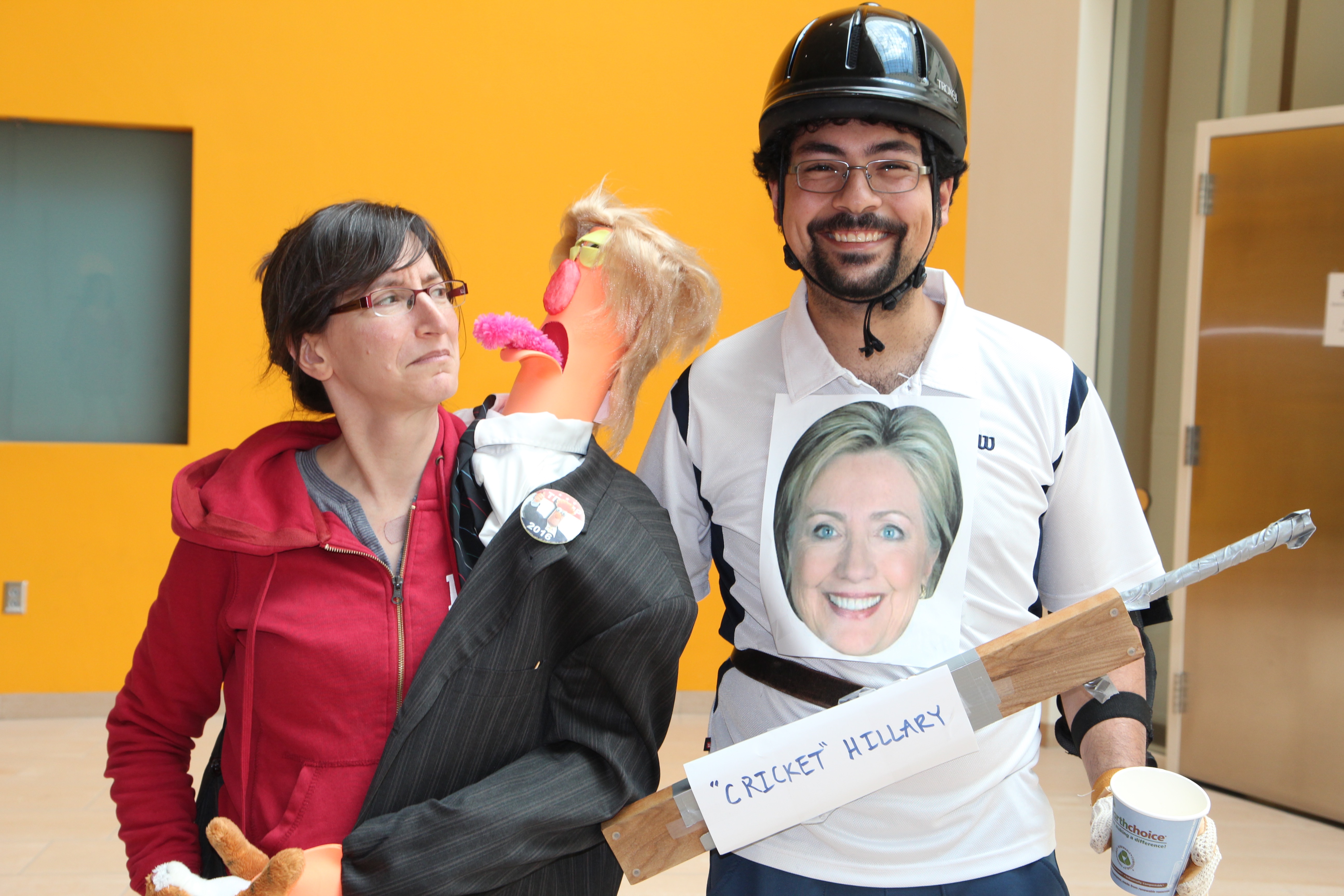
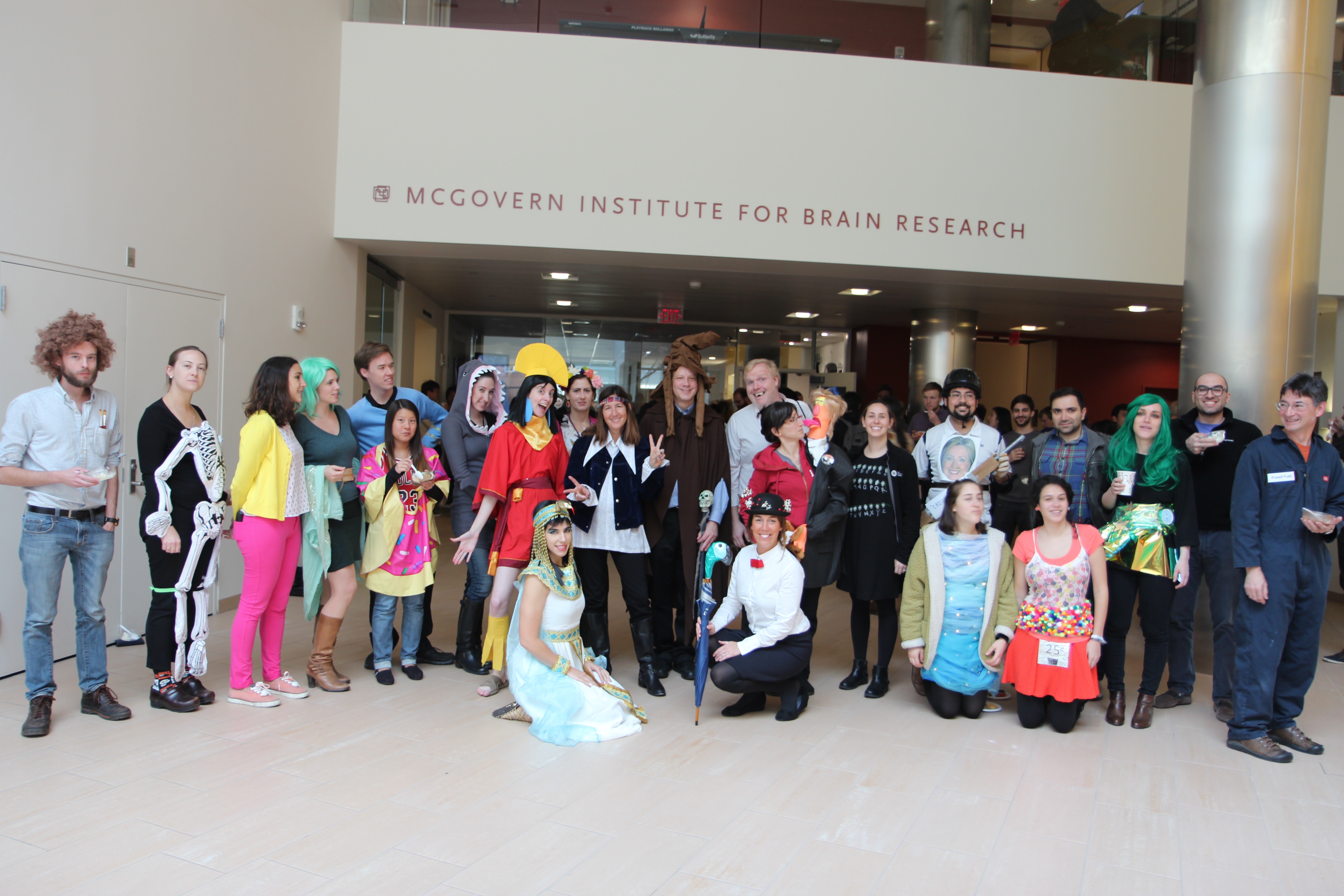

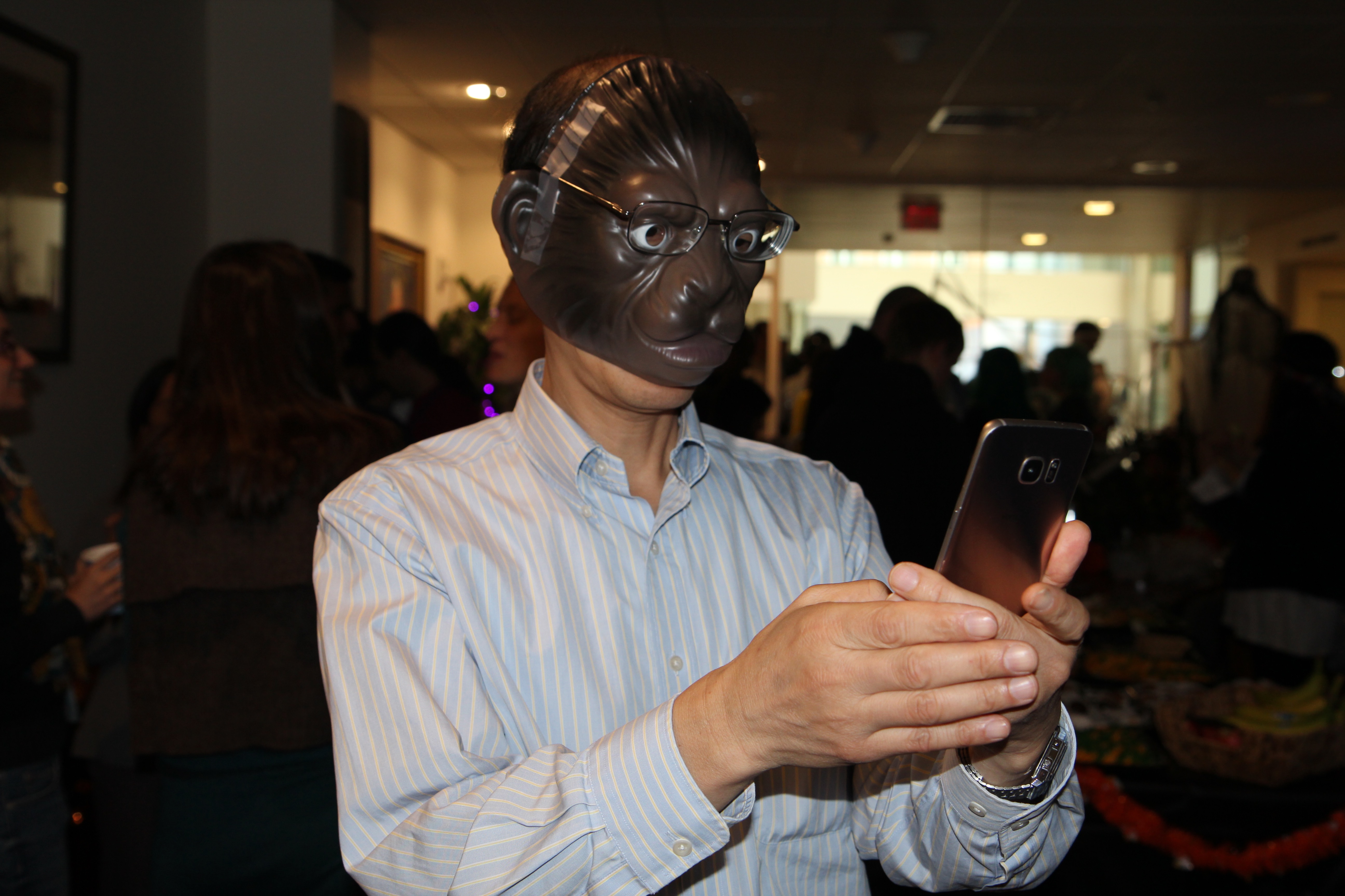


And the winner of this year’s costume contest (by popular vote) goes to Ruth Rosenholtz for her depiction of presidential candidate Donald Trump! See below for a full gallery of images from our annual Halloween party.
The newest addition to MIT’s Public Art Collection is now on permanent display at the McGovern Institute for Brain Research at 550 Main Street in Kendall Square, Cambridge, Massachusetts. “SCIENTIA,” a monumental bronze sculpture by Ursula von Rydingsvard is a gift from Lore Harp McGovern and represents the 52nd piece of public art on campus. The new work will be dedicated in a public ceremony on Friday, Oct. 28 at 5 p.m., followed by a free artist talk at 6 p.m. in the Singleton Auditorium (Room 46-3002).
“’SCIENTIA’ represents that art and science are not separate entities,” says Lore Harp McGovern, co-founder of the McGovern Institute and a member of the Council for the Arts at MIT. “Art defines our humanity, advances our curiosity, and forces us to ask big questions — questions the McGovern Institute for Brain Research is trying to answer. ‘SCIENTIA’ invites you in.”
Von Rydingsvard’s “SCIENTIA” is among her most ambitious sculptures to date, at approximately 24 feet tall and over 17,000 pounds. In creating the work, the artist first produced a wood model in her studio using 4×4-inch cedar beams milled for the construction industry. Using circular saws and a range of cutting tools, she sliced, marked, and shaped the wood elements, then stacked them to create layers that were glued and screwed into place. The full-scale wood model was then transported to Polich Tallix Fine Art Foundry (founded by Richard Polich SM ’65), where the majority of the sculpture was sand cast while the delicate filigree sections were cast using the lost-wax technique. Von Rydingsvard patinated the bronze surface by hand with chemicals and a blow torch.
For over 30 years, von Rydingsvard has been making monumental sculptures that reveal the trace of the human hand and resemble objects and environments that echo the artist’s family heritage in pre-industrial Poland. The artist’s childhood was marked by the strain of living in eight different refugee camps over the course of five years. Her earliest recollections — of displacement and subsistence through humble means — infuse her work with emotional potency. Von Rydingsvard has built towering cedar structures, creating intricate networks of individual beams, shaped by sharp and lyrical cuts and fused together to form rich, dynamic surfaces. While abstract at its core, von Rydingsvard’s work takes visual cues from the landscape, the human body, and utilitarian objects — such as the artist’s collection of household vessels — and demonstrates an interest in the point where the human-made meets nature.
“Ursula von Rydingsvard’s commissioned piece for the McGovern is a fantastic addition to MIT’s great public art collection,” List Visual Arts Center Director Paul C. Ha says. “This powerful sculpture will inspire many and will be one of the signature pieces in our collection. We’re grateful for Ms. McGovern’s thoughtfulness and her generosity in helping us acquire this magnificent piece for MIT.”
MIT neuroscientists have discovered that brain cells called glial cells play a critical role in controlling appetite and feeding behavior. In a study of mice, the researchers found that activating these cells stimulates overeating, and that when the cells are suppressed, appetite is also suppressed.
The findings could offer scientists a new target for developing drugs against obesity and other appetite-related disorders, the researchers say. The study is also the latest in recent years to implicate glial cells in important brain functions. Until about 10 years ago, glial cells were believed to play more of a supporting role for neurons.
“In the last few years, abnormal glial cell activities have been strongly implicated in neurodegenerative disorders. There is more and more evidence to point to the importance of glial cells in modulating neuronal function and in mediating brain disorders,” says Guoping Feng, the James W. and Patricia Poitras Professor of Neuroscience. Feng is also a member of MIT’s McGovern Institute for Brain Research and the Stanley Center for Psychiatric Research at the Broad Institute.
Feng is one of the senior authors of the study, which appears in the Oct. 18 edition of the journal eLife. The other senior author is Weiping Han, head of the Laboratory of Metabolic Medicine at the Singapore Bioimaging Consortium in Singapore. Naiyan Chen, a postdoc at the Singapore Bioimaging Consortium and the McGovern Institute, is the lead author.
Turning on appetite
It has long been known that the hypothalamus, an almond-sized structure located deep within the brain, controls appetite as well as energy expenditure, body temperature, and circadian rhythms including sleep cycles. While performing studies on glial cells in other parts of the brain, Chen noticed that the hypothalamus also appeared to have a lot of glial cell activity.
“I was very curious at that point what glial cells would be doing in the hypothalamus, since glial cells have been shown in other brain areas to have an influence on regulation of neuronal function,” she says.
Within the hypothalamus, scientists have identified two key groups of neurons that regulate appetite, known as AgRP neurons and POMC neurons. AgRP neurons stimulate feeding, while POMC neurons suppress appetite.
Until recently it has been difficult to study the role of glial cells in controlling appetite or any other brain function, because scientists haven’t developed many techniques for silencing or stimulating these cells, as they have for neurons. Glial cells, which make up about half of the cells in the brain, have many supporting roles, including cushioning neurons and helping them form connections with one another.
In this study, the research team used a new technique developed at the University of North Carolina to study a type of glial cell known as an astrocyte. Using this strategy, researchers can engineer specific cells to produce a surface receptor that binds to a chemical compound known as CNO, a derivative of clozapine. Then, when CNO is given, it activates the glial cells.
The MIT team found that turning on astrocyte activity with just a single dose of CNO had a significant effect on feeding behavior.
“When we gave the compound that specifically activated the receptors, we saw a robust increase in feeding,” Chen says. “Mice are not known to eat very much in the daytime, but when we gave drugs to these animals that express a particular receptor, they were eating a lot.”
The researchers also found that in the short term (three days), the mice did not gain extra weight, even though they were eating more.
“This raises the possibility that glial cells may also be modulating neurons that control energy expenditures, to compensate for the increased food intake,” Chen says. “They might have multiple neuronal partners and modulate multiple energy homeostasis functions all at the same time.”
When the researchers silenced activity in the astrocytes, they found that the mice ate less than normal.
Suzanne Dickson, a professor of neuroendocrinology at the University of Gothenburg in Sweden described the study as part of a “paradigm shift” toward the idea that glial cells have a less passive role than previously believed.
“We tend to think of glial cells as providing a support network for neuronal processes and that their activation is also important in certain forms of brain trauma or inflammation,” says Dickson, who was not involved in the research. “This study adds to the emerging evidence base that glial cells may also exert specific effects to control nerve cell function in normal physiology.”
Unknown interactions
Still unknown is how the astrocytes exert their effects on neurons. Some recent studies have suggested that glial cells can secrete chemical messengers such as glutamate and ATP; if so, these “gliotransmitters” could influence neuron activity.
Another hypothesis is that instead of secreting chemicals, astrocytes exert their effects by controlling the uptake of neurotransmitters from the space surrounding neurons, thereby affecting neuron activity indirectly.
Feng now plans to develop new research tools that could help scientists learn more about astrocyte-neuron interactions and how astrocytes contribute to modulation of appetite and feeding. He also hopes to learn more about whether there are different types of astrocytes that may contribute differently to feeding behavior, especially abnormal behavior.
“We really know very little about how astrocytes contribute to the modulation of appetite, eating, and metabolism,” he says. “In the future, dissecting out these functional difference will be critical for our understanding of these disorders.”
We have a reasonable understanding of the rules behind the genome’s protein-coding components. We can look at a DNA sequence and point with confidence to where a gene’s coding region begins, where it ends, and pieces of its geography.
For the remaining 98 percent of the genome — the part that dictates which genes a cell reads — it’s a different story. What knowledge we have of the rules governing this “dark matter” comes from from studying and manipulating individual bits of noncoding DNA one at a time. The rulebook that governs how the noncoding genome works, however, has remained out of reach.
“Ninety percent of the genetic variations that affect human disease are in the noncoding regions,” said Broad founding director Eric Lander. “But we haven’t had any way to tell, in a systematic way, which regulators affect which genes.”
In a pair of newly published Science papers, two research teams at the Broad show how methods leveraging CRISPR gene editing could help grasp those rules.
Using two complementary approaches, the teams — one from the Lander lab, the other from that of Broad Core Institute Member and McGovern Institute for Brain Research investigator Feng Zhang — used CRISPR as a tool to systematically probe thousands of noncoding DNA sequences simultaneously (much as Zhang and others did previously with coding DNA). In the process, both identified several interesting genetic regulators, including ones millions of bases away from the genes they control.
“We’d like to be able to catalog the noncoding elements that control every gene’s expression in every cell type,” said Jesse Engreitz, a postdoctoral fellow in the Lander lab and senior author on one of the papers. “This is a massive problem in biology, and it’s a rate-limiting step for connecting many genetic associations to their fundamental molecular mechanisms and to human disease.”
Variations on a theme
Both teams used pooled CRISPR screens (which scan and edit large swaths of the genome simultaneously using a molecular scalpel called the Cas9 enzyme and thousands of guide RNAs, which target Cas9 to specific sequences) to perturb noncoding DNA. But they did so in different ways.
Zhang, Neville Sanjana (a Zhang lab alum and now a core member of the New York Genome Center), and Jason Wright (another Zhang alum, now at Homology Medicines) used Cas9 to make precise edits to overlapping stretches of noncoding DNA — in their case, in regions surrounding three genes (NF1, NF2, and CUL3) whose functional loss has been linked to drug resistance in a form of melanoma.
“This approach lets us induce a wide diversity of mutations,” Sanjana explained. “We don’t have to speculate how a given sequence might best be disrupted.”
Engreitz, Lander, and graduate student Charles Fulco, on the other hand, employed a CRISPR interference system, using an inactive or “dead” form of Cas9 fused to a protein fragment called a KRAB domain to silence their target sequences (around MYC and GATA1, the genes for two important transcription factors).
“This system provides a good quantitative estimate of a given noncoding region’s regulatory influence,” Engreitz said. “It both shows you where the dials are that control a given gene, and tells you how much each dial matters.”
Each team then used a functional readout (increased drug resistance in melanoma cells for Sanjana, Wright, and Zhang; a drop in cell growth for Fulco, Lander, and Engreitz) and deep sequencing to see which of their guide RNAs impacted expression of their genes of interest and map the regulators those guide RNAs affected.
The two teams’ findings, confirmed with an array of additional techniques (e.g., chromatin profiling, 3D conformational capture, transcription factor profiling), point to the potential for tracing the noncoding genome’s regulatory wiring leveraging CRISPR tools. Fulco, Lander, and Engreitz found and ranked the relative importance of seven MYC and three GATA1 enhancers (short pieces of noncoding DNA that boost a gene’s chances of being read). Sanjana, Wright, and Zhang’s screen pinpointed numerous enhancers and transcription factor binding sites just for CUL3 alone.
Studying sequences in their natural habitat
While similar in principle to traditional reporter assays (where scientists couple interesting sequences to reporter genes in plasmids), these pooled CRISPR screens have a distinct difference: they probe the sequences directly, in their native habitat.
“The screens interrogate the sequences in their endogenous context,” Sanjana emphasized. “Reporter assays can be very helpful, but they lack the 3D conformation or local chromatin environment of the native genomic context. Here, the regulatory sequences undergo all of their normal interactions.”
“For example, we could see long-range loops between gene promoters and noncoding sites thousands of bases away,” he continued. “We would have missed these interesting 3D interactions entirely if we just looked at these regulatory elements in isolation.”
One limitation, Engreitz noted, is that neither CRISPR approach, in its current form, addresses the genome’s inherent redundancy. “Maybe it’s not enough to break one enhancer to really understand how a gene is controlled. Maybe you have to break more than one,” he said. “We can’t do that yet.”
But Engreitz, Sanjana, and Lander are all optimistic about the potential for using CRISPR-based approaches to reveal the noncoding genome’s underlying order.
“One interesting challenge with the noncoding genome is that while it is huge, the individual functional elements within it can be quite small,” Sanjana said. “In the future, it will be important to think about how we can develop new approaches that interrogate larger regions while maintaining high resolution.”
Engreitz agreed, adding, “There’s a potential that as we map more of these connections we’re going to learn the rules that let us predict them for the rest of the noncoding genome.”
“These approaches, using libraries of guide RNAs to bring CRISPR in to cut or bring in inhibitors, let you directly see the effects of large areas of noncoding DNA on different genes,” Lander said. “I think this is going to crack open systematic maps of gene regulation.”
Papers cited:
Fulco CP et al. Systematic mapping of functional enhancer-promoter connections with CRISPR interference. Science. September 29, 2016. DOI: 10.1126/science.aag2445
Sanjana NE et al. High-resolution interrogation of functional elements in the noncoding genome. Science. September 29, 2016. DOI: 10.1126/science.aaf7613
People who are too frightened of flying to board an airplane, or too scared of spiders to venture into the basement, can seek a kind of treatment called exposure therapy. In a safe environment, they repeatedly face cues such as photos of planes or black widows, as a way to stamp out their fearful response — a process known as extinction.
Unfortunately, the effects of exposure therapy are not permanent, and many people experience a relapse. MIT scientists have now identified a way to enhance the long-term benefit of extinction in rats, offering a way to improve the therapy in people suffering from phobias and more complicated conditions such as post-traumatic stress disorder (PTSD).
Work conducted in the laboratory of Ki Goosens, a research affiliate of the McGovern Institute for Brain Research, has pinpointed a neural circuit that becomes active during exposure therapy in the rats. In a study published Sept. 27 in eLife, the researchers showed that they could stretch the therapy’s benefits for at least two months by boosting the circuit’s activity during treatment.
“When you give extinction training to humans or rats, and you wait long enough, you observe a phenomenon called spontaneous recovery, in which the fear that was originally learned comes back,” Goosens explains. “It’s one of the barriers to this type of therapy. You spend all this time going through it, but then it’s not a permanent fix for your problem.”
According to statistics from the National Institute of Mental Health, 18 percent of U.S. adults are diagnosed with a fear or anxiety disorder each year, with 22 percent of those patients experiencing severe symptoms.
How to quench a fear
The neural circuit identified by the scientists connects a part of the brain involved in fear memory, called the basolateral amygdala (BLA), with another region called the nucleus accumbens (NAc), that helps the brain process rewarding events. Goosens and her colleagues call it the BLA-NAc circuit.
Researchers have been considering a link between fear and reward for some time, Goosens says. “The amygdala is a part of the brain that is tightly linked with fear memory but it’s also been linked to positive reward learning as well, and the accumbens is a key reward area in the brain,” she explains. “What we’ve been thinking about is whether extinction is rewarding. When you’re expecting something bad and you don’t get it, does your brain treat that like it’s a good thing?”
To find out if there was a specific brain circuit involved, the researchers first trained rats to fear a certain noise by pairing it with foot shock. They later gave the rats extinction training, during which the noise was presented in the absence of foot shock, and they looked at markers of neural activity in the brain. The results revealed the BLA-NAc reward circuit was recruited by the brain during exposure therapy, as the rats gave up their fear of the bad noise.
Once Goosens and her colleagues had identified the circuit, they looked for ways to boost its activity. First, they paired a sugary drink with the fear-related sound during extinction training, hoping to associate the sound with a reward. This type of training, called counterconditioning, associates fear-eliciting cues with rewarding events or memories, instead of with neutral events as in most extinction training.
Rats that received the counterconditioning were significantly less likely to spontaneously revert to their fearful states, compared to those that received regular extinction training for up to 55 days later, the scientists found.
They also found that the benefits of extinction could be prolonged with optogenetic stimulation, in which the circuit was genetically modified so that it could be stimulated directly with tiny bursts of light from an optical fiber.
The ongoing benefit that came from stimulating the circuit was one of the most surprising — and welcome — findings from the study, Goosens says. “The effect that we saw was one that really emerged months later, and we want to know what’s happening over those two months. What is the circuit doing to suppress the recovery of fear over that period of time? We still don’t understand what that is.”
Another interesting finding from the study was that the circuit was active during both fear learning and fear extinction, says lead author Susana Correia, a former research scientist in the Goosens lab who is now a scientist at Ironwood Pharmaceuticals. “Understanding if these are molecularly different subcircuits within this projection could allow the development of a pharmaceutical approach to target the fear extinction pathway and to improve cognitive therapy,” Correia says.
Immediate and future impacts on therapy
Some therapists are already using counterconditioning in treating PTSD, and Goosens suggests that the rat study might encourage further exploration of this technique in human therapy.
And while it isn’t likely that humans will receive direct optogenetic therapy any time soon, Goosens says there is a benefit to knowing exactly which circuits are involved in extinction.
In neurofeedback studies, for instance, brain scan technologies such as fMRI or EEG could be used to help a patient learn to activate specific parts of their brain, including the BLA-NAc reward circuit, during exposure therapy.
Studies like this one, Goosens says, offer a “target for a personalized medicine approach where feedback is used during therapy to enhance the effectiveness of that therapy.”
Other MIT authors on the paper include technical assistant Anna McGrath, undergraduate Allison Lee, and McGovern principal investigator and Institute Professor Ann Graybiel.
The study was funded by the U.S. Army Research Office, the Defense Advanced Research Projects Agency (DARPA), and the National Institute of Mental Health.
Our perception of the world arises within the brain, based on sensory information that is sometimes ambiguous, allowing more than one interpretation. Familiar demonstrations of this point include the famous Necker cube and the “duck-rabbit” drawing (right) in which two different interpretations flip back and forth over time.
Another example is binocular rivalry, in which the two eyes are presented with different images that are perceived in alternation. Several years ago, this phenomenon caught the eye of Caroline Robertson, who is now a Harvard Fellow working in the lab of McGovern Investigator Nancy Kanwisher. Back when she was a graduate student at Cambridge University, Robertson realized that binocular rivalry might be used to probe the basis of autism, among the most mysterious of all brain disorders.
Robertson’s idea was based on the hypothesis that autism involves an imbalance between excitation and inhibition within the brain. Although widely supported by indirect evidence, this has been very difficult to test directly in human patients. Robertson realized that binocular rivalry might provide a way to perform such a test. The perceptual switches that occur during rivalry are thought to involve competition between different groups of neurons in the visual cortex, each group reinforcing its own interpretation via excitatory connections while suppressing the alternative interpretation through inhibitory connections. Thus, if the balance is altered in the brains of people with autism, the frequency of switching might also be different, providing a simple and easily measurable marker of the disease state.
To test this idea, Robertson recruited adults with and without autism, and presented them with two distinct and differently colored images in each eye. As expected, their perceptions switched back and forth between the two images, with short periods of mixed perception in between. This was true for both groups, but when she measured the timing of these switches, Robertson found that individuals with autism do indeed see the world in a measurably different way than people without the disorder. Individuals with autism cycle between the left and right images more slowly, with the intervening periods of mixed perception lasting longer than in people without autism. The more severe their autistic symptoms, as determined by a standard clinical behavioral evaluation, the greater the difference.
Robertson had found a marker for autism that is more objective than current methods that involve one person assessing the behavior of another. The measure is immediate and relies on brain activity that happens automatically, without people thinking about it. “Sensation is a very simple place to probe,” she says.
When she arrived in Kanwisher’s lab, Robertson wanted to use brain imaging to probe the basis for the perceptual phenomenon that she had discovered. With Kanwisher’s encouragement, she began by repeating the behavioral experiment with a new group of subjects, to check that her previous results were not a fluke. Having confirmed that the finding was real, she then scanned the subjects using an imaging method called Magnetic Resonance Spectroscopy (MRS), in which an MRI scanner is reprogrammed to measure concentrations of neurotransmitters and other chemicals in the brain. Kanwisher had never used MRS before, but when Robertson proposed the experiment, she was happy to try it. “Nancy’s the kind of mentor who could support the idea of using a new technique and guide me to approach it rigorously,” says Robertson.
For each of her subjects, Robertson scanned their brains to measure the amounts of two key neurotransmitters, glutamate, which is the main excitatory transmitter in the brain, and GABA, which is the main source of inhibition. When she compared the brain chemistry to the behavioral results in the binocular rivalry task, she saw something intriguing and unexpected. In people without autism, the amount of GABA in the visual cortex was correlated with the strength of the suppression, consistent with the idea that GABA enables signals from one eye to inhibit those from the other eye. But surprisingly, there was no such correlation in the autistic individuals—suggesting that GABA was somehow unable to exert its normal suppressive effect. It isn’t yet clear exactly what is going wrong in the brains of these subjects, but it’s an early flag, says Robertson. “The next step is figuring out which part of the pathway is disrupted.”
Robertson’s approach starts from the top-down, working backward from a measurable behavior to look for brain differences, but it isn’t the only way in. Another approach is to start with genes that are linked to autism in humans, and to understand how they affect neurons and brain circuits. This is the bottom-up approach of McGovern Investigator Guoping Feng, who studies a gene called Shank3 that codes for a protein that helps build synapses, the connections through which neurons send signals to each other. Several years ago Feng knocked out Shank3 in mice, and found that the mice exhibited behaviors reminiscent of human autism, including repetitive grooming, anxiety, and impaired social interaction and motor control.
These earlier studies involved a variety of different mutations that disabled the Shank3 gene. But when postdoc Yang Zhou joined Feng’s lab, he brought a new perspective. Zhou had come from a medical background and wanted to do an experiment more directly connected to human disease. So he suggested making a mouse version of a Shank3 mutation seen in human patients, and testing its effects.
Zhou’s experiment would require precise editing of the mouse Shank3 gene, previously a difficult and time-consuming task. But help was at hand, in the form of a collaboration with McGovern Investigator Feng Zhang, a pioneer in the development of genome-editing methods.
Using Zhang’s techniques, Zhou was able to generate mice with two different mutations: one that had been linked to human autism, and another that had been discovered in a few patients with schizophrenia.
The researchers found that mice with the autism-related mutation exhibited behavioral changes at a young age that paralleled behaviors seen in children with autism. They also found early changes in synapses within a brain region called the striatum. In contrast, mice with the schizophrenia-related gene appeared normal until adolescence, and then began to exhibit changes in behavior and also changes in the prefrontal cortex, a brain region that is implicated in human schizophrenia. “The consequences of the two different Shank3 mutations were quite different in certain aspects, which was very surprising to us,” says Zhou.
The fact that different mutations in just one gene can produce such different results illustrates exactly how complex these neuropsychiatric disorders can be. “Not only do we need to study different genes, but we also have to understand different mutations and which brain regions have what defects,” says Feng, who received funding from the Poitras Center for Affective Disorders research and the Simons Center for the Social Brain. Robertson and Kanwisher were also supported by the Simons Center.
The brain alterations that lead to autism are thought to arise early in development, long before the condition is diagnosed, raising concerns that it may be difficult to reverse the effects once the damage is done. With the Shank3 knockout mice, Feng and his team were able to approach this question in a new way, asking what would happen if the missing gene were to be restored in adulthood.
To find the answer, lab members Yuan Mei and Patricia Monteiro, along with Zhou, studied another strain of mice, in which the Shank3 gene was switched off but could be reactivated at any time by adding a drug to their diet. When adult mice were tested six weeks after the gene was switched back on, they no longer showed repetitive grooming behaviors, and they also showed normal levels of social interaction with other mice, despite having grown up without a functioning Shank3 gene. Examination of their brains confirmed that many of the synaptic alterations were also rescued when the gene was restored.
Not every symptom was reversed by this treatment; even after six weeks or more of restored Shank3 expression, the mice continued to show heightened anxiety and impaired motor control. But even these deficits could be prevented if the Shank3 gene was restored earlier in life, soon after birth.
The results are encouraging because they indicate a surprising degree of brain plasticity, persisting into adulthood. If the results can be extrapolated to human patients, they suggest that even in adulthood, autism may be at least partially reversible if the right treatment can be found. “This shows us the possibility,” says Zhou. “If we could somehow put back the gene in patients who are missing it, it could help improve their life quality.”
Robertson and Feng are approaching the challenge of autism from different starting points, but already there are signs of convergence. Feng is finding early signs that his Shank3 mutant mice may have an altered balance of inhibitory and excitatory circuits, consistent with what Robertson and Kanwisher have found in humans.
Feng is continuing to study these mice, and he also hopes to study the effects of a similar mutation in non-human primates, whose brains and behaviors are more similar to those of humans than rodents. Robertson, meanwhile, is planning to establish a version of the binocular rivalry test in animal models, where it is possible to alter the balance between inhibition and excitation experimentally (for example, via a genetic mutation or a drug treatment). If this leads to changes in binocular rivalry, it would strongly support the link to the perceptual changes seen in humans.
One challenge, says Robertson, will be to develop new methods to measure the perceptions of mice and other animals. “The mice can’t tell us what they are seeing,” she says. “But it would also be useful in humans, because it would allow us to study young children and patients who are non-verbal.”
The imbalance hypothesis is a promising lead, but no single explanation is likely to encompass all of autism, according to McGovern director Bob Desimone. “Autism is a notoriously heterogeneous condition,” he explains. “We need to try multiple approaches in order to maximize the chance of success.”
McGovern researchers are doing exactly that, with projects underway that range from scanning children to developing new molecular and microscopic methods for examining brain changes in animal disease models. Although genetic studies provide some of the strongest clues, Desimone notes that there is also evidence for environmental contributions to autism and other brain disorders. “One that’s especially interesting to us is a maternal infection and inflammation, which in mice at least can affect brain development in ways we’re only beginning to understand.”
The ultimate goal, says Desimone, is to connect the dots and to understand how these diverse human risk factors affect brain function. “Ultimately, we want to know what these different pathways have in common,” he says. “Then we can come up with rational strategies for the development of new treatments.”
MIT neuroscientists have discovered connections deep within the brain that appear to form a communication pathway between areas that control emotion, decision-making, and movement. The researchers suspect that these connections, which they call striosome-dendron bouquets, may be involved in controlling how the brain makes decisions that are influenced by emotion or anxiety.
This circuit may also be one of the targets of the neural degeneration seen in Parkinson’s disease, says Ann Graybiel, an Institute Professor at MIT, member of the McGovern Institute for Brain Research, and the senior author of the study.
Graybiel and her colleagues were able to find these connections using a technique developed at MIT known as expansion microscopy, which enables scientists to expand brain tissue before imaging it. This produces much higher-resolution images than would otherwise be possible with conventional microscopes.
That technique was developed in the lab of Edward Boyden, an associate professor of biological engineering and brain and cognitive sciences at the MIT Media Lab, who is also an author of this study. Jill Crittenden, a research scientist at the McGovern Institute, is the lead author of the paper, which appears in the Proceedings of the National Academy of Sciences the week of Sept. 19.
Tracing a circuit
In this study, the researchers focused on a small region of the brain known as the striatum, which is part of the basal ganglia — a cluster of brain centers associated with habit formation, control of voluntary movement, emotion, and addiction. Malfunctions of the basal ganglia have been associated with Parkinson’s and Huntington’s diseases, as well as autism, obsessive-compulsive disorder, and Tourette’s syndrome.
Much of the striatum is uncharted territory, but Graybiel’s lab has previously identified clusters of cells there known as striosomes. She also found that these clusters receive very specific input from parts of the brain’s prefrontal cortex involved in processing emotions, and showed that this communication pathway is necessary for making decisions that require an anxiety-provoking cost-benefit analysis, such as choosing whether to take a job that pays more but forces a move away from family and friends.
Her studies also suggested that striosomes relay information to cells within a region called the substantia nigra, one of the brain’s main dopamine-producing centers. Dopamine has many functions in the brain, including roles in initiating movement and regulating mood.
To figure out how these regions might be communicating, Graybiel, Crittenden, and their colleagues used expansion microscopy to image the striosomes and discovered extensive connections between those clusters of cells and dopamine-producing cells of the substantia nigra. The dopamine-producing cells send down many tiny extensions known as dendrites that become entwined with axons that come up to meet them from the striosomes, forming a bouquet-like structure.
“With expansion microscopy, we could finally see direct connections between these cells by unraveling their unusual rope-like bundles of axons and dendrites,” Crittenden says. “What’s really exciting to us is we can see that it’s small discrete clusters of dopamine cells with bundles that are being targeted.”
Hard decisions
This finding expands the known decision-making circuit so that it encompasses the prefrontal cortex, striosomes, and a subset of dopamine-producing cells. Together, the striosomes may be acting as a gatekeeper that absorbs sensory and emotional information coming from the cortex and integrates it to produce a decision on how to react, which is initiated by the dopamine-producing cells, the researchers say.
To explore that possibility, the researchers plan to study mice in which they can selectively activate or shut down the striosome-dendron bouquet as the mice are prompted to make decisions requiring a cost-benefit analysis.
The researchers also plan to investigate whether these connections are disrupted in mouse models of Parkinson’s disease. MRI studies and postmortem analysis of brains of Parkinson’s patients have shown that death of dopamine cells in the substantia nigra is strongly correlated with the disease, but more work is needed to determine if this subset overlaps with the dopamine cells that form the striosome-dendron bouquets.
TITLE: “From Cells to Cognition: Understanding Brain Function in Health and Disease”
DATE: Tuesday November 8, 2016
TIME: 8:30am – 5:00pm
LOCATION: MIT Bldg 46-3002 (Singleton Auditorium)
QUESTIONS? Naomi Berkowitz | naomiber@mit.edu | 617.715.5396
Registration is now closed.
PROGRAM
8:30 am
Continental Breakfast Served in Atrium
9:00 am – 9:10 am
ROBERT DESIMONE | McGovern Institute
, MIT
Welcoming Remarks
Session I (Chair: TBA)
9:10 am – 9:50 am
BAILONG XIAO | IDG/McGovern Institute, Tsinghua University
In touch with the mechanosensitive Piezo channels: structure, ion permeation and mechano-gating
9:50 am – 10:30 am
JING YANG | IDG/McGovern Institute, Peking University
The mechanism underlying neuronal response to axonal injury
10:30 am – 10:45 am
Break
10:45 am – 11:25 am
JUN YAO | IDG/McGovern Institute, Tsinghua University
Ca2+ dependent synaptic vesicle cycling
11:25 am – 12:05 pm
YONG ZHANG | IDG/McGovern Institute, Peking University
Visualizing AMPA receptor synaptic plasticity in vivo
12:05 pm – 01:30 pm
Poster Session and Lunch in Atrium
Session II (Chair: TBA)
1:30 pm – 2:10 pm
GUOPING FENG | McGovern Institute, MIT
Thalamic reticular nucleus dysfunction in neurodevelopmental disorders
2:10 pm – 2:50 pm
HUAN LUO | IDG/McGovern Institute, Peking University
Serial-like sampling of visual objects during sustained attention
2:50 pm – 3:30 pm
XIAOHONG WAN | IDG/McGovern Institute Beijing Normal University
Investigating neural mechanisms of metacognition
3:30 pm – 3:45 pm
Break
3:45 pm – 4:25 pm
YINA MA | IDG/McGovern Institute, Beijing Normal University
Oxytocin and social adaptation
4:25 pm – 5:05 pm
BO HONG | IDG/McGovern Institute, Tsinghua University
Spatio-temporal organization and assembly of human cortical networks
5:05 pm – 6:05 pm
Reception 5th floor overlooking Atrium
Supported by Hugo Shong and the McGovern Institutes
A Chinese translation of an animation depicting the CRISPR-Cas9 method for genome editing – a powerful new technology with many applications in biomedical research, including the potential to treat human genetic disease. Feng Zhang, a leader in the development of this technology, is a faculty member at MIT, an investigator at the McGovern Institute for Brain Research, and a core member of the Broad Institute. Further information can be found on Prof. Zhang’s website at http://zlab.mit.edu.
Images and footage courtesy of Sputnik Animation, the Broad Institute of MIT and Harvard, Justin Knight and pond5.
When asked to visualize your childhood home, you can probably picture not only the house you lived in, but also the buildings next door and across the street. MIT neuroscientists have now identified two brain regions that are involved in creating these panoramic memories.
These brain regions help us to merge fleeting views of our surroundings into a seamless, 360-degree panorama, the researchers say.
“Our understanding of our environment is largely shaped by our memory for what’s currently out of sight,” says Caroline Robertson, a postdoc at MIT’s McGovern Institute for Brain Research and a junior fellow of the Harvard Society of Fellows. “What we were looking for are hubs in the brain where your memories for the panoramic environment are integrated with your current field of view.”
Robertson is the lead author of the study, which appears in the Sept. 8 issue of the journal Current Biology. Nancy Kanwisher, the Walter A. Rosenblith Professor of Brain and Cognitive Sciences and a member of the McGovern Institute, is the paper’s lead author.
Building memories
As we look at a scene, visual information flows from our retinas into the brain, which has regions that are responsible for processing different elements of what we see, such as faces or objects. The MIT team suspected that areas involved in processing scenes — the occipital place area (OPA), the retrosplenial complex (RSC), and parahippocampal place area (PPA) — might also be involved in generating panoramic memories of a place such as a street corner.
If this were true, when you saw two images of houses that you knew were across the street from each other, they would evoke similar patterns of activity in these specialized brain regions. Two houses from different streets would not induce similar patterns.
“Our hypothesis was that as we begin to build memory of the environment around us, there would be certain regions of the brain where the representation of a single image would start to overlap with representations of other views from the same scene,” Robertson says.
The researchers explored this hypothesis using immersive virtual reality headsets, which allowed them to show people many different panoramic scenes. In this study, the researchers showed participants images from 40 street corners in Boston’s Beacon Hill neighborhood. The images were presented in two ways: Half the time, participants saw a 100-degree stretch of a 360-degree scene, but the other half of the time, they saw two noncontinuous stretches of a 360-degree scene.
After showing participants these panoramic environments, the researchers then showed them 40 pairs of images and asked if they came from the same street corner. Participants were much better able to determine if pairs came from the same corner if they had seen the two scenes linked in the 100-degree image than if they had seen them unlinked.
Brain scans revealed that when participants saw two images that they knew were linked, the response patterns in the RSC and OPA regions were similar. However, this was not the case for image pairs that the participants had not seen as linked. This suggests that the RSC and OPA, but not the PPA, are involved in building panoramic memories of our surroundings, the researchers say.
Priming the brain
In another experiment, the researchers tested whether one image could “prime” the brain to recall an image from the same panoramic scene. To do this, they showed participants a scene and asked them whether it had been on their left or right when they first saw it. Before that, they showed them either another image from the same street corner or an unrelated image. Participants performed much better when primed with the related image.
“After you have seen a series of views of a panoramic environment, you have explicitly linked them in memory to a known place,” Robertson says. “They also evoke overlapping visual representations in certain regions of the brain, which is implicitly guiding your upcoming perceptual experience.”
The research was funded by the National Science Foundation Science and Technology Center for Brains, Minds, and Machines; and the Harvard Milton Fund.