To evaluate school quality, states require students to take standardized tests; in many cases, passing those tests is necessary to receive a high-school diploma. These high-stakes tests have also been shown to predict students’ future educational attainment and adult employment and income.
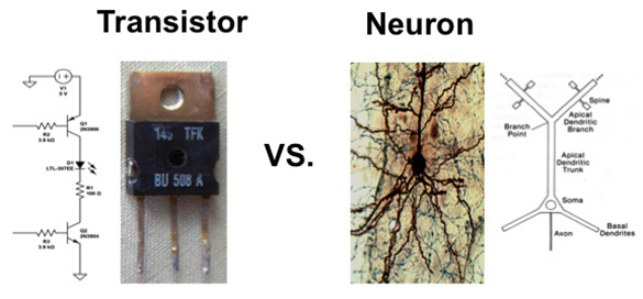
Such tests are designed to measure the knowledge and skills that students have acquired in school — what psychologists call “crystallized intelligence.” However, schools whose students have the highest gains on test scores do not produce similar gains in “fluid intelligence” — the ability to analyze abstract problems and think logically — according to a new study from MIT neuroscientists working with education researchers at Harvard University and Brown University.
In a study of nearly 1,400 eighth-graders in the Boston public school system, the researchers found that some schools have successfully raised their students’ scores on the Massachusetts Comprehensive Assessment System (MCAS). However, those schools had almost no effect on students’ performance on tests of fluid intelligence skills, such as working memory capacity, speed of information processing, and ability to solve abstract problems.
“Our original question was this: If you have a school that’s effectively helping kids from lower socioeconomic environments by moving up their scores and improving their chances to go to college, then are those changes accompanied by gains in additional cognitive skills?” says John Gabrieli, the Grover M. Hermann Professor of Health Sciences and Technology, professor of brain and cognitive sciences, and senior author of a forthcoming Psychological Science paper describing the findings.
Instead, the researchers found that educational practices designed to raise knowledge and boost test scores do not improve fluid intelligence. “It doesn’t seem like you get these skills for free in the way that you might hope, despite learning a lot by being a good student,” says Gabrieli, who is also a member of MIT’s McGovern Institute for Brain Research.
Measuring cognition
This study grew out of a larger effort to find measures beyond standardized tests that can predict long-term success for students. “As we started that study, it struck us that there’s been surprisingly little evaluation of different kinds of cognitive abilities and how they relate to educational outcomes,” Gabrieli says.
The data for the Psychological Science study came from students attending traditional, charter, and exam schools in Boston. Some of those schools have had great success improving their students’ MCAS scores — a boost that studies have found also translates to better performance on the SAT and Advanced Placement tests.
The researchers calculated how much of the variation in MCAS scores was due to the school that students attended. For MCAS scores in English, schools accounted for 24 percent of the variation, and they accounted for 34 percent of the math MCAS variation. However, the schools accounted for very little of the variation in fluid cognitive skills — less than 3 percent for all three skills combined.
In one example of a test of fluid reasoning, students were asked to choose which of six pictures completed the missing pieces of a puzzle — a task requiring integration of information such as shape, pattern, and orientation.
“It’s not always clear what dimensions you have to pay attention to get the problem correct. That’s why we call it fluid, because it’s the application of reasoning skills in novel contexts,” says Amy Finn, an MIT postdoc and lead author of the paper.
Even stronger evidence came from a comparison of about 200 students who had entered a lottery for admittance to a handful of Boston’s oversubscribed charter schools, many of which achieve strong improvement in MCAS scores. The researchers found that students who were randomly selected to attend high-performing charter schools did significantly better on the math MCAS than those who were not chosen, but there was no corresponding increase in fluid intelligence scores.
However, the researchers say their study is not about comparing charter schools and district schools. Rather, the study showed that while schools of both types varied in their impact on test scores, they did not vary in their impact on fluid cognitive skills.
“What’s nice about this study is it seems to narrow down the possibilities of what educational interventions are achieving,” says Daniel Willingham, a professor of psychology at the University of Virginia who was not part of the research team. “We’re usually primarily concerned with outcomes in schools, but the underlying mechanisms are also important.”
The researchers plan to continue tracking these students, who are now in 10th grade, to see how their academic performance and other life outcomes evolve. They have also begun to participate in a new study of high school seniors to track how their standardized test scores and cognitive abilities influence their rates of college attendance and graduation.
Implications for education
Gabrieli notes that the study should not be interpreted as critical of schools that are improving their students’ MCAS scores. “It’s valuable to push up the crystallized abilities, because if you can do more math, if you can read a paragraph and answer comprehension questions, all those things are positive,” he says.
He hopes that the findings will encourage educational policymakers to consider adding practices that enhance cognitive skills. Although many studies have shown that students’ fluid cognitive skills predict their academic performance, such skills are seldom explicitly taught.
“Schools can improve crystallized abilities, and now it might be a priority to see if there are some methods for enhancing the fluid ones as well,” Gabrieli says.
Some studies have found that educational programs that focus on improving memory, attention, executive function, and inductive reasoning can boost fluid intelligence, but there is still much disagreement over what programs are consistently effective.
The research was a collaboration with the Center for Education Policy Research at Harvard University, Transforming Education, and Brown University, and was funded by the Bill and Melinda Gates Foundation and the National Institutes of Health.