A bundle of spiny apical dendrites, reconstructed from a series of ultra-thin slices of mouse cortex. Image: Daniel Berger and Sebastian Seung (MIT); based on data from Jeff Lichtman and colleagues (Harvard).
Category: Uncategorized
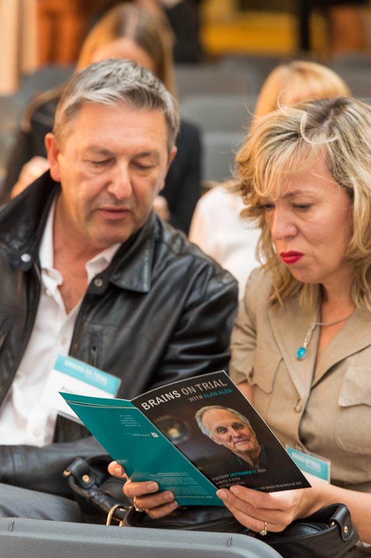
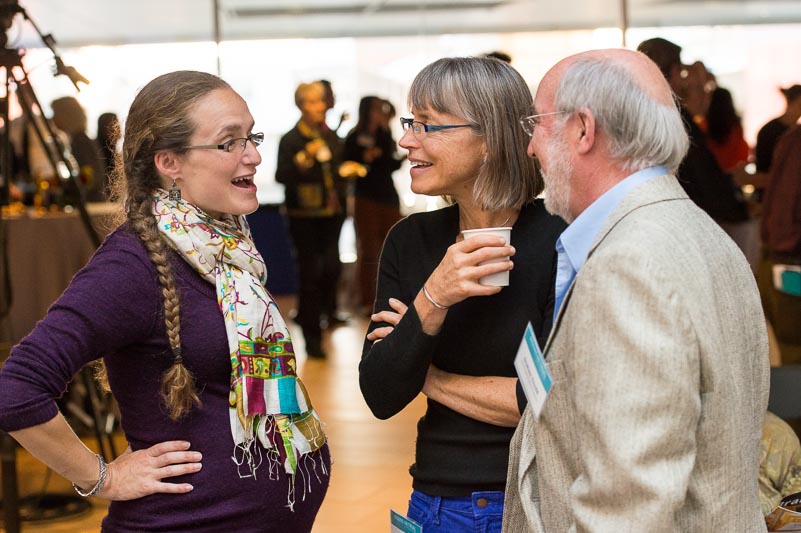
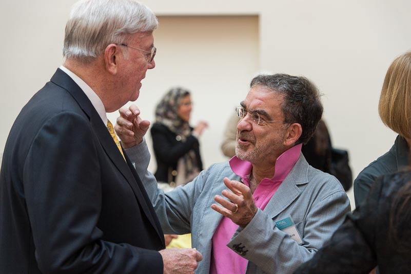
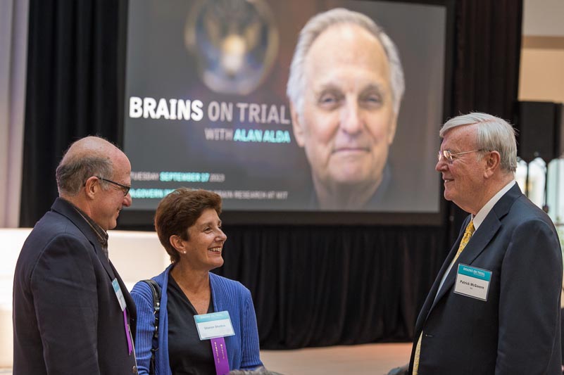
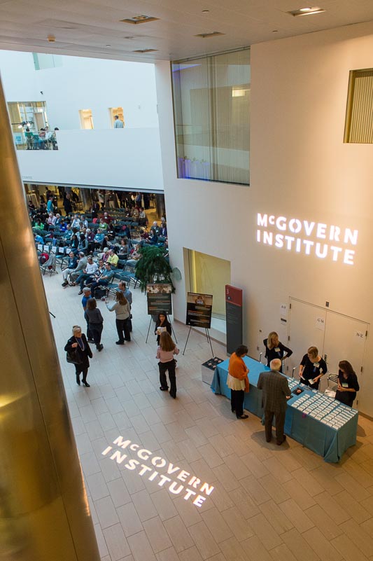
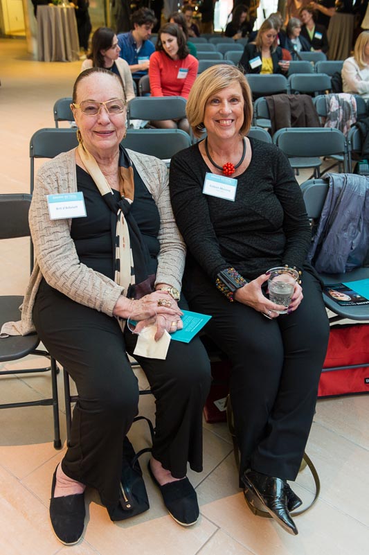
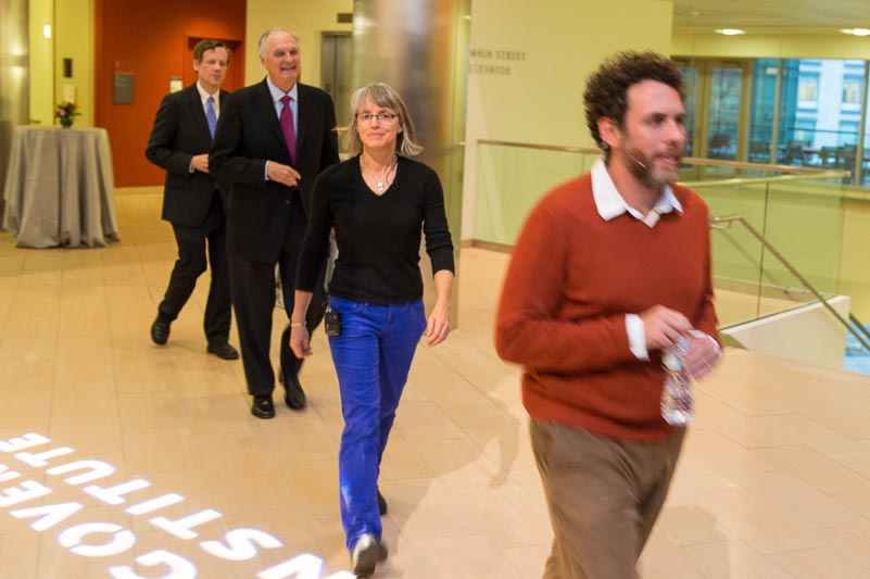
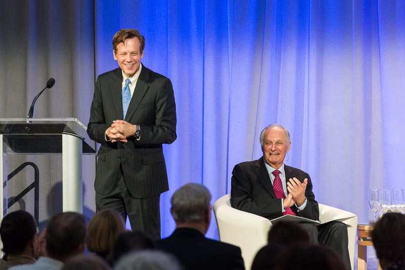
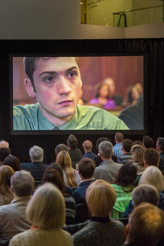
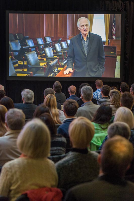
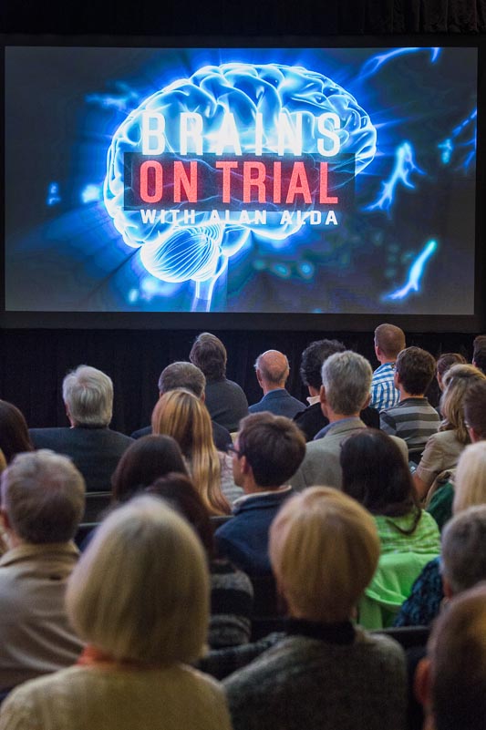
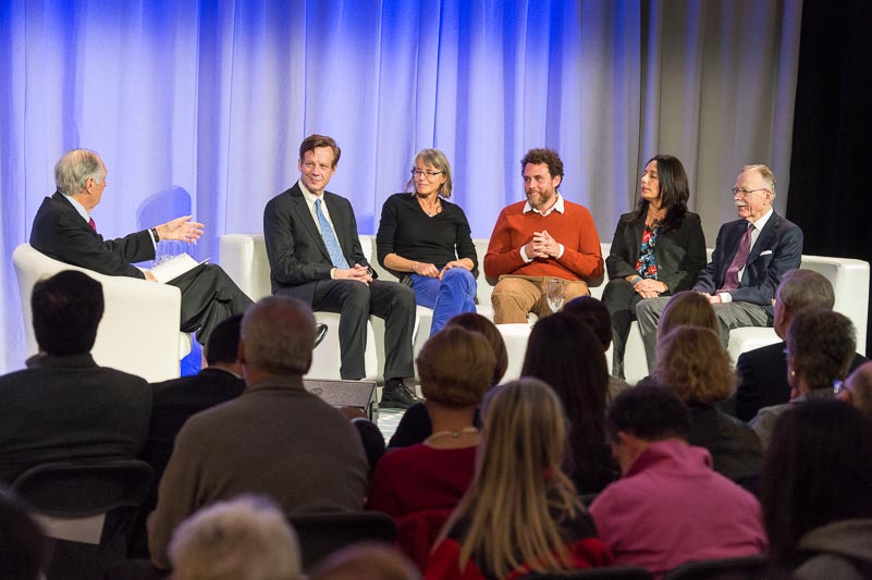
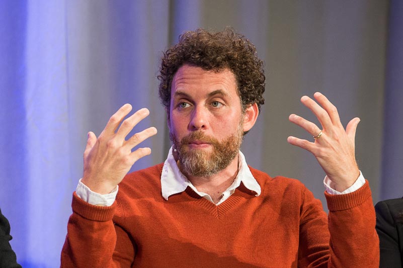
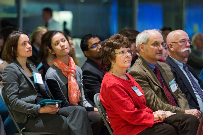
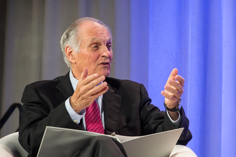
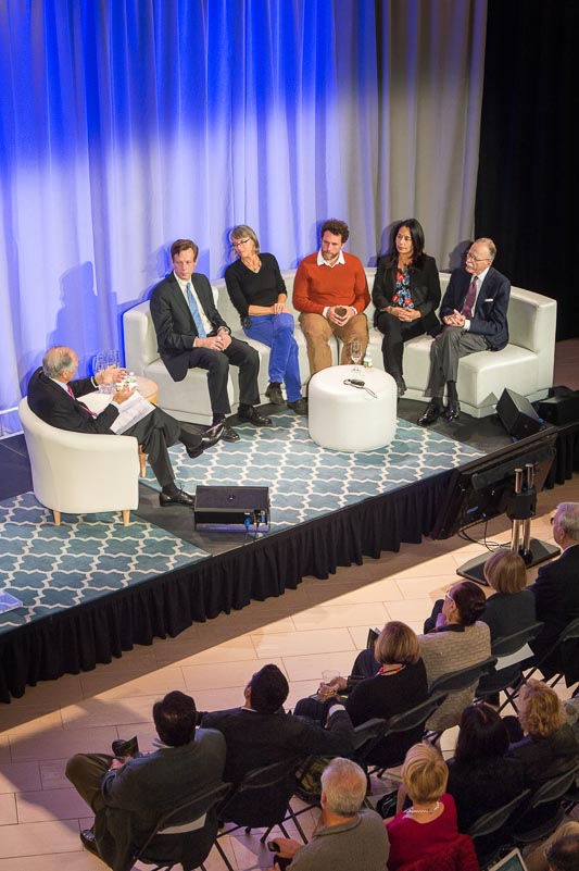
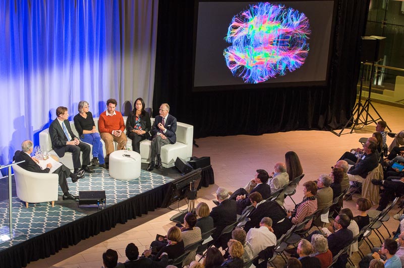
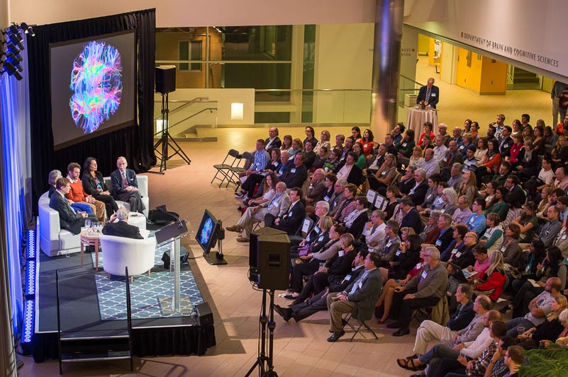
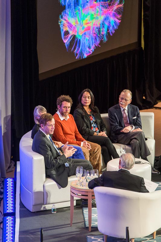
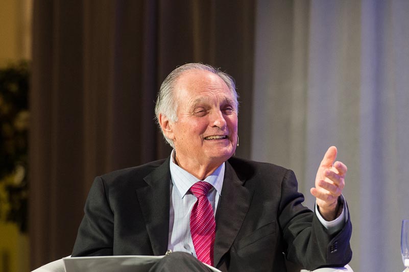
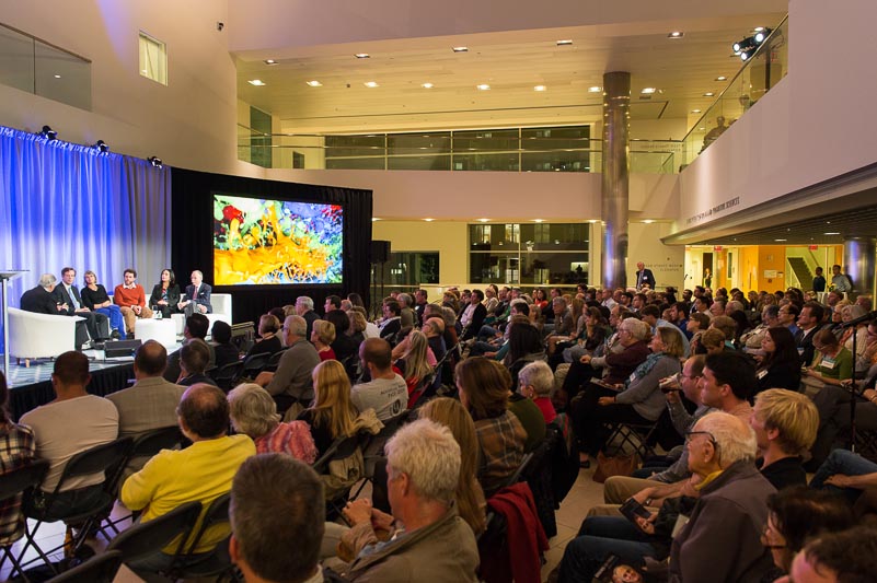
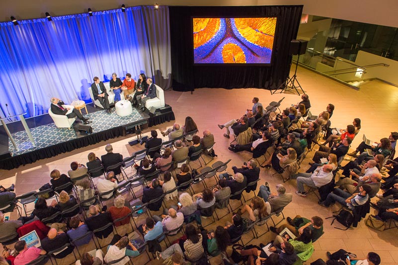
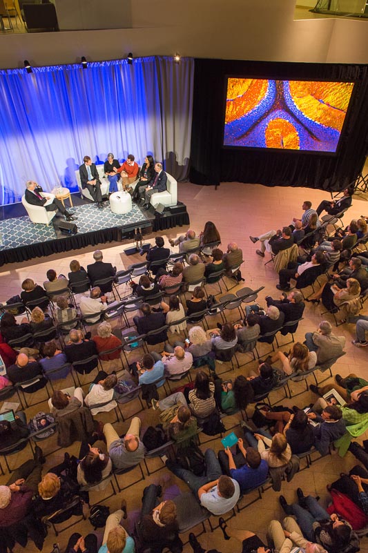
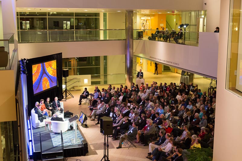
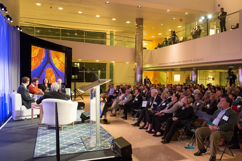
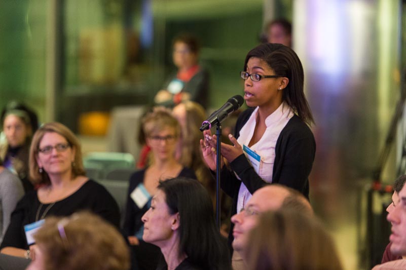
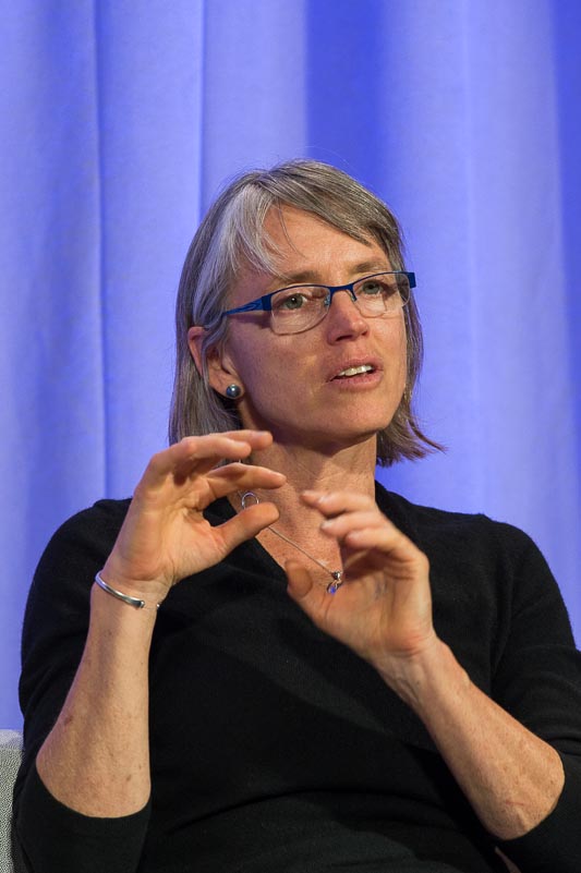
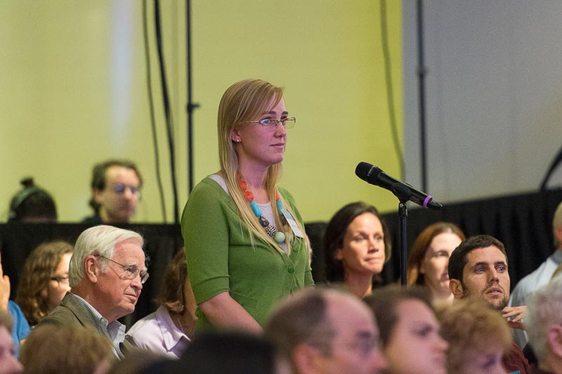
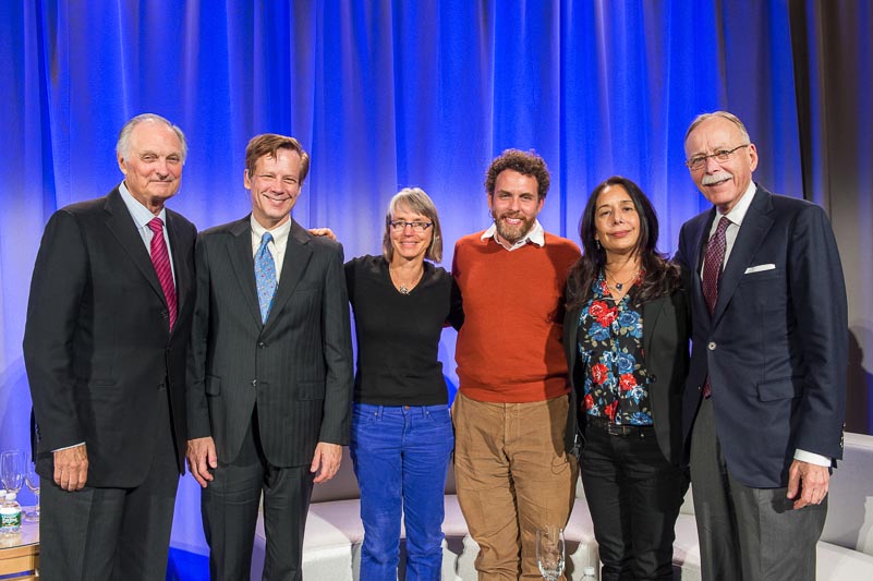
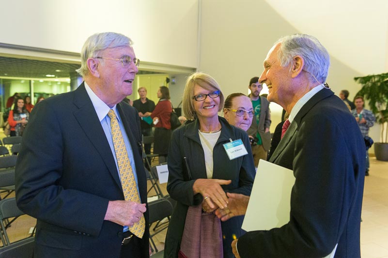
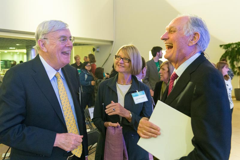
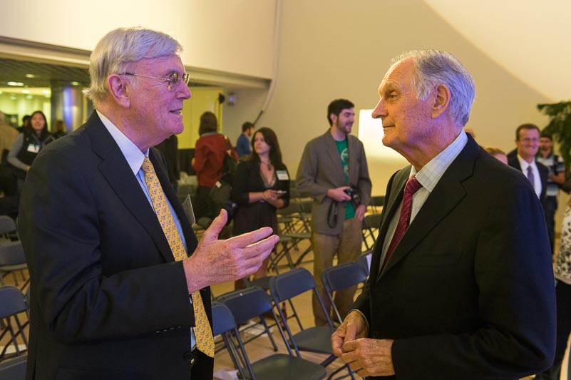
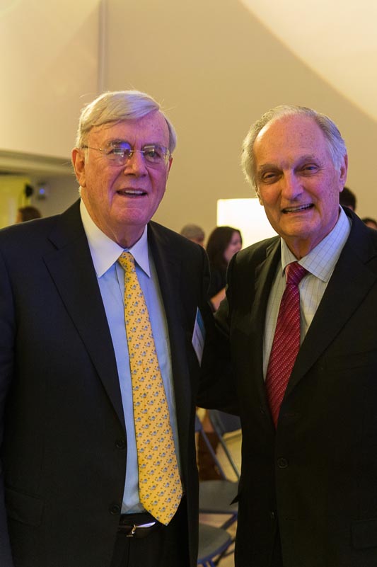
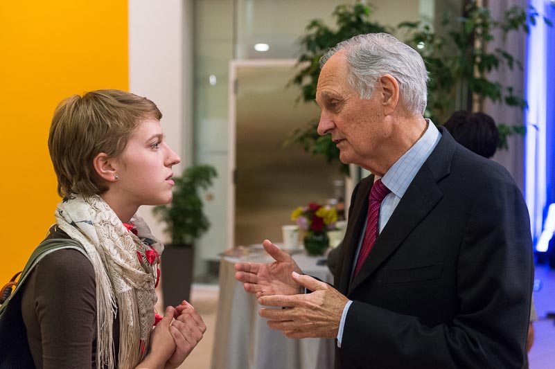
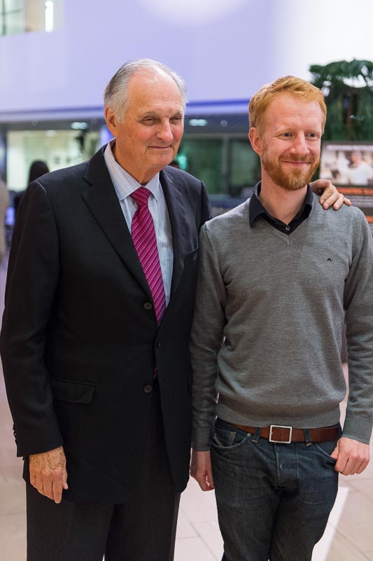
Brains on Trial with Alan Alda
What if we could peer into the brain to determine guilt or innocence? Could advances in neuroscience help reform our criminal justice system?
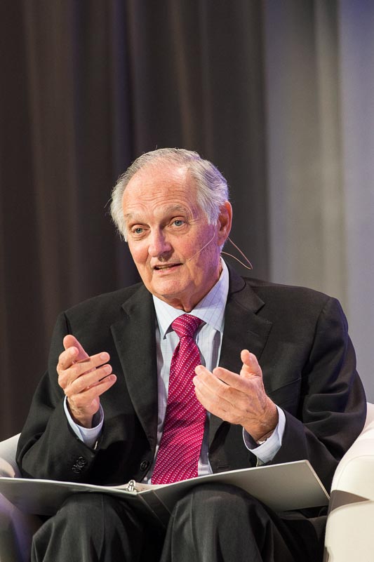
On Tuesday, September 17th, the McGovern Institute hosted a discussion with a distinguished group of legal and neuroscience experts who debated these and related questions. Alan Alda moderated the panel of experts, showed clips from his 2-part PBS special, “Brains on Trial,” and engaged the audience in a Q&A session.
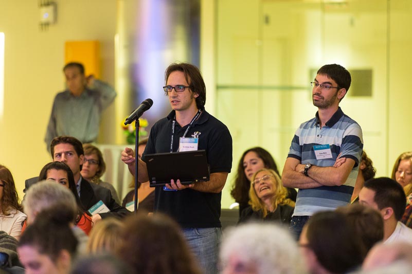
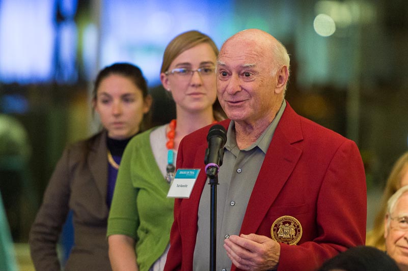
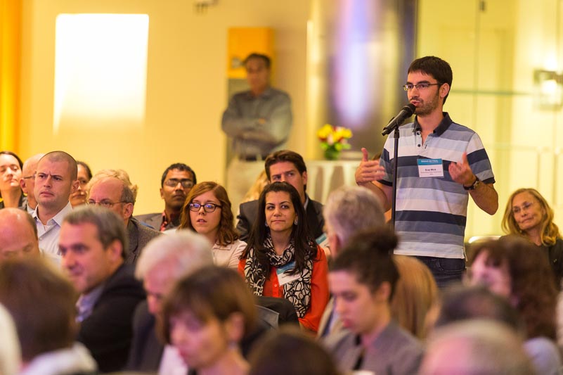
See below for a photo gallery of the event. All photos courtesy of Justin Knight.


Brains on Trial with Alan Alda
What if we could peer into the brain to determine guilt or innocence? Could advances in neuroscience help reform our criminal justice system?
On Tuesday, September 17th, the McGovern Institute hosted a discussion with a distinguished group of legal and neuroscience experts who debated these and related questions. Alan Alda moderated the panel of experts, showed clips from his 2-part PBS special, “Brains on Trial,” and engaged the audience in a Q&A session.
Feng Zhang named to Popular Science Brilliant 10
Popular Science magazine has named two MIT junior faculty members — Pedro Reis and Feng Zhang — to its 2013 Brilliant 10 list of young stars in science and technology. The list will appear in the magazine’s October issue.
“Popular Science prides itself on revealing the innovations and ideas that are laying today’s groundwork for tomorrow’s breakthroughs, and the Brilliant 10 is one of the most exciting ways we do that,” says Jake Ward, editor-in-chief. “This collection of 10 brilliant young researchers is our chance to honor the most promising work — and the most hardworking people — in science and technology today. This year’s winners are particularly distinguished and I’m proud to welcome them all as members of the 2013 Brilliant 10.”
Pedro Reis, the Esther and Harold E. Edgerton Assistant Professor of Civil and Environmental Engineering and Mechanical Engineering, studies the mechanics of slender structures, with a particular focus on devising new ways of turning mechanical failure into functionality.
Over the past few years, Reis, 35, has published a number of eclectic and impactful papers in prominent journals. In 2009 he reported on the delamination of thin films adhered to soft foundations, which is relevant for stretchable electronics. He explained why adhesive films tear into triangular shapes, a problem that applies to both the everyday peeling of adhesive tape from a roll and the manufacturing of tapered graphene nanoribbons. Motivated by the closing of aquatic flowers, he recently discovered a new mechanism for passively pipetting liquids using a petal-shaped object. And last year inspired by a toy, Reis introduced the Buckliball, a new class of structures that uses buckling to provide origami-like folding capabilities to curved structures with potential uses for encapsulation and soft robotics.
In other work undertaken just for fun, Reis and colleagues reported in 2010 that when cats lap fluids (milk or water, for example), they take advantage of a perfect balance between gravity and inertia.
Feng Zhang, 31, is the W.M. Keck Career Development Professor in Biomedical Engineering, an assistant professor in the department of Brain and Cognitive Sciences, a member of the McGovern Institute for Brain Research and a core member of the Broad Institute. He received the award for his work on genome editing. Earlier this year he reported a powerful new way to make targeted mutations in genomic DNA, based on a bacterial system known as CRISPR. The new method will greatly accelerate the development of animal models of human genetic diseases, and may eventually make it possible to correct genetic mutations in patients. Zhang, a pioneer in optogenetics, has also recently invented a new method for controlling gene expression with light, in which light-sensitive plant proteins are engineered to create an “optical switch” that can turn other genes on or off at will.
This is the 12th annual Brilliant 10 list. Ten MIT researchers were included on previous lists.
Nanodiamonds
The Boyden lab is exploring the use of fluorescent nanodiamonds as a new class of optical probes for neuroscience research. Photo: Justin Knight
Dyslexia ‘seen in brain scans’ of preschool children
John Gabrieli’s lab has found that differences in a key language structure can be seen even before children start learning to read.
The study was picked up by various news outlets including the BBC, CBS, WBUR, US News and World Report, the UK Daily Mail, Fox News, and more.
Visualizing the Brain
Zeynep Saygin, a postdoc in Nancy Kanwisher’s lab uses a technology known as diffusion-weighted MR imaging to reveal long-range connections in the brain.
Tracking the roots of reading ability
Researchers in the Gabrieli lab have found that differences in a key language structure can be seen even before children start learning to read.
Brain scans may help diagnose dyslexia
About 10 percent of the U.S. population suffers from dyslexia, a condition that makes learning to read difficult. Dyslexia is usually diagnosed around second grade, but the results of a new study from MIT could help identify those children before they even begin reading, so they can be given extra help earlier.
The study, done with researchers at Boston Children’s Hospital, found a correlation between poor pre-reading skills in kindergartners and the size of a brain structure that connects two language-processing areas.
Previous studies have shown that in adults with poor reading skills, this structure, known as the arcuate fasciculus, is smaller and less organized than in adults who read normally. However, it was unknown if these differences cause reading difficulties or result from lack of reading experience.
“We were very interested in looking at children prior to reading instruction and whether you would see these kinds of differences,” says John Gabrieli, the Grover M. Hermann Professor of Health Sciences and Technology, professor of brain and cognitive sciences and a member of MIT’s McGovern Institute for Brain Research.
Gabrieli and Nadine Gaab, an assistant professor of pediatrics at Boston Children’s Hospital, are the senior authors of a paper describing the results in the Aug. 14 issue of the Journal of Neuroscience. Lead authors of the paper are MIT postdocs Zeynep Saygin and Elizabeth Norton.
The path to reading
The new study is part of a larger effort involving approximately 1,000 children at schools throughout Massachusetts and Rhode Island. At the beginning of kindergarten, children whose parents give permission to participate are assessed for pre-reading skills, such as being able to put words together from sounds.
“From that, we’re able to provide — at the beginning of kindergarten — a snapshot of how that child’s pre-reading abilities look relative to others in their classroom or other peers, which is a real benefit to the child’s parents and teachers,” Norton says.
The researchers then invite a subset of the children to come to MIT for brain imaging. The Journal of Neuroscience study included 40 children who had their brains scanned using a technique known as diffusion-weighted imaging, which is based on magnetic resonance imaging (MRI).
This type of imaging reveals the size and organization of the brain’s white matter — bundles of nerves that carry information between brain regions. The researchers focused on three white-matter tracts associated with reading skill, all located on the left side of the brain: the arcuate fasciculus, the inferior longitudinal fasciculus (ILF) and the superior longitudinal fasciculus (SLF).
When comparing the brain scans and the results of several different types of pre-reading tests, the researchers found a correlation between the size and organization of the arcuate fasciculus and performance on tests of phonological awareness — the ability to identify and manipulate the sounds of language.
Phonological awareness can be measured by testing how well children can segment sounds, identify them in isolation, and rearrange them to make new words. Strong phonological skills have previously been linked with ease of learning to read. “The first step in reading is to match the printed letters with the sounds of letters that you know exist in the world,” Norton says.
The researchers also tested the children on two other skills that have been shown to predict reading ability — rapid naming, which is the ability to name a series of familiar objects as quickly as you can, and the ability to name letters. They did not find any correlation between these skills and the size or organization of the white-matter structures scanned in this study.
Early intervention
The left arcuate fasciculus connects Broca’s area, which is involved in speech production, and Wernicke’s area, which is involved in understanding written and spoken language. A larger and more organized arcuate fasciculus could aid in communication between those two regions, the researchers say.
Gabrieli points out that the structural differences found in the study don’t necessarily reflect genetic differences; environmental influences could also be involved. “At the moment when the children arrive at kindergarten, which is approximately when we scan them, we don’t know what factors lead to these brain differences,” he says.
The researchers plan to follow three waves of children as they progress to second grade and evaluate whether the brain measures they have identified predict poor reading skills.
“We don’t know yet how it plays out over time, and that’s the big question: Can we, through a combination of behavioral and brain measures, get a lot more accurate at seeing who will become a dyslexic child, with the hope that that would motivate aggressive interventions that would help these children right from the start, instead of waiting for them to fail?” Gabrieli says.
For at least some dyslexic children, offering extra training in phonological skills can help them improve their reading skills later on, studies have shown.
The research was funded by the National Institutes of Health, the Poitras Center for Affective Disorders Research, the Ellison Medical Foundation and the Halis Family Foundation.
Brains on Trial: Neuroscience and the Law
What if we could peer into the brain to determine guilt or innocence? Could advances in neuroscience help reform our criminal justice system?
We invite you to join the discussion with a distinguished group of legal and neuroscience experts who will debate these and related questions on Tuesday, September 17th. Alan Alda will moderate the panel of experts, show clips from his 2-part PBS special, “Brains on Trial,” and engage the audience in a Q&A session. We hope you will join us!
REGISTER NOW
BRAINS ON TRIAL DISCUSSION WITH ALAN ALDA
DATE: Tuesday September 17, 2013
TIME: 6:00 – 8:30
LOCATION: McGovern Institute for Brain Research at MIT (MIT Bldg 46, Third Floor Atrium)
QUESTIONS? brainsontrial@mit.edu or 617.324.2077
MODERATOR
 Alan Alda, a seven-time Emmy Award–winner, played Hawkeye Pierce on the classic television series, M*A*S*H, and appeared in continuing roles on ER, The West Wing, and 30 Rock. His long-time interest in science and in promoting a greater public understanding of science led to his hosting the award-winning PBS series Scientific American Frontiers for eleven years, on which he interviewed hundreds of scientists from around the world. He has 33 Emmy nominations as actor, writer, and director, and is a Television Hall of Fame inductee. He has also appeared on the Broadway stage, where he received three Tony nominations.
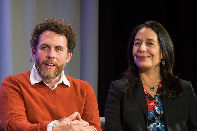
PANELISTS
 Robert Desimone is director of the McGovern Institute and the Doris and Don Berkey Professor in MIT’s Department of Brain and Cognitive Sciences. He served as network co-director of the Macarthur Law and Neuroscience Project from 2008-2010. Prior to joining the McGovern Institute in 2004, he was director of the Intramural Research Program at the National Institutes of Mental Health. He is a member of the National Academy of Sciences and the American Academy of Arts and Sciences and a recipient of numerous awards, including the Troland Prize of the National Academy of Sciences.
 Joshua D. Greene is the John and Ruth Hazel Associate Professor of the Social Sciences in the Department of Psychology at Harvard University. He is an experimental psychologist, neuroscientist, and philosopher. He studies the psychology and neuroscience of morality, focusing on the interplay between emotion and reasoning in moral decision-making. In 2012 he was awarded the Stanton Prize by the Society for Philosophy and Psychology. He is the author of the forthcoming book Moral Tribes: Emotion, Reason, and the Gap Between Us and Them.
 Nancy Kanwisher is the Walter A. Rosenblith Professor of Cognitive Neuroscience in the Department of Brain and Cognitive Sciences and a founding member of the McGovern Institute. She joined the MIT faculty in 1997, and prior to that was a faculty member at UCLA and Harvard University. In 1999, she received the National Academy of Sciences Troland Research Award. The Kanwisher lab uses brain imaging to study the functional organization of the human brain.
 Bea Luna is a professor of psychiatry at the University of Pittsburgh School of Medicine. She is the director of the Laboratory of Neurocognitive Development, where she leads projects investigating the brain basis of typical and abnormal adolescent development of voluntary behaviors and motivation.
 Stephen J. Morse is the Ferdinand Wakeman Hubbell Professor of Law, Professor of Psychology and Law in Psychiatry, and Associate Director of the Center for Neuroscience and Society at the University of Pennsylvania. He is a veteran in the fields of psychology and law and was instrumental in building the MacArthur Law and Neuroscience Project. Being both a scientist and a legal expert, he understands and writes extensively on the relevance of neuroscience to law. He also serves as a member of the MacArthur Foundation Research Network on Law and neuroscience.
This event is based on a two-part PBS special, “Brains on Trial with Alan Alda,” scheduled for broadcast on September 11 and 18 at 10PM. Watch the preview below:
Brains on Trial with Alan Alda takes a fictitious crime – a convenience store robbery that goes horribly wrong – and builds from it a gripping courtroom drama. As the trial unfolds it takes us into the brains of the major participants – defendant, witnesses, jurors, judge – while Alan Alda visits the laboratories of some dozen neuroscientists exploring how brains work when they become entangled with the law. The research he discovers poses the controversial question: How does our rapidly expanding ability to peer into people’s minds and decode their thoughts and feelings affect trials like the one we are watching in the future? And should it?