Whether speaking Turkish or Norwegian, the brain’s language network looks the same

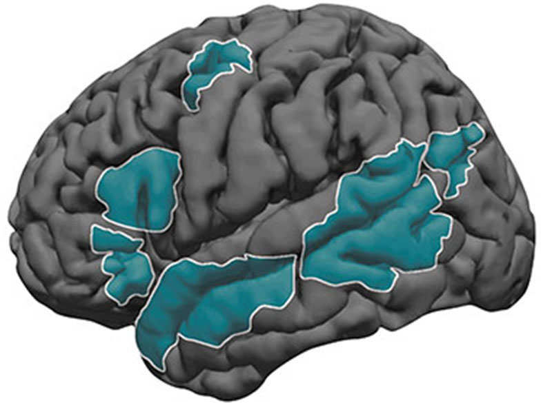
Over several decades, neuroscientists have created a well-defined map of the brain’s “language network,” or the regions of the brain that are specialized for processing language. Found primarily in the left hemisphere, this network includes regions within Broca’s area, as well as in other parts of the frontal and temporal lobes.

However, the vast majority of those mapping studies have been done in English speakers as they listened to or read English texts. MIT neuroscientists have now performed brain imaging studies of speakers of 45 different languages. The results show that the speakers’ language networks appear to be essentially the same as those of native English speakers.

The findings, while not surprising, establish that the location and key properties of the language network appear to be universal. The work also lays the groundwork for future studies of linguistic elements that would be difficult or impossible to study in English speakers because English doesn’t have those features.

“This study is very foundational, extending some findings from English to a broad range of languages,” says Evelina Fedorenko, the Frederick A. and Carole J. Middleton Career Development Associate Professor of Neuroscience at MIT and a member of MIT’s McGovern Institute for Brain Research. “The hope is that now that we see that the basic properties seem to be general across languages, we can ask about potential differences between languages and language families in how they are implemented in the brain, and we can study phenomena that don’t really exist in English.”

Fedorenko is the senior author of the study, which appears today in Nature Neuroscience. Saima Malik-Moraleda, a PhD student in the Speech and Hearing Bioscience and Technology program at Harvard University, and Dima Ayyash, a former research assistant, are the lead authors of the paper.

Mapping language networks

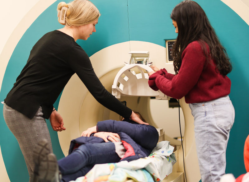
The precise locations and shapes of language areas differ across individuals, so to find the language network, researchers ask each person to perform a language task while scanning their brains with functional magnetic resonance imaging (fMRI). Listening to or reading sentences in one’s native language should activate the language network. To distinguish this network from other brain regions, researchers also ask participants to perform tasks that should not activate it, such as listening to an unfamiliar language or solving math problems.

Several years ago, Fedorenko began designing these “localizer” tasks for speakers of languages other than English. While most studies of the language network have used English speakers as subjects, English does not include many features commonly seen in other languages. For example, in English, word order tends to be fixed, while in other languages there is more flexibility in how words are ordered. Many of those languages instead use the addition of morphemes, or segments of words, to convey additional meaning and relationships between words.

“There has been growing awareness for many years of the need to look at more languages, if you want make claims about how language works, as opposed to how English works,” Fedorenko says. “We thought it would be useful to develop tools to allow people to rigorously study language processing in the brain in other parts of the world. There’s now access to brain imaging technologies in many countries, but the basic paradigms that you would need to find the language-responsive areas in a person are just not there.”

For the new study, the researchers performed brain imaging of two speakers of 45 different languages, representing 12 different language families. Their goal was to see if key properties of the language network, such as location, left lateralization, and selectivity, were the same in those participants as in people whose native language is English.

The researchers decided to use “Alice in Wonderland” as the text that everyone would listen to, because it is one of the most widely translated works of fiction in the world. They selected 24 short passages and three long passages, each of which was recorded by a native speaker of the language. Each participant also heard nonsensical passages, which should not activate the language network, and was asked to do a variety of other cognitive tasks that should not activate it.

The team found that the language networks of participants in this study were found in approximately the same brain regions, and had the same selectivity, as those of native speakers of English.

“Language areas are selective,” Malik-Moraleda says. “They shouldn’t be responding during other tasks such as a spatial working memory task, and that was what we found across the speakers of 45 languages that we tested.”

Additionally, language regions that are typically activated together in English speakers, such as the frontal language areas and temporal language areas, were similarly synchronized in speakers of other languages.

The researchers also showed that among all of the subjects, the small amount of variation they saw between individuals who speak different languages was the same as the amount of variation that would typically be seen between native English speakers.

Similarities and differences

While the findings suggest that the overall architecture of the language network is similar across speakers of different languages, that doesn’t mean that there are no differences at all, Fedorenko says. As one example, researchers could now look for differences in speakers of languages that predominantly use morphemes, rather than word order, to help determine the meaning of a sentence.

“There are all sorts of interesting questions you can ask about morphological processing that don’t really make sense to ask in English, because it has much less morphology,” Fedorenko says.

Another possibility is studying whether speakers of languages that use differences in tone to convey different word meanings would have a language network with stronger links to auditory brain regions that encode pitch.

Right now, Fedorenko’s lab is working on a study in which they are comparing the ‘temporal receptive fields’ of speakers of six typologically different languages, including Turkish, Mandarin, and Finnish. The temporal receptive field is a measure of how many words the language processing system can handle at a time, and for English, it has been shown to be six to eight words long.

“The language system seems to be working on chunks of just a few words long, and we’re trying to see if this constraint is universal across these other languages that we’re testing,” Fedorenko says.

The researchers are also working on creating language localizer tasks and finding study participants representing additional languages beyond the 45 from this study.

The research was funded by the National Institutes of Health and research funds from MIT’s Department of Brain and Cognitive Sciences, the McGovern Institute, and the Simons Center for the Social Brain. Malik-Moraleda was funded by a la Caixa Fellowship and a Friends of McGovern fellowship.

A voice for change — in Spanish

Jessica Chomik-Morales had a bicultural childhood. She was born in Boca Raton, Florida, where her parents had come seeking a better education for their daughter than she would have access to in Paraguay. But when she wasn’t in school, Chomik-Morales was back in that small, South American country with her family. One of the consequences of growing up in two cultures was an early interest in human behavior. “I was always in observer mode,” Chomik-Morales says, recalling how she would tune in to the nuances of social interactions in order to adapt and fit in.

Today, that fascination with human behavior is driving Chomik-Morales as she works with MIT professor of cognitive science Laura Schulz and Walter A. Rosenblith Professor of Cognitive Neuroscience and McGovern Institute for Brain Research investigator Nancy Kanwisher as a post-baccalaureate research scholar, using functional brain imaging to investigate how the brain recognizes and understands causal relationships. Since arriving at MIT last fall, she’s worked with study volunteers to collect functional MRI (fMRI) scans and used computational approaches to interpret the images. She’s also refined her own goals for the future.

Jessica Chomik-Morales (right) with postdoctoral associate Héctor De Jesús-Cortés. Photo: Steph Stevens

She plans to pursue a career in clinical neuropsychology, which will merge her curiosity about the biological basis of behavior with a strong desire to work directly with people. “I’d love to see what kind of questions I could answer about the neural mechanisms driving outlier behavior using fMRI coupled with cognitive assessment,” she says. And she’s confident that her experience in MIT’s two-year post-baccalaureate program will help her get there. “It’s given me the tools I need, and the techniques and methods and good scientific practice,” she says. “I’m learning that all here. And I think it’s going to make me a more successful scientist in grad school.”

The road to MIT

Chomik-Morales’s path to MIT was not a straightforward trajectory through the U.S. school system. When her mom, and later her dad, were unable to return to the U.S., she started eight grade in the capital city of Asunción. It did not go well. She spent nearly every afternoon in the principal’s office, and soon her father was encouraging her to return to the United States. “You are an American,” he told her. “You have a right to the educational system there.”

Back in Florida, Chomik-Morales became a dedicated student, even while she worked assorted jobs and shuffled between the homes of families who were willing to host her. “I had to grow up,” she says. “My parents are sacrificing everything just so I can have a chance to be somebody. People don’t get out of Paraguay often, because there aren’t opportunities and it’s a very poor country. I was given an opportunity, and if I waste that, then that is disrespect not only to my parents, but to my lineage, to my country.”

As she graduated from high school and went on to earn a degree in cognitive neuroscience at Florida Atlantic University, Chomik-Morales found herself experiencing things that were completely foreign to her family. Though she spoke daily with her mom via WhatsApp, it was hard to share what she was learning in school or what she was doing in the lab. And while they celebrated her academic achievements, Chomik-Morales knew they didn’t really understand them. “Neither of my parents went to college,” she says. “My mom told me that she never thought twice about learning about neuroscience. She had this misconception that it was something that she would never be able to digest.”

Chomik-Morales believes that the wonders of neuroscience are for everybody. But she also knows that Spanish speakers like her mom have few opportunities to hear the kinds of accessible, engaging stories that might draw them in. So she’s working to change that. With support from the McGovern Institute, the National Science Foundation funded Science and Technology Center for Brains, Minds, and Machines, Chomik-Morales is hosting and producing a weekly podcast called “Mi Última Neurona” (“My Last Neuron”), which brings conversations with neuroscientists to Spanish speakers around the world.

Listeners hear how researchers at MIT and other institutions are exploring big concepts like consciousness and neurodegeneration, and learn about the approaches they use to study the brain in humans, animals, and computational models. Chomik-Morales wants listeners to get to know neuroscientists on a personal level too, so she talks with her guests about their career paths, their lives outside the lab, and often, their experiences as immigrants in the United States.

After recording an interview with Chomik-Morales that delved into science, art, and the educational system in his home country of Peru, postdoc Arturo Deza thinks “Mi Última Neurona” has the potential to inspire Spanish speakers in Latin America, as well immigrants in other countries. “Even if you’re not a scientist, it’s really going to captivate you and you’re going to get something out of it,” he says. To that point, Chomik-Morales’s mother has quickly become an enthusiastic listener, and even begun seeking out resources to learn more about the brain on her own.

Chomik-Morales hopes the stories her guests share on “Mi Última Neurona” will inspire a future generation of Hispanic neuroscientists. She also wants listeners to know that a career in science doesn’t have to mean leaving their country behind. “Gain whatever you need to gain from outside, and then, if it’s what you desire, you’re able to go back and help your own community,” she says. With “Mi Última Neurona,” she adds, she feels she is giving back to her roots.

Unexpected synergy

This story originally appeared in the Spring 2022 issue of BrainScan.

***

Recent results from cognitive neuroscientist Nancy Kanwisher’s lab have left her pondering the role of music in human evolution. “Music is this big mystery,” she says. “Every human society that’s been studied has music. No other animals have music in the way that humans do. And nobody knows why humans have music at all. This has been a puzzle for centuries.”

MIT neuroscientist and McGovern Investigator Nancy Kanwisher. Photo: Jussi Puikkonen/KNAW

Some biologists and anthropologists have reasoned that since there’s no clear evolutionary advantage for humans’ unique ability to create and respond to music, these abilities must have emerged when humans began to repurpose other brain functions. To appreciate song, they’ve proposed, we draw on parts of the brain dedicated to speech and language. It makes sense, Kanwisher says: music and language are both complex, uniquely human ways of communicating. “It’s very sensible to think that there might be common machinery,” she says. “But there isn’t.”

That conclusion is based on her team’s 2015 discovery of neurons in the human brain that respond only to music. They first became clued in to these music-sensitive cells when they asked volunteers to listen to a diverse panel of sounds inside an MRI scanner. Functional brain imaging picked up signals suggesting that some neurons were specialized to detect only music but the broad map of brain activity generated by an fMRI couldn’t pinpoint those cells.

Singing in the brain

Kanwisher’s team wanted to know more but neuroscientists who study the human brain can’t always probe its circuitry with the exactitude of their colleagues who study the brains of mice or rats. They can’t insert electrodes into human brains to monitor the neurons they’re interested in. Neurosurgeons, however, sometimes do — and thus, collaborating with neurosurgeons has created unique opportunities for Kanwisher and other McGovern investigators to learn about the human brain.

Kanwisher’s team collaborated with clinicians at Albany Medical Center to work with patients who are undergoing monitoring prior to surgical treatment for epilepsy. Before operating, a neurosurgeon must identify the spot in their patient’s brain that is triggering seizures. This means inserting electrodes into the brain to monitor specific areas over a few days or weeks. The electrodes they implant pinpoint activity far more precisely, both spatially and temporally, than an MRI. And with patients’ permission, researchers like Kanwisher can take advantage of the information they collect.

“The intracranial recording from human brains that’s possible from collaboration with neurosurgeons is extremely precious to us,” Kanwisher says. “All of the research is kind of opportunistic, on whatever the surgeons are doing for clinical reasons. But sometimes we get really lucky and the electrodes are right in an area where we have long-standing scientific questions that those data can answer.”

Song-selective neural population (yellow) in the “inflated” human brain. Image: Sam Norman-Haignere

The unexpected discovery of song-specific neurons, led by postdoctoral researcher Sam Norman-Haignere, who is now an assistant professor at the University of Rochester Medical Center, emerged from such a collaboration. The team worked with patients at Albany Medical Center whose presurgical monitoring encompassed the auditory-processing part of the brain that they were curious about. Sure enough, certain electrodes picked up activity only when patients were listening to music. The data indicated that in some of those locations, it didn’t matter what kind of music was playing: the cells fired in response to a range of sounds that included flute solos, heavy metal, and rap. But other locations became active exclusively in response to vocal music. “We did not have that hypothesis at all, Kanwisher says. “It reallytook our breath away,” she says.

When that discovery is considered along with findings from McGovern colleague Ev Fedorenko, who has shown that the brain’s language-processing regions do not respond to music, Kanwisher says it’s now clear that music and language are segregated in the human brain. The origins of our unique appreciation for music, however, remain a mystery.

Clinical advantage

Clinical collaborations are also important to researchers in Ann Graybiels lab, who rely largely on model organisms like mice and rats to investigate the fine details of neural circuits. Working with clinicians helps keep them focused on answering questions that matter to patients.

In studying how the brain makes decisions, the Graybiel lab has zeroed in on connections that are vital for making choices that carry both positive and negative consequences. This is the kind of decision-making that you might call on when considering whether to accept a job that pays more but will be more demanding than your current position, for example. In experiments with rats, mice, and monkeys, they’ve identified different neurons dedicated to triggering opposing actions “approach” or “avoid” in these complex decision-making tasks. They’ve also found evidence that both age and stress change how the brain deals with these kinds of decisions.

In work led by former Graybiel lab research scientist Ken-ichi Amemori, they have worked with psychiatrist Diego Pizzagalli at McLean Hospital to learn what happens in the human brain when people make these complex decisions.

By monitoring brain activity as people made decisions inside an MRI scanner, the team identified regions that lit up when people chose to “approach” or “avoid.” They also found parallel activity patterns in monkeys that performed the same task, supporting the relevance of animal studies to understanding this circuitry.

In people diagnosed with major depression, however, the brain responded to approach-avoidance conflict somewhat differently. Certain areas were not activated as strongly as they were in people without depression, regardless of whether subjects ultimately chose to “approach” or “avoid.” The team suspects that some of these differences might reflect a stronger tendency toward avoidance, in which potential rewards are less influential for decision-making, while an individual is experiencing major depression.

The brain activity associated with approach-avoidance conflict in humans appears to align with what Graybiel’s team has seen in mice, although clinical imaging cannot reveal nearly as much detail about the involved circuits. Graybiel says that gives her confidence that what they are learning in the lab, where they can manipulate and study neural circuits with precision, is important. “I think there’s no doubt that this is relevant to humans,” she says. “I want to get as far into the mechanisms as possible, because maybe we’ll hit something that’s therapeutically valuable, or maybe we will really get an intuition about how parts of the brain work. I think that will help people.”

An optimized solution for face recognition

The human brain seems to care a lot about faces. It’s dedicated a specific area to identifying them, and the neurons there are so good at their job that most of us can readily recognize thousands of individuals. With artificial intelligence, computers can now recognize faces with a similar efficiency—and neuroscientists at MIT’s McGovern Institute have found that a computational network trained to identify faces and other objects discovers a surprisingly brain-like strategy to sort them all out.

The finding, reported March 16, 2022, in Science Advances, suggests that the millions of years of evolution that have shaped circuits in the human brain have optimized our system for facial recognition.

“The human brain’s solution is to segregate the processing of faces from the processing of objects,” explains Katharina Dobs, who led the study as a postdoctoral researcher in McGovern investigator Nancy Kanwisher’s lab. The artificial network that she trained did the same. “And that’s the same solution that we hypothesize any system that’s trained to recognize faces and to categorize objects would find,” she adds.

“These two completely different systems have figured out what a—if not the—good solution is. And that feels very profound,” says Kanwisher.

Functionally specific brain regions

More than twenty years ago, Kanwisher’s team discovered a small spot in the brain’s temporal lobe that responds specifically to faces. This region, which they named the fusiform face area, is one of many brain regions Kanwisher and others have found that are dedicated to specific tasks, such as the detection of written words, the perception of vocal songs, and understanding language.

Kanwisher says that as she has explored how the human brain is organized, she has always been curious about the reasons for that organization. Does the brain really need special machinery for facial recognition and other functions? “‘Why questions’ are very difficult in science,” she says. But with a sophisticated type of machine learning called a deep neural network, her team could at least find out how a different system would handle a similar task.

Dobs, who is now a research group leader at Justus Liebig University Giessen in Germany, assembled hundreds of thousands of images with which to train a deep neural network in face and object recognition. The collection included the faces of more than 1,700 different people and hundreds of different kinds of objects, from chairs to cheeseburgers. All of these were presented to the network, with no clues about which was which. “We never told the system that some of those are faces, and some of those are objects. So it’s basically just one big task,” Dobs says. “It needs to recognize a face identity, as well as a bike or a pen.”

Visualization of the preferred stimulus for example face-ranked filters. While filters in early layers (e.g., Conv5) were maximally activated by simple features, filters responded to features that appear somewhat like face parts (e.g., nose and eyes) in mid-level layers (e.g., Conv9) and appear to represent faces in a more holistic manner in late convolutional layers. Image: Kanwisher lab

As the program learned to identify the objects and faces, it organized itself into an information-processing network with that included units specifically dedicated to face recognition. Like the brain, this specialization occurred during the later stages of image processing. In both the brain and the artificial network, early steps in facial recognition involve more general vision processing machinery, and final stages rely on face-dedicated components.

It’s not known how face-processing machinery arises in a developing brain, but based on their findings, Kanwisher and Dobs say networks don’t necessarily require an innate face-processing mechanism to acquire that specialization. “We didn’t build anything face-ish into our network,” Kanwisher says. “The networks managed to segregate themselves without being given a face-specific nudge.”

Kanwisher says it was thrilling seeing the deep neural network segregate itself into separate parts for face and object recognition. “That’s what we’ve been looking at in the brain for twenty-some years,” she says. “Why do we have a separate system for face recognition in the brain? This tells me it is because that is what an optimized solution looks like.”

Now, she is eager to use deep neural nets to ask similar questions about why other brain functions are organized the way they are. “We have a new way to ask why the brain is organized the way it is,” she says. “How much of the structure we see in human brains will arise spontaneously by training networks to do comparable tasks?”

New MRI probe can reveal more of the brain’s inner workings

Using a novel probe for functional magnetic resonance imaging (fMRI), MIT biological engineers have devised a way to monitor individual populations of neurons and reveal how they interact with each other.

Similar to how the gears of a clock interact in specific ways to turn the clock’s hands, different parts of the brain interact to perform a variety of tasks, such as generating behavior or interpreting the world around us. The new MRI probe could potentially allow scientists to map those networks of interactions.

“With regular fMRI, we see the action of all the gears at once. But with our new technique, we can pick up individual gears that are defined by their relationship to the other gears, and that’s critical for building up a picture of the mechanism of the brain,” says Alan Jasanoff, an MIT professor of biological engineering, brain and cognitive sciences, and nuclear science and engineering.

Using this technique, which involves genetically targeting the MRI probe to specific populations of cells in animal models, the researchers were able to identify neural populations involved in a circuit that responds to rewarding stimuli. The new MRI probe could also enable studies of many other brain circuits, the researchers say.

Jasanoff, who is also an associate investigator at the McGovern Institute, is the senior author of the study, which appears today in Nature Neuroscience. The lead authors of the paper are recent MIT PhD recipient Souparno Ghosh and former MIT research scientist Nan Li.

Tracing connections

Traditional fMRI imaging measures changes to blood flow in the brain, as a proxy for neural activity. When neurons receive signals from other neurons, it triggers an influx of calcium, which causes a diffusible gas called nitric oxide to be released. Nitric oxide acts in part as a vasodilator that increases blood flow to the area.

Imaging calcium directly can offer a more precise picture of brain activity, but that type of imaging usually requires fluorescent chemicals and invasive procedures. The MIT team wanted to develop a method that could work across the brain without that type of invasiveness.

“If we want to figure out how brain-wide networks of cells and brain-wide mechanisms function, we need something that can be detected deep in tissue and preferably across the entire brain at once,” Jasanoff says. “The way that we chose to do that in this study was to essentially hijack the molecular basis of fMRI itself.”

The researchers created a genetic probe, delivered by viruses, that codes for a protein that sends out a signal whenever the neuron is active. This protein, which the researchers called NOSTIC (nitric oxide synthase for targeting image contrast), is an engineered form of an enzyme called nitric oxide synthase. The NOSTIC protein can detect elevated calcium levels that arise during neural activity; it then generates nitric oxide, leading to an artificial fMRI signal that arises only from cells that contain NOSTIC.

The probe is delivered by a virus that is injected into a particular site, after which it travels along axons of neurons that connect to that site. That way, the researchers can label every neural population that feeds into a particular location.

“When we use this virus to deliver our probe in this way, it causes the probe to be expressed in the cells that provide input to the location where we put the virus,” Jasanoff says. “Then, by performing functional imaging of those cells, we can start to measure what makes input to that region take place, or what types of input arrive at that region.”

Turning the gears

In the new study, the researchers used their probe to label populations of neurons that project to the striatum, a region that is involved in planning movement and responding to reward. In rats, they were able to determine which neural populations send input to the striatum during or immediately following a rewarding stimulus — in this case, deep brain stimulation of the lateral hypothalamus, a brain center that is involved in appetite and motivation, among other functions.

One question that researchers have had about deep brain stimulation of the lateral hypothalamus is how wide-ranging the effects are. In this study, the MIT team showed that several neural populations, located in regions including the motor cortex and the entorhinal cortex, which is involved in memory, send input into the striatum following deep brain stimulation.

“It’s not simply input from the site of the deep brain stimulation or from the cells that carry dopamine. There are these other components, both distally and locally, that shape the response, and we can put our finger on them because of the use of this probe,” Jasanoff says.

During these experiments, neurons also generate regular fMRI signals, so in order to distinguish the signals that are coming specifically from the genetically altered neurons, the researchers perform each experiment twice: once with the probe on, and once following treatment with a drug that inhibits the probe. By measuring the difference in fMRI activity between these two conditions, they can determine how much activity is present in probe-containing cells specifically.

The researchers now hope to use this approach, which they call hemogenetics, to study other networks in the brain, beginning with an effort to identify some of the regions that receive input from the striatum following deep brain stimulation.

“One of the things that’s exciting about the approach that we’re introducing is that you can imagine applying the same tool at many sites in the brain and piecing together a network of interlocking gears, which consist of these input and output relationships,” Jasanoff says. “This can lead to a broad perspective on how the brain works as an integrated whole, at the level of neural populations.”

The research was funded by the National Institutes of Health and the MIT Simons Center for the Social Brain.

Singing in the brain

Press Mentions

For the first time, MIT neuroscientists have identified a population of neurons in the human brain that lights up when we hear singing, but not other types of music.

These neurons, found in the auditory cortex, appear to respond to the specific combination of voice and music, but not to either regular speech or instrumental music. Exactly what they are doing is unknown and will require more work to uncover, the researchers say.

“The work provides evidence for relatively fine-grained segregation of function within the auditory cortex, in a way that aligns with an intuitive distinction within music,” says Sam Norman-Haignere, a former MIT postdoc who is now an assistant professor of neuroscience at the University of Rochester Medical Center.

The work builds on a 2015 study in which the same research team used functional magnetic resonance imaging (fMRI) to identify a population of neurons in the brain’s auditory cortex that responds specifically to music. In the new work, the researchers used recordings of electrical activity taken at the surface of the brain, which gave them much more precise information than fMRI.

“There’s one population of neurons that responds to singing, and then very nearby is another population of neurons that responds broadly to lots of music. At the scale of fMRI, they’re so close that you can’t disentangle them, but with intracranial recordings, we get additional resolution, and that’s what we believe allowed us to pick them apart,” says Norman-Haignere.

Norman-Haignere is the lead author of the study, which appears today in the journal Current Biology. Josh McDermott, an associate professor of brain and cognitive sciences, and Nancy Kanwisher, the Walter A. Rosenblith Professor of Cognitive Neuroscience, both members of MIT’s McGovern Institute for Brain Research and Center for Brains, Minds and Machines (CBMM), are the senior authors of the study.

Neural recordings

In their 2015 study, the researchers used fMRI to scan the brains of participants as they listened to a collection of 165 sounds, including different types of speech and music, as well as everyday sounds such as finger tapping or a dog barking. For that study, the researchers devised a novel method of analyzing the fMRI data, which allowed them to identify six neural populations with different response patterns, including the music-selective population and another population that responds selectively to speech.

In the new study, the researchers hoped to obtain higher-resolution data using a technique known as electrocorticography (ECoG), which allows electrical activity to be recorded by electrodes placed inside the skull. This offers a much more precise picture of electrical activity in the brain compared to fMRI, which measures blood flow in the brain as a proxy of neuron activity.

“With most of the methods in human cognitive neuroscience, you can’t see the neural representations,” Kanwisher says. “Most of the kind of data we can collect can tell us that here’s a piece of brain that does something, but that’s pretty limited. We want to know what’s represented in there.”

Electrocorticography cannot be typically be performed in humans because it is an invasive procedure, but it is often used to monitor patients with epilepsy who are about to undergo surgery to treat their seizures. Patients are monitored over several days so that doctors can determine where their seizures are originating before operating. During that time, if patients agree, they can participate in studies that involve measuring their brain activity while performing certain tasks. For this study, the MIT team was able to gather data from 15 participants over several years.

For those participants, the researchers played the same set of 165 sounds that they used in the earlier fMRI study. The location of each patient’s electrodes was determined by their surgeons, so some did not pick up any responses to auditory input, but many did. Using a novel statistical analysis that they developed, the researchers were able to infer the types of neural populations that produced the data that were recorded by each electrode.

“When we applied this method to this data set, this neural response pattern popped out that only responded to singing,” Norman-Haignere says. “This was a finding we really didn’t expect, so it very much justifies the whole point of the approach, which is to reveal potentially novel things you might not think to look for.”

That song-specific population of neurons had very weak responses to either speech or instrumental music, and therefore is distinct from the music- and speech-selective populations identified in their 2015 study.

Music in the brain

In the second part of their study, the researchers devised a mathematical method to combine the data from the intracranial recordings with the fMRI data from their 2015 study. Because fMRI can cover a much larger portion of the brain, this allowed them to determine more precisely the locations of the neural populations that respond to singing.

“This way of combining ECoG and fMRI is a significant methodological advance,” McDermott says. “A lot of people have been doing ECoG over the past 10 or 15 years, but it’s always been limited by this issue of the sparsity of the recordings. Sam is really the first person who figured out how to combine the improved resolution of the electrode recordings with fMRI data to get better localization of the overall responses.”

The song-specific hotspot that they found is located at the top of the temporal lobe, near regions that are selective for language and music. That location suggests that the song-specific population may be responding to features such as the perceived pitch, or the interaction between words and perceived pitch, before sending information to other parts of the brain for further processing, the researchers say.

The researchers now hope to learn more about what aspects of singing drive the responses of these neurons. They are also working with MIT Professor Rebecca Saxe’s lab to study whether infants have music-selective areas, in hopes of learning more about when and how these brain regions develop.

The research was funded by the National Institutes of Health, the U.S. Army Research Office, the National Science Foundation, the NSF Science and Technology Center for Brains, Minds, and Machines, the Fondazione Neurone, the Howard Hughes Medical Institute, and the Kristin R. Pressman and Jessica J. Pourian ’13 Fund at MIT.

Assessing connections in the brain’s reading network

When we read, information zips between language processing centers in different parts of the brain, traveling along neural highways in the white matter. This coordinated activity allows us to decipher words and comprehend their meaning. Many neuroscientists suspect that variations in white matter may underlie differences in reading ability, and hope that by determining which white matter tracts are involved, they will be able to guide the development of more effective interventions for children who struggle with reading skills.

In a January 14, 2022, online publication in the journal NeuroImage, scientists at MIT’s McGovern Institute report on the largest brain imaging study to date to evaluate the relationship between white matter structure and reading ability. Their findings suggest that if white matter deficiencies are a significant cause of reading disability, new strategies will be needed to pin them down.

White matter is composed of bundles of insulated nerve fibers. It can be thought of as the internet of the brain, says senior author John Gabrieli, the Grover Hermann Professor of Health Sciences and Technology at MIT. “It’s the connectivity: the way that the brain communicates at some distance to orchestrate higher-level thoughts, and abilities like reading,” explains Gabrieli, who is also a professor of brain and cognitive sciences and an investigator at the McGovern Institute.

The left inferior cerebellar peduncle, a white matter tract that connects the cerebellum to the brainstem and spinal cord. Image: Steven Meisler

Long-distance connections

To visualize white matter and study its structure, neuroscientists use an imaging technique called diffusion-weighted imaging (DWI). Images are collected in an MRI scanner by tracking the movements of water molecules in the brain. A key measure used to interpret these images is fractional anisotropy (FA), which varies with many physical features of nerve fibers, such as their density, diameter, and degree of insulation. Although FA does not measure any of these properties directly, it is considered an indicator of structural integrity within white matter tracts.

Several studies have found the FA of one or more white matter tracts to be lower in children with low reading scores or dyslexia than in children with stronger reading abilities. But those studies are small—usually involving only a few dozen children—and their findings are inconsistent. So it has been difficult to attribute reading problems to poor connections between specific parts of the brain.

Hoping to glean more conclusive results, Gabrieli and Steven Meisler, a graduate student in the Harvard Program in Speech and Hearing Bioscience and Technology who is completing his doctoral work in the Gabrieli lab, turned to a large collection of high-quality brain images available through the Child Mind Institute’s Healthy Brain Network. Using DWI images collected from 686 children and state-of-the-art methods of analysis, they assessed the FA of 20 white matter tracts that are thought to be important for reading.

The children represented in the dataset had diverse reading abilities, but surprisingly, when they compared children with and without reading disability, Meisler and Gabrieli found no significant differences in the FA of any of the 20 tracts. Nor did they find any correlation between white matter FA and children’s overall reading scores.

More detailed analysis did link reading ability to the FA of two particular white matter tracts. The researchers only detected the correlation when they narrowed their analysis to children older than eight, who are usually reading to learn, rather than learning to read. Within this group, they found two white matter tracts whose FA was lower in children who struggled with a specific reading skill: reading “pseudowords.” The ability to read nonsense words is used to assess knowledge of the relationship between letters and sounds, since real words can be recognized instead through experience and memory.

The right superior longitudinal fasciculus, a white matter tract that connects frontal brain regions to parietal areas. The research team found that fractional anisotropy (FA) of the right superior longitudinal fasciculus and the left inferior cerebellar peduncles (shown above) correlated positively with pseudoword reading ability among children ages 9 and older. Image: Steven Meisler

The first of these tracts connects language processing centers in the frontal and parietal brain regions. The other contains fibers that connect that the brainstem with the cerebellum, and may help control the eye movements needed to see and track words. The FA differences that Meisler and Gabrieli linked to reading scores were small, and it’s not yet clear what they mean. Since less cohesive structure in these two tracts was linked to lower pseudoword-reading scores only in older children, it may be a consequence of living with a reading disability rather than a cause, Meisler says.

The findings don’t rule out a role for white matter structure in reading disability, but they do suggest that researchers will need a different approach to find relevant features. “Our results suggest that FA does not relate to reading abilities as much as previously thought,” Meisler says. In future studies, he says, researchers will likely need to take advantage of more advanced methods of image analysis to assess features that more directly reflect white matter’s ability to serve as a conduit of information.

The craving state

This story originally appeared in the Winter 2022 issue of BrainScan.

***

For people struggling with substance use disorders — and there are about 35 million of them worldwide — treatment options are limited. Even among those who seek help, relapse is common. In the United States, an epidemic of opioid addiction has been declared a public health emergency.

A 2019 survey found that 1.6 million people nationwide had an opioid use disorder, and the crisis has surged since the start of the COVID-19 pandemic. The Centers for Disease Control and Prevention estimates that more than 100,000 people died of drug overdose between April 2020 and April 2021 — nearly 30 percent more overdose deaths than occurred during the same period the previous year.

In the United States, an epidemic of opioid addiction has been declared a public health emergency.

A deeper understanding of what addiction does to the brain and body is urgently needed to pave the way to interventions that reliably release affected individuals from its grip. At the McGovern Institute, researchers are turning their attention to addiction’s driving force: the deep, recurring craving that makes people prioritize drug use over all other wants and needs.

McGovern Institute co-founder, Lore Harp McGovern.

“When you are in that state, then it seems nothing else matters,” says McGovern Investigator Fan Wang. “At that moment, you can discard everything: your relationship, your house, your job, everything. You only want the drug.”

With a new addiction initiative catalyzed by generous gifts from Institute co-founder Lore Harp McGovern and others, McGovern scientists with diverse expertise have come together to begin clarifying the neurobiology that underlies the craving state. They plan to dissect the neural transformations associated with craving at every level — from the drug-induced chemical changes that alter neuronal connections and activity to how these modifications impact signaling brain-wide. Ultimately, the McGovern team hopes not just to understand the craving state, but to find a way to relieve it — for good.

“If we can understand the craving state and correct it, or at least relieve a little bit of the pressure,” explains Wang, who will help lead the addiction initiative, “then maybe we can at least give people a chance to use their top-down control to not take the drug.”

The craving cycle

For individuals suffering from substance use disorders, craving fuels a cyclical pattern of escalating drug use. Following the euphoria induced by a drug like heroin or cocaine, depression sets in, accompanied by a drug craving motivated by the desire to relieve that suffering. And as addiction progresses, the peaks and valleys of this cycle dip lower: the pleasant feelings evoked by the drug become weaker, while the negative effects a person experiences in its absence worsen. The craving remains, and increasing use of the drug are required to relieve it.

By the time addiction sets in, the brain has been altered in ways that go beyond a drug’s immediate effects on neural signaling.

These insidious changes leave individuals susceptible to craving — and the vulnerable state endures. Long after the physical effects of withdrawal have subsided, people with substance use disorders can find their craving returns, triggered by exposure to a small amount of the drug, physical or social cues associated with previous drug use, or stress. So researchers will need to determine not only how different parts of the brain interact with one another during craving and how individual cells and the molecules within them are affected by the craving state — but also how things change as addiction develops and progresses.

Circuits, chemistry and connectivity

One clear starting point is the circuitry the brain uses to control motivation. Thanks in part to decades of research in the lab of McGovern Investigator Ann Graybiel, neuroscientists know a great deal about how these circuits learn which actions lead to pleasure and which lead to pain, and how they use that information to establish habits and evaluate the costs and benefits of complex decisions.

Graybiel’s work has shown that drugs of abuse strongly activate dopamine-responsive neurons in a part of the brain called the striatum, whose signals promote habit formation. By increasing the amount of dopamine that neurons release, these drugs motivate users to prioritize repeated drug use over other kinds of rewards, and to choose the drug in spite of pain or other negative effects. Her group continues to investigate the naturally occurring molecules that control these circuits, as well as how they are hijacked by drugs of abuse.

Distribution of opioid receptors targeted by morphine (shown in blue) in two regions in the dorsal striatum and nucleus accumbens of the mouse brain. Image: Ann Graybiel

In Fan Wang’s lab, work investigating the neural circuits that mediate the perception of physical pain has led her team to question the role of emotional pain in craving. As they investigated the source of pain sensations in the brain, they identified neurons in an emotion-regulating center called the central amygdala that appear to suppress physical pain in animals. Now, Wang wants to know whether it might be possible to modulate neurons involved in emotional pain to ameliorate the negative state that provokes drug craving.

These animal studies will be key to identifying the cellular and molecular changes that set the brain up for recurring cravings. And as McGovern scientists begin to investigate what happens in the brains of rodents that have been trained to self-administer addictive drugs like fentanyl or cocaine, they expect to encounter tremendous complexity.

McGovern Associate Investigator Polina Anikeeva, whose lab has pioneered new technologies that will help the team investigate the full spectrum of changes that underlie craving, says it will be important to consider impacts on the brain’s chemistry, firing patterns, and connectivity. To that end, multifunctional research probes developed in her lab will be critical to monitoring and manipulating neural circuits in animal models.

Imaging technology developed by investigator Ed Boyden will also enable nanoscale protein visualization brain-wide. An important goal will be to identify a neural signature of the craving state. With such a signal, researchers can begin to explore how to shut off that craving — possibly by directly modulating neural signaling.

Targeted treatments

“One of the reasons to study craving is because it’s a natural treatment point,” says McGovern Associate Investigator Alan Jasanoff. “And the dominant kind of approaches that people in our team think about are approaches that relate to neural circuits — to the specific connections between brain regions and how those could be changed.” The hope, he explains, is that it might be possible to identify a brain region whose activity is disrupted during the craving state, then use clinical brain stimulation methods to restore normal signaling — within that region, as well as in other connected parts of the brain.

To identify the right targets for such a treatment, it will be crucial to understand how the biology uncovered in laboratory animals reflects what’s happens in people with substance use disorders. Functional imaging in John Gabrieli’s lab can help bridge the gap between clinical and animal research by revealing patterns of brain activity associated with the craving state in both humans and rodents. A new technique developed in Jasanoff’s lab makes it possible to focus on the activity between specific regions of an animal’s brain. “By doing that, we hope to build up integrated models of how information passes around the brain in craving states, and of course also in control states where we’re not experiencing craving,” he explains.

In delving into the biology of the craving state, McGovern scientists are embarking on largely unexplored territory — and they do so with both optimism and urgency. “It’s hard to not appreciate just the size of the problem, and just how devastating addiction is,” says Anikeeva. “At this point, it just seems almost irresponsible to not work on it, especially when we do have the tools and we are interested in the general brain regions that are important for that problem. I would say that there’s almost a civic duty.”

McGovern Institute Director receives highest honor from the Society for Neuroscience

The Society for Neuroscience will present its highest honor, the Ralph W. Gerard Prize in Neuroscience, to McGovern Institute Director Robert Desimone at its annual meeting today.

The Gerard Prize is named for neuroscientist Ralph W. Gerard who helped establish the Society for Neuroscience, and honors “outstanding scientists who have made significant contributions to neuroscience throughout their careers.” Desimone will share the $30,000 prize with Vanderbilt University neuroscientist Jon Kaas.

Desimone is being recognized for his career contributions to understanding cortical function in the visual system. His seminal work on attention spans decades, including the discovery of a neural basis for covert attention in the temporal cortex and the creation of the biased competition model, suggesting that attention is biased towards material relevant to the task. More recent work revealed how synchronized brain rhythms help enhance visual processing. Desimone also helped discover both face cells and neural populations that identify objects even when the size or location of the object changes. His long list of contributions includes mapping the extrastriate visual cortex, publishing the first report of columns for motion processing outside the primary visual cortex, and discovering how the temporal cortex retains memories. Desimone’s work has moved the field from broad strokes of input and output to a more nuanced understanding of cortical function that allows the brain to make sense of the environment.

At its annual meeting, beginning today, the Society will honor Desimone and other leading researchers who have made significant contributions to neuroscience — including the understanding of cognitive processes, drug addiction, neuropharmacology, and theoretical models — with this year’s Outstanding Achievement Awards.

“The Society is honored to recognize this year’s awardees, whose groundbreaking research has revolutionized our understanding of the brain, from the level of the synapse to the structure and function of the cortex, shedding light on how vision, memory, perception of touch and pain, and drug
addiction are organized in the brain,” SfN President Barry Everitt, said. “This exceptional group of neuroscientists has made fundamental discoveries, paved the way for new therapeutic approaches, and introduced new tools that will lay the foundation for decades of research to come.”

A connectome for cognition

The lateral prefrontal cortex is a particularly well-connected part of the brain. Neurons there communicate with processing centers throughout the rest of the brain, gathering information and sending commands to implement executive control over behavior. Now, scientists at MIT’s McGovern Institute have mapped these connections and revealed an unexpected order within them: The lateral prefrontal cortex, they’ve found, contains maps of other major parts of the brain’s cortex.

The researchers, led by postdoctoral researcher Rui Xu and McGovern Institute Director Robert Desimone, report that the lateral prefrontal cortex contains a set of maps that represent the major processing centers in the other parts of the cortex, including the temporal and parietal lobes. Their organization likely supports the lateral prefrontal cortex’s roles managing complex functions such as attention and working memory, which require integrating information from multiple sources and coordinating activity elsewhere in the brain. The findings are published November 4, 2021, in the journal Neuron.

Topographic maps

The layout of the maps, which allows certain regions of the lateral prefrontal cortex to directly interact with multiple areas across the brain, indicates that this part of the brain is particularly well positioned for its role. “This function of integrating and then sending back control signals to appropriate levels in the processing hierarchies of the brain is clearly one of the reasons that prefrontal cortex is so important for cognition and executive control,” says Desimone.

In many parts of the brain, neurons’ physical organization has been found to reflect the information represented there. For example, individual neurons’ positions within the visual cortex mirror the layout of the cells in the retina from which they receive input, such that the spatial pattern of neuronal activity in this part of the brain provides an approximate view of the image seen by the eyes. For example, if you fixate on the first letter of a word, the next letters in the word will map to sequential locations in the visual cortex. Likewise, the arm and hand are mapped to adjacent locations in the somatic cortex, where the brain receives sensory information from the skin.

Topographic maps such as these, which have been found primarily in brain regions involved in sensory and motor processing, offer clues about how information is stored and processed in the brain. Neuroscientists have hoped that topographic maps within the lateral prefrontal cortex will provide insight into the complex cognitive processes that are carried out there—but such maps have been elusive.

Previous anatomical studies had given little indication how different parts of the brain communicate preferentially to specific locations within the prefrontal cortex to give rise to regional specialization of cognitive functions. Recently, however, the Desimone lab identified two areas within the lateral prefrontal cortex of monkeys with specific roles in focusing an animal’s visual attention. Knowing that some spots within the lateral prefrontal cortex were wired for specific functions, they wondered if others were, too. They decided they needed a detailed map of the connections emanating from this part of the brain, and devised a plan to plot connectivity from hundreds of points within the lateral prefrontal cortex.

Cortical connectome

To generate a wiring diagram, or connectome, Xu used functional MRI to monitor activity throughout a monkey’s brain as he stimulated specific points within its lateral prefrontal cortex. He moved systematically through the brain region, stimulating points spaced as close as one millimeter apart, and noting which parts of the brain lit up in response. Ultimately, the team collected data from about 100 sites for each of two monkeys.

As the data accumulated, clear patterns emerged. Different regions within the lateral prefrontal cortex formed orderly connections with each of five processing centers throughout the brain. Points within each of these maps connected to sites with the same relative positions in the distant processing centers. Because some parts of the lateral prefrontal cortex are wired to interact with more than one processing centers, these maps overlap, positioning the prefrontal cortex to integrate information from different sources.

The team found significant overlap, for example, between the maps of the temporal cortex, a part of the brain that uses visual information to recognize objects, and the parietal cortex, which computes the spatial relationships between objects. “It is mapping objects and space together in a way that would integrate the two systems,” explains Desimone. “And then on top of that, it has other maps of other brain systems that are partially overlapping with that—so they’re all sort of coming together.”

Desimone and Xu say the new connectome will help guide further investigations of how the prefrontal cortex orchestrates complex cognitive processes. “I think this really gives us a direction for the future, because we now need to understand the cognitive concepts that are mapped there,” Desimone says.

Already, they say, the connectome offers encouragement that a deeper understanding of complex cognition is within reach. “This topographic connectivity gives the lateral prefrontal some specific advantage to serve its function,” says Xu. “This suggests that lateral prefrontal cortex has a fine organization, just like the more studied parts of the brain, so the approaches that have been used to study these other regions may also benefit the studies of high-level cognition.”