Real-time feedback helps adolescents with depression quiet the mind

Real-time feedback about brain activity can help adolescents with depression or anxiety quiet their minds, according to a new study from MIT scientists. The researchers, led by McGovern research affiliate Susan Whitfield-Gabrieli, have used functional magnetic resonance imaging (fMRI) to show patients what’s happening in their brain as they practice mindfulness inside the scanner and to encourage them to focus on the present. They report in the journal Molecular Psychiatry that doing so settles down neural networks that are associated with symptoms of depression.

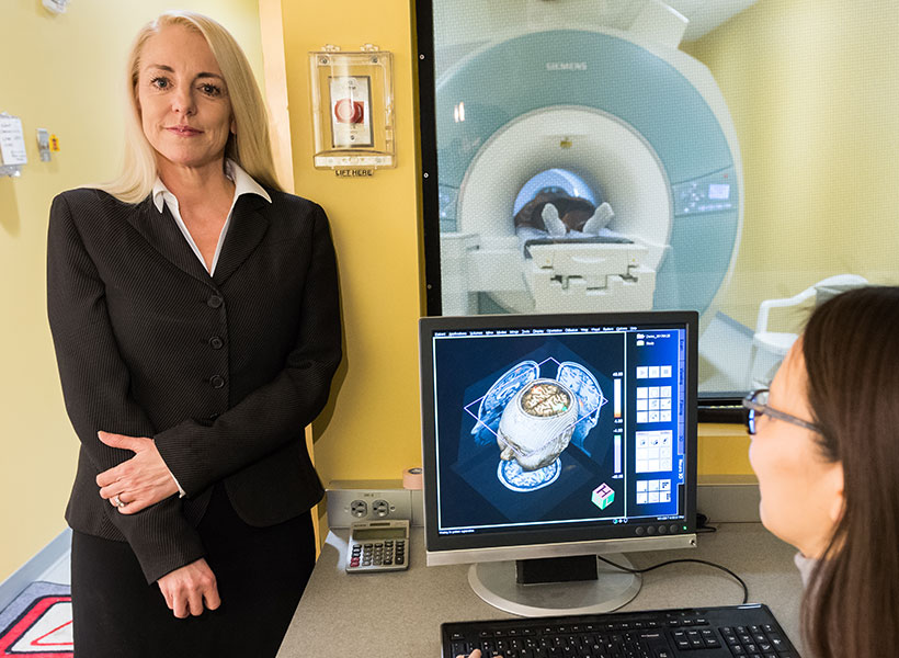
McGovern research affiliate Susan Whitfield-Gabrieli in the Martinos Imaging Center.

“We know this mindfulness meditation is really good for kids and teens, and we think this real-time fMRI neurofeedback is really a way to engage them and provide a visual representation of how they’re doing,” says Whitfield-Gabrieli. “And once we train people how to do mindfulness meditation, they can do it on their own at any time, wherever they are.”

The approach could be a valuable tool to alleviate or prevent depression in young people, which has been on the rise in recent years and escalated alarmingly during the Covid-19 pandemic. “This has gone from bad to catastrophic, in my perspective,” Whitfield-Gabrieli says. “We have to think out of the box and come up some really innovative ways to help.”

Default mode network

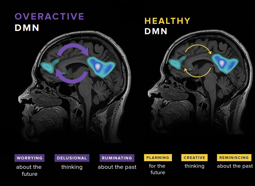
Mindfulness meditation, in which practitioners focus their awareness on the present moment, can modulate activity within the brain’s default mode network, which is so named because it is most active when a person is not focused on any particular task. Two hubs within the default mode network, the medial prefrontal cortex and the posterior cingulate cortex, are of particular interest to Whitfield-Gabrieli and her colleagues, due to a potential role in the symptoms of depression and anxiety.

“These two core hubs are very engaged when we’re thinking about the past or the future and we’re not really engaged in the present moment,” she explains. “If we’re in a healthy state of mind, we may be reminiscing about the past or planning for the future. But if we’re depressed, that reminiscing may turn into rumination or obsessively rehashing the past. If we’re particularly anxious, we may be obsessively worrying about the future.”

Whitfield-Gabrieli explains that these key hubs are often hyperconnected in people with anxiety and depression. The more tightly correlated the activity of the two regions are, the worse a person’s symptoms are likely to be. Mindfulness, she says, can help interrupt that hyperconnectivity.

“Mindfulness really helps to focus on the now, which just precludes all of this mind wandering and repetitive negative thinking,” she explains. In fact, she and her colleagues have found that mindfulness practice can reduce stress and improve attention in children. But she acknowledges that it can be difficult to engage young people and help them focus on the practice.

Tuning the mind

To help people visualize the benefits of their mindfulness practice, the researchers developed a game that can be played while an MRI scanner tracks a person’s brain activity. On a screen inside the scanner, the participant sees a ball and two circles. The circle at the top of the screen represents a desirable state in which the activity of the brain’s default mode network has been reduced, and the activity of a network the brain uses to focus on attention-demanding tasks—the frontal parietal network—has increased. An initial fMRI scan identifies these networks in each individual’s brain, creating a customized mental map on which the game is based.

“They’re training their brain to tune their mind. And they love it.” – Susan Whitfield-Gabrieli

As the person practices mindfulness meditation, which they learn prior to entering the scanner, the default mode network in the brain quiets while the frontal parietal mode activates. When the scanner detects this change, the ball moves and eventually enters its target. With an initial success, the target shrinks, encouraging even more focus. When the participant’s mind wanders from their task, the default mode network activation increases (relative to the frontal parietal network) and the ball moves down towards the second circle, which represents an undesirable state. “Basically, they’re just moving this ball with their brain,” Whitfield-Gabrieli says. “They’re training their brain to tune their mind. And they love it.”

Nine individuals between the ages of 17 and 19 with a history of major depression or anxiety disorders tried this new approach to mindfulness training, and for each of them, Whitfield-Gabrieli’s team saw a reduction in connectivity within the default mode network. Now they are working to determine whether an electroencephalogram, in which brain activity is measured with noninvasive electrodes, can be used to provide similar neurofeedback during mindfulness training—an approach that could be more accessible for broad clinical use.

Whitfield-Gabrieli notes that hyperconnectivity in the default mode network is also associated with psychosis, and she and her team have found that mindfulness meditation with real-time fMRI feedback can help reduce symptoms in adults with schizophrenia. Future studies are planned to investigate how the method impacts teens’ ability to establish a mindfulness practice and its potential effects on depression symptoms.

Partnership with MIT Museum explores relationship between neuroscience and society

What does a healthy relationship between neuroscience and society look like? How do we set the conditions for that relationship to flourish? Researchers and staff at the McGovern Institute and the MIT Museum have been exploring these questions with a five-month planning grant from the Dana Foundation.

Between October 2022 and March 2023, the team tested the potential for an MIT Center for Neuroscience and Society through a series of MIT-sponsored events that were attended by students and faculty of nearby Cambridge Public Schools. The goal of the project was to learn more about what happens when the distinct fields of neuroscience, ethics, and public engagement are brought together to work side-by-side.

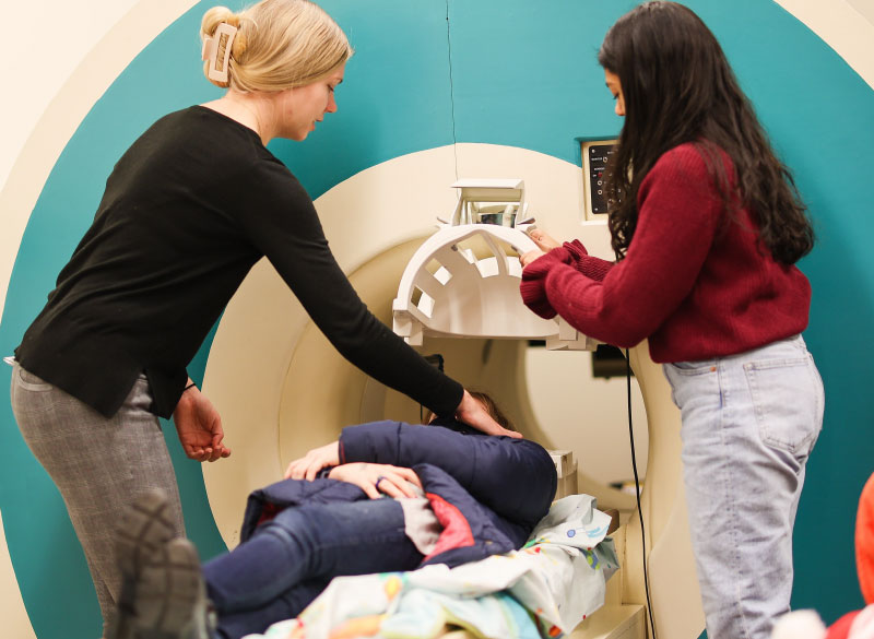
Researchers assist volunteer in mock MRI scanner
Gabrieli lab members Sadie Zacharek (left) and Shruti Nishith (right) demonstrate how the MRI mock scanner works with a student volunteer from the Cambridge Public Schools. Photo: Emma Skakel, MIT Museum

Middle schoolers visit McGovern

Over four days in February, more than 90 sixth graders from Rindge Avenue Upper Campus (RAUC) in Cambridge, Massachusetts, visited the McGovern Institute and participated in hands-on experiments and discussions about the ethical, legal, and social implications of neuroscience research. RAUC is one of four middle schools in the city of Cambridge with an economically, racially, and culturally diverse student population. The middle schoolers interacted with an MIT team led by McGovern Scientific Advisor Jill R. Crittenden, including seventeen McGovern neuroscientists, three MIT Museum outreach coordinators, and neuroethicist Stephanie Bird, a member of the Dana Foundation planning grant team.

“It is probably the only time in my life I will see a real human brain.” – RAUC student

The students participated in nine activities each day, including trials of brain-machine interfaces, close-up examinations of preserved human brains, a tour of McGovern’s imaging center in which students watched as their teacher’s brain was scanned, and a visit to the MIT Museum’s interactive Artificial Intelligence Gallery.

Imagine-IT, a brain-machine interface designed by a team of middle school students during a visit to the McGovern Institute.

To close out their visit, students worked in groups alongside experts to invent brain-computer interfaces designed to improve or enhance human abilities. At each step, students were introduced to ethical considerations through consent forms, questions regarding the use of animal and human brains, and the possible impacts of their own designs on individuals and society.

“I admit that prior to these four days, I would’ve been indifferent to the inclusion of children’s voices in a discussion about technically complex ethical questions, simply because they have not yet had any opportunity to really understand how these technologies work,” says one researcher involved in the visit. “But hearing the students’ questions and ideas has changed my perspective. I now believe it is critically important that all age groups be given a voice when discussing socially relevant issues, such as the ethics of brain computer interfaces or artificial intelligence.”


For more information on the proposed MIT Center for Neuroscience and Society, visit the MIT Museum website.

Studies of unusual brains reveal critical insights into brain organization, function

EG (a pseudonym) is an accomplished woman in her early 60s: she is a college graduate and has an advanced professional degree. She has a stellar vocabulary—in the 98th percentile, according to tests—and has mastered a foreign language (Russian) to the point that she sometimes dreams in it.

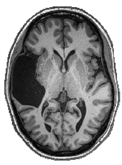
She also has, likely since birth, been missing her left temporal lobe, a part of the brain known to be critical for language.

In 2016, EG contacted McGovern Institute Investigator Evelina Fedorenko, who studies the computations and brain regions that underlie language processing, to see if her team might be interested in including her in their research.

“EG didn’t know about her missing temporal lobe until age 25, when she had a brain scan for an unrelated reason,” says Fedorenko, the Frederick A. (1971) and Carole J. Middleton Career Development Associate Professor of Neuroscience at MIT. “As with many cases of early brain damage, she had no linguistic or cognitive deficits, but brains like hers are invaluable for understanding how cognitive functions reorganize in the tissue that remains.”

“I told her we definitely wanted to study her brain.” – Ev Fedorenko

Previous studies have shown that language processing relies on an interconnected network of frontal and temporal regions in the left hemisphere of the brain. EG’s unique brain presented an opportunity for Fedorenko’s team to explore how language develops in the absence of the temporal part of these core language regions.

Greta Tuckute, a graduate student in the Fedorenko lab, is the first author of the Neuropsychologia study. Photo: Caitlin Cunningham

Their results appeared recently in the journal Neuropsychologia. They found, for the first time, that temporal language regions appear to be critical for the emergence of frontal language regions in the same hemisphere — meaning, without a left temporal lobe, EG’s intact frontal lobe did not develop a capacity for language.

They also reveal much more: EG’s language system resides happily in her right hemisphere. “Our findings provide both visual and statistical proof of the brain’s remarkable plasticity, its ability to reorganize, in the face of extensive early damage,” says Greta Tuckute, a graduate student in the Fedorenko lab and first author of the paper.

In an introduction to the study, EG herself puts the social implications of the findings starkly. “Please do not call my brain abnormal, that creeps me out,” she . “My brain is atypical. If not for accidentally finding these differences, no one would pick me out of a crowd as likely to have these, or any other differences that make me unique.”

How we process language

The frontal and temporal lobes are part of the cerebrum, the largest part of the brain. The cerebrum controls many functions, including the five senses, language, working memory, personality, movement, learning, and reasoning. It is divided into two hemispheres, the left and the right, by a deep longitudinal fissure. The two hemispheres communicate via a thick bundle of nerve fibers called the corpus callosum. Each hemisphere comprises four main lobes—frontal, parietal, temporal, and occipital. Core parts of the language network reside in the frontal and temporal lobes.

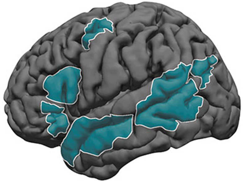
Core parts of the language network (shown in teal) reside in the left frontal and temporal lobes. Image: Ev Fedorenko

In most individuals, the language system develops in both the right and left hemispheres, with the left side dominant from an early age. The frontal lobe develops slower than the temporal lobe. Together, the interconnected frontal and temporal language areas enable us to understand and produce words, phrases, and sentences.

How, then, did EG, with no left temporal lobe, come to speak, comprehend, and remember verbal information (even a foreign language!) with such proficiency?

Simply put, the right hemisphere took over: “EG has a completely well-functioning neurotypical-like language system in her right hemisphere,” says Tuckute. “It is incredible that a person can use a single hemisphere—and the right hemisphere at that, which in most people is not the dominant hemisphere where language is processed—and be perfectly fine.”

Journey into EG’s brain

In the study, the researchers conducted two scans of EG’s brain using functional magnetic resonance imaging (fMRI), one in 2016 and one in 2019, and had her complete a range of behaviorial tests. fMRI measures the level of blood oxygenation across the brain and can be used to make inferences about where neural activity is taking place. The researchers also scanned the brains of 151 “neurotypical” people. The large number of participants, combined with robust task paradigms and rigorous statistical analyses made it possible to draw conclusions from a single case such as EG.

Magnetic resonance image of EG’s brain showing missing left temporal lobe. Image: Fedorenko Lab

Fedorenko is a staunch advocate of the single case study approach—common in medicine but not currently in neuroscience. “Unusual brains—and unusual individuals more broadly—can provide critical insights into brain organization and function that we simply cannot gain by looking at more typical brains.” Studying individual brains with fMRI, however, requires paradigms that work robustly at the single-brain level. This is not true of most paradigms used in the field, which require averaging many brains together to obtain an effect. Developing individual-level fMRI paradigms for language research has been the focus of Fedorenko’s early work, although the main reason for doing so had nothing to do with studying atypical brains: individual-level analyses are simply better—they are more sensitive and their results are more interpretable and meaningful.

“Looking at high-quality data in an individual participant versus looking at a group-level map is akin to using a high-precision microscope versus looking with a naked myopic eye, when all you see is a blur,” she wrote in an article published in Current Opinion in Behaviorial Sciences in 2021. Having developed and validated such paradigms, though, is now allowing Fedorenko and her group to probe interesting brains.

While in the scanner, each participant performed a task that Fedorenko began developing more than a decade ago. They were presented with a series of words that form real, meaningful sentences, and with a series of “nonwords”—strings of letters that are pronounceable but without meaning. In typical brains, language areas respond more strongly when participants read sentences compared to when they read nonword sequences.

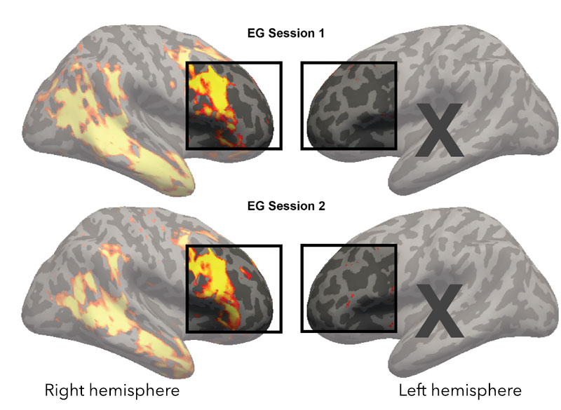
Similarly, in response to the real sentences, the language regions in EG’s right frontal and temporal lobes lit up—they were bursting with activity—while the left frontal lobe regions remained silent. In the neurotypical participants, the language regions in both the left and right frontal and temporal lobes lit up, with the left areas outshining the right.

fMRI showing EG’s language activation on the brain surface. The right frontal lobe shows robust activations, while the left frontal lobe does not have any language responsive areas. Image: Fedorenko lab

“EG showed a very strong response in the right temporal and frontal regions that process language,” says Tuckute. “And if you look at the controls, whose language dominant hemisphere is in the left, EG’s response in her right hemisphere was similar—or even higher—compared to theirs, just on the opposite side.”

Leaving no stone unturned, the researchers next asked whether the lack of language responses in EG’s left frontal lobe might be due to a general lack of response to cognitive tasks rather than just to language. So they conducted a non-language, working-memory task: they had EG and the neurotypical participants perform arithmetic addition problems while in the scanner. In typical brains, this task elicits responses in frontal and parietal areas in both hemisphers.

Not only did regions of EG’s right frontal lobe light up in response to the task, those in her left frontal lobe did, too. “Both EG’s language-dominant (right) hemisphere, and her non-language-dominant (left) hemisphere showed robust responses to this working-memory task ,” says Tuckute. “So, yes, there’s definitely cognitive processing going on there. This selective lack of language responses in EG’s left frontal lobe led us to conclude that, for language, you need the temporal language region to ‘wire up’ the frontal language region.”

Next steps

In science, the answer to one question opens the door to untold more. “In EG, language took over a large chunk of the right frontal and temporal lobes,” says Fedorenko. “So what happens to the functions that in neurotypical individuals generally live in the right hemisphere?”

Many of those, she says, are social functions. The team has already tested EG on social tasks and is currently exploring how those social functions cohabit with the language ones in her right hemisphere. How can they all fit? Do some of the social functions have to migrate to other parts of the brain? They are also working with EG’s family: they have now scanned EG’s three siblings (one of whom is missing most of her right temporal lobe; the other two are neurotypical) and her father (also neurotypical).

The “Interesting Brains Project” website details current projects, findings, and ways to participate.

The project has now grown to include many other individuals with interesting brains, who contacted Fedorenko after some of this work was covered by news outlets. A website for this project can be found here. The project promises to provide unique insights into how our plastic brains reorganize and adapt to various circumstances.


Self-assembling proteins can store cellular “memories”

As cells perform their everyday functions, they turn on a variety of genes and cellular pathways. MIT engineers have now coaxed cells to inscribe the history of these events in a long protein chain that can be imaged using a light microscope.

Cells programmed to produce these chains continuously add building blocks that encode particular cellular events. Later, the ordered protein chains can be labeled with fluorescent molecules and read under a microscope, allowing researchers to reconstruct the timing of the events.

This technique could help shed light on the steps that underlie processes such as memory formation, response to drug treatment, and gene expression.

“There are a lot of changes that happen at organ or body scale, over hours to weeks, which cannot be tracked over time,” says Edward Boyden, the Y. Eva Tan Professor in Neurotechnology, a professor of biological engineering and brain and cognitive sciences at MIT, a Howard Hughes Medical Institute investigator, and a member of MIT’s McGovern Institute for Brain Research and Koch Institute for Integrative Cancer Research.

If the technique could be extended to work over longer time periods, it could also be used to study processes such as aging and disease progression, the researchers say.

Boyden is the senior author of the study, which appears today in Nature Biotechnology. Changyang Linghu, a former J. Douglas Tan Postdoctoral Fellow at the McGovern Institute, who is now an assistant professor at the University of Michigan, is the lead author of the paper.

Cellular history

Biological systems such as organs contain many different kinds of cells, all of which have distinctive functions. One way to study these functions is to image proteins, RNA, or other molecules inside the cells, which provide hints to what the cells are doing. However, most methods for doing this offer only a glimpse of a single moment in time, or don’t work well with very large populations of cells.

“Biological systems are often composed of a large number of different types of cells. For example, the human brain has 86 billion cells,” Linghu says. “To understand those kinds of biological systems, we need to observe physiological events over time in these large cell populations.”

To achieve that, the research team came up with the idea of recording cellular events as a series of protein subunits that are continuously added to a chain. To create their chains, the researchers used engineered protein subunits, not normally found in living cells, that can self-assemble into long filaments.

The researchers designed a genetically encoded system in which one of these subunits is continuously produced inside cells, while the other is generated only when a specific event occurs. Each subunit also contains a very short peptide called an epitope tag — in this case, the researchers chose tags called HA and V5. Each of these tags can bind to a different fluorescent antibody, making it easy to visualize the tags later on and determine the sequence of the protein subunits.

For this study, the researchers made production of the V5-containing subunit contingent on the activation of a gene called c-fos, which is involved in encoding new memories. HA-tagged subunits make up most of the chain, but whenever the V5 tag shows up in the chain, that means that c-fos was activated during that time.

“We’re hoping to use this kind of protein self-assembly to record activity in every single cell,” Linghu says. “It’s not only a snapshot in time, but also records past history, just like how tree rings can permanently store information over time as the wood grows.”

Recording events

In this study, the researchers first used their system to record activation of c-fos in neurons growing in a lab dish. The c-fos gene was activated by chemically induced activation of the neurons, which caused the V5 subunit to be added to the protein chain.

To explore whether this approach could work in the brains of animals, the researchers programmed brain cells of mice to generate protein chains that would reveal when the animals were exposed to a particular drug. Later, the researchers were able to detect that exposure by preserving the tissue and analyzing it with a light microscope.

The researchers designed their system to be modular, so that different epitope tags can be swapped in, or different types of cellular events can be detected, including, in principle, cell division or activation of enzymes called protein kinases, which help control many cellular pathways.

The researchers also hope to extend the recording period that they can achieve. In this study, they recorded events for several days before imaging the tissue. There is a tradeoff between the amount of time that can be recorded and the time resolution, or frequency of event recording, because the length of the protein chain is limited by the size of the cell.

“The total amount of information it could store is fixed, but we could in principle slow down or increase the speed of the growth of the chain,” Linghu says. “If we want to record for a longer time, we could slow down the synthesis so that it will reach the size of the cell within, let’s say two weeks. In that way we could record longer, but with less time resolution.”

The researchers are also working on engineering the system so that it can record multiple types of events in the same chain, by increasing the number of different subunits that can be incorporated.

The research was funded by the Hock E. Tan and K. Lisa Yang Center for Autism Research, John Doerr, the National Institutes of Health, the National Science Foundation, the U.S. Army Research Office, and the Howard Hughes Medical Institute.

New sensor uses MRI to detect light deep in the brain

Using a specialized MRI sensor, MIT researchers have shown that they can detect light deep within tissues such as the brain.

Imaging light in deep tissues is extremely difficult because as light travels into tissue, much of it is either absorbed or scattered. The MIT team overcame that obstacle by designing a sensor that converts light into a magnetic signal that can be detected by MRI (magnetic resonance imaging).

This type of sensor could be used to map light emitted by optical fibers implanted in the brain, such as the fibers used to stimulate neurons during optogenetic experiments. With further development, it could also prove useful for monitoring patients who receive light-based therapies for cancer, the researchers say.

“We can image the distribution of light in tissue, and that’s important because people who use light to stimulate tissue or to measure from tissue often don’t quite know where the light is going, where they’re stimulating, or where the light is coming from. Our tool can be used to address those unknowns,” says Alan Jasanoff, an MIT professor of biological engineering, brain and cognitive sciences, and nuclear science and engineering.

Jasanoff, who is also an associate investigator at MIT’s McGovern Institute for Brain Research, is the senior author of the study, which appears today in Nature Biomedical Engineering. Jacob Simon PhD ’21 and MIT postdoc Miriam Schwalm are the paper’s lead authors, and Johannes Morstein and Dirk Trauner of New York University are also authors of the paper.

A light-sensitive probe

Scientists have been using light to study living cells for hundreds of years, dating back to the late 1500s, when the light microscope was invented. This kind of microscopy allows researchers to peer inside cells and thin slices of tissue, but not deep inside an organism.

“One of the persistent problems in using light, especially in the life sciences, is that it doesn’t do a very good job penetrating many materials,” Jasanoff says. “Biological materials absorb light and scatter light, and the combination of those things prevents us from using most types of optical imaging for anything that involves focusing in deep tissue.”

To overcome that limitation, Jasanoff and his students decided to design a sensor that could transform light into a magnetic signal.

“We wanted to create a magnetic sensor that responds to light locally, and therefore is not subject to absorbance or scattering. Then this light detector can be imaged using MRI,” he says.

Jasanoff’s lab has previously developed MRI probes that can interact with a variety of molecules in the brain, including dopamine and calcium. When these probes bind to their targets, it affects the sensors’ magnetic interactions with the surrounding tissue, dimming or brightening the MRI signal.

To make a light-sensitive MRI probe, the researchers decided to encase magnetic particles in a nanoparticle called a liposome. The liposomes used in this study are made from specialized light-sensitive lipids that Trauner had previously developed. When these lipids are exposed to a certain wavelength of light, the liposomes become more permeable to water, or “leaky.” This allows the magnetic particles inside to interact with water and generate a signal detectable by MRI.

The particles, which the researchers called liposomal nanoparticle reporters (LisNR), can switch from permeable to impermeable depending on the type of light they’re exposed to. In this study, the researchers created particles that become leaky when exposed to ultraviolet light, and then become impermeable again when exposed to blue light. The researchers also showed that the particles could respond to other wavelengths of light.

“This paper shows a novel sensor to enable photon detection with MRI through the brain. This illuminating work introduces a new avenue to bridge photon and proton-driven neuroimaging studies,” says Xin Yu, an assistant professor radiology at Harvard Medical School, who was not involved in the study.

Mapping light

The researchers tested the sensors in the brains of rats — specifically, in a part of the brain called the striatum, which is involved in planning movement and responding to reward. After injecting the particles throughout the striatum, the researchers were able to map the distribution of light from an optical fiber implanted nearby.

The fiber they used is similar to those used for optogenetic stimulation, so this kind of sensing could be useful to researchers who perform optogenetic experiments in the brain, Jasanoff says.

“We don’t expect that everybody doing optogenetics will use this for every experiment — it’s more something that you would do once in a while, to see whether a paradigm that you’re using is really producing the profile of light that you think it should be,” Jasanoff says.

In the future, this type of sensor could also be useful for monitoring patients receiving treatments that involve light, such as photodynamic therapy, which uses light from a laser or LED to kill cancer cells.

The researchers are now working on similar probes that could be used to detect light emitted by luciferases, a family of glowing proteins that are often used in biological experiments. These proteins can be used to reveal whether a particular gene is activated or not, but currently they can only be imaged in superficial tissue or cells grown in a lab dish.

Jasanoff also hopes to use the strategy used for the LisNR sensor to design MRI probes that can detect stimuli other than light, such as neurochemicals or other molecules found in the brain.

“We think that the principle that we use to construct these sensors is quite broad and can be used for other purposes too,” he says.

The research was funded by the National Institutes of Health, the G. Harold and Leila Y. Mathers Foundation, a Friends of the McGovern Fellowship from the McGovern Institute for Brain Research, the MIT Neurobiological Engineering Training Program, and a Marie Curie Individual Fellowship from the European Commission.

Season’s Greetings from the McGovern Institute

This year’s holiday video (shown above) was inspired by Ev Fedorenko’s July 2022 Nature Neuroscience paper, which found similar patterns of brain activation and language selectivity across speakers of 45 different languages.

Universal language network

Ev Fedorenko uses the widely translated book “Alice in Wonderland” to test brain responses to different languages. Photo: Caitlin Cunningham

Over several decades, neuroscientists have created a well-defined map of the brain’s “language network,” or the regions of the brain that are specialized for processing language. Found primarily in the left hemisphere, this network includes regions within Broca’s area, as well as in other parts of the frontal and temporal lobes. Although roughly 7,000 languages are currently spoken and signed across the globe, the vast majority of those mapping studies have been done in English speakers as they listened to or read English texts.

To truly understand the cognitive and neural mechanisms that allow us to learn and process such diverse languages, Fedorenko and her team scanned the brains of speakers of 45 different languages while they listened to Alice in Wonderland in their native language. The results show that the speakers’ language networks appear to be essentially the same as those of native English speakers — which suggests that the location and key properties of the language network appear to be universal.

The many languages of McGovern

English may be the primary language used by McGovern researchers, but more than 35 other languages are spoken by scientists and engineers at the McGovern Institute. Our holiday video features 30 of these researchers saying Happy New Year in their native (or learned) language. Below is the complete list of languages included in our video. Expand each accordion to learn more about the speaker of that particular language and the meaning behind their new year’s greeting.

Not every reader’s struggle is the same

Many children struggle to learn to read, and studies have shown that students from a lower socioeconomic status (SES) background are more likely to have difficulty than those from a higher SES background.

MIT neuroscientists have now discovered that the types of difficulties that lower-SES students have with reading, and the underlying brain signatures, are, on average, different from those of higher-SES students who struggle with reading.

In a new study, which included brain scans of more than 150 children as they performed tasks related to reading, researchers found that when students from higher SES backgrounds struggled with reading, it could usually be explained by differences in their ability to piece sounds together into words, a skill known as phonological processing.

However, when students from lower SES backgrounds struggled, it was best explained by differences in their ability to rapidly name words or letters, a task associated with orthographic processing, or visual interpretation of words and letters. This pattern was further confirmed by brain activation during phonological and orthographic processing.

These differences suggest that different types of interventions may needed for different groups of children, the researchers say. The study also highlights the importance of including a wide range of SES levels in studies of reading or other types of academic learning.

“Within the neuroscience realm, we tend to rely on convenience samples of participants, so a lot of our understanding of the neuroscience components of reading in general, and reading disabilities in particular, tends to be based on higher-SES families,” says Rachel Romeo, a former graduate student in the Harvard-MIT Program in Health Sciences and Technology and the lead author of the study. “If we only look at these nonrepresentative samples, we can come away with a relatively biased view of how the brain works.”

Romeo is now an assistant professor in the Department of Human Development and Quantitative Methodology at the University of Maryland. John Gabrieli, the Grover Hermann Professor of Health Sciences and Technology and a professor of brain and cognitive sciences at MIT, is the senior author of the paper, which appears today in the journal Developmental Cognitive Neuroscience.

Components of reading

For many years, researchers have known that children’s scores on standardized assessments of reading are correlated with socioeconomic factors such as school spending per student or the number of children at the school who qualify for free or reduced-price lunches.

Studies of children who struggle with reading, mostly done in higher-SES environments, have shown that the aspect of reading they struggle with most is phonological awareness: the understanding of how sounds combine to make a word, and how sounds can be split up and swapped in or out to make new words.

“That’s a key component of reading, and difficulty with phonological processing is often one of the hallmarks of dyslexia or other reading disorders,” Romeo says.

In the new study, the MIT team wanted to explore how SES might affect phonological processing as well as another key aspect of reading, orthographic processing. This relates more to the visual components of reading, including the ability to identify letters and read words.

To do the study, the researchers recruited first and second grade students from the Boston area, making an effort to include a range of SES levels. For the purposes of this study, SES was assessed by parents’ total years of formal education, which is commonly used as a measure of the family’s SES.

“We went into this not necessarily with any hypothesis about how SES might relate to the two types of processing, but just trying to understand whether SES might be impacting one or the other more, or if it affects both types the same,” Romeo says.

The researchers first gave each child a series of standardized tests designed to measure either phonological processing or orthographic processing. Then, they performed fMRI scans of each child while they carried out additional phonological or orthographic tasks.

The initial series of tests allowed the researchers to determine each child’s abilities for both types of processing, and the brain scans allowed them to measure brain activity in parts of the brain linked with each type of processing.

The results showed that at the higher end of the SES spectrum, differences in phonological processing ability accounted for most of the differences between good readers and struggling readers. This is consistent with the findings of previous studies of reading difficulty. In those children, the researchers also found greater differences in activity in the parts of the brain responsible for phonological processing.

However, the outcomes were different when the researchers analyzed the lower end of the SES spectrum. There, the researchers found that variance in orthographic processing ability accounted for most of the differences between good readers and struggling readers. MRI scans of these children revealed greater differences in brain activity in parts of the brain that are involved in orthographic processing.

Optimizing interventions

There are many possible reasons why a lower SES background might lead to difficulties in orthographic processing, the researchers say. It might be less exposure to books at home, or limited access to libraries and other resources that promote literacy. For children from this background who struggle with reading, different types of interventions might benefit them more than the ones typically used for children who have difficulty with phonological processing.

In a 2017 study, Gabrieli, Romeo, and others found that a summer reading intervention that focused on helping students develop the sensory and cognitive processing necessary for reading was more beneficial for students from lower-SES backgrounds than children from higher-SES backgrounds. Those findings also support the idea that tailored interventions may be necessary for individual students, they say.

“There are two major reasons we understand that cause children to struggle as they learn to read in these early grades. One of them is learning differences, most prominently dyslexia, and the other one is socioeconomic disadvantage,” Gabrieli says. “In my mind, schools have to help all these kinds of kids become the best readers they can, so recognizing the source or sources of reading difficulty ought to inform practices and policies that are sensitive to these differences and optimize supportive interventions.”

Gabrieli and Romeo are now working with researchers at the Harvard University Graduate School of Education to evaluate language and reading interventions that could better prepare preschool children from lower SES backgrounds to learn to read. In her new lab at the University of Maryland, Romeo also plans to further delve into how different aspects of low SES contribute to different areas of language and literacy development.

“No matter why a child is struggling with reading, they need the education and the attention to support them. Studies that try to tease out the underlying factors can help us in tailoring educational interventions to what a child needs,” she says.

The research was funded by the Ellison Medical Foundation, the Halis Family Foundation, and the National Institutes of Health.

Studies of autism tend to exclude women, researchers find

In recent years, researchers who study autism have made an effort to include more women and girls in their studies. However, despite these efforts, most studies of autism consistently enroll small numbers of female subjects or exclude them altogether, according to a new study from MIT.

The researchers found that a screening test commonly used to determine eligibility for studies of autism consistently winnows out a much higher percentage of women than men, creating a “leaky pipeline” that results in severe underrepresentation of women in studies of autism.

This lack of representation makes it more difficult to develop useful interventions or provide accurate diagnoses for girls and women, the researchers say.

“I think the findings favor having a more inclusive approach and widening the lens to end up being less biased in terms of who participates in research,” says John Gabrieli, the Grover Hermann Professor of Health Sciences and Technology and a professor of brain and cognitive sciences at MIT. “The more we understand autism in men and women and nonbinary individuals, the better services and more accurate diagnoses we can provide.”

Gabrieli, who is also a member of MIT’s McGovern Institute for Brain Research, is the senior author of the study, which appears in the journal Autism Research. Anila D’Mello, a former MIT postdoc who is now an assistant professor at the University of Texas Southwestern, is the lead author of the paper. MIT Technical Associate Isabelle Frosch, Research Coordinator Cindy Li, and Research Specialist Annie Cardinaux are also authors of the paper.

Gabrieli lab researchers Annie Cardinaux (left), Anila D’Mello (center), Cindy Li (right), and Isabelle Frosch (not pictured) have
uncovered sex biases in ASD research. Photo: Steph Stevens

Screening out females

Autism spectrum disorders are diagnosed based on observation of traits such as repetitive behaviors and difficulty with language and social interaction. Doctors may use a variety of screening tests to help them make a diagnosis, but these screens are not required.

For research studies of autism, it is routine to use a screening test called the Autism Diagnostic Observation Schedule (ADOS) to determine eligibility for the study. This test, which assesses social interaction, communication, play, and repetitive behaviors, provides a quantitative score in each category, and only participants who reach certain scores qualify for inclusion in studies.

While doing a study exploring how quickly the brains of autistic adults adapt to novel events in the environment, scientists in Gabrieli’s lab began to notice that the ADOS appeared to have unequal effects on male and female participation in research. As the study progressed, D’Mello noticed some significant brain differences between the male and female subjects in the study.

To investigate these differences further, D’Mello tried to find more female participants using an MIT database of autistic adults who have expressed interest in participating in research studies. However, when she sorted through the subjects, she found that only about half of the women in the database had met the ADOS cutoff scores typically required for inclusion in autism studies, compared to 80 percent of the males.

“We realized then that there’s a discrepancy and that the ADOS is essentially screening out who eventually participated in research,” D’Mello says. “We were really surprised at how many males we retained and how many females we lost to the ADOS.”

To see if this phenomenon was more widespread, the researchers looked at six publicly available datasets, which include more than 40,000 adults who have been diagnosed as autistic. For some of these datasets, participants were screened with ADOS to determine their eligibility to participate in studies, while for others, a “community diagnosis” — diagnosis from a doctor or other health care provider — was sufficient.

The researchers found that in datasets that required ADOS screening for eligibility, the ratio of male to female participants ended up being around 8:1, while in those that required only a community diagnosis the ratios ranged from about 2:1 to 1:1.

Previous studies have found differences between behavioral patterns in autistic men and women, but the ADOS test was originally developed using a largely male sample, which may explain why it often excludes women from research studies, D’Mello says.

“There were few females in the sample that was used to create this assessment, so it might be that it’s not great at picking up the female phenotype, which may differ in certain ways — primarily in domains like social communication,” she says.

Effects of exclusion

Failure to include more women and girls in studies of autism may contribute to shortcomings in the definitions of the disorder, the researchers say.

“The way we think about it is that the field evolved perhaps an implicit bias in how autism is defined, and it was driven disproportionately by analysis of males, and recruitment of males, and so on,” Gabrieli says. “So, the definition doesn’t fit as well, on average, with the different expression of autism that seems to be more common in females.”

This implicit bias has led to documented difficulties in receiving a diagnosis for girls and women, even when their symptoms are the same as those presented by autistic boys and men.

“Many females might be missed altogether in terms of diagnoses, and then our study shows that in the research setting, what is already a small pool gets whittled down at a much larger rate than that of males,” D’Mello says.

Excluding girls and women from this kind of research study can lead to treatments that don’t work as well for them, and it contributes to the perception that autism doesn’t affect women as much as men.

“The goal is that research should directly inform treatment, therapies, and public perception,” D’Mello says. “If the research is saying that there aren’t females with autism, or that the brain basis of autism only looks like the patterns established in males, then you’re not really helping females as much as you could be, and you’re not really getting at the truth of what the disorder might be.”

The researchers now plan to further explore some of the gender and sex-based differences that appear in autism, and how they arise. They also plan to expand the gender categories that they include. In the current study, the surveys that each participant filled out asked them to choose male or female, but the researchers have updated their questionnaire to include nonbinary and transgender options.

The research was funded by the Hock E. Tan and K. Lisa Yang Center for Autism Research, the Simons Center for the Social Brain at MIT, and the National Institutes of Mental Health.

These neurons have food on the brain

A gooey slice of pizza. A pile of crispy French fries. Ice cream dripping down a cone on a hot summer day. When you look at any of these foods, a specialized part of your visual cortex lights up, according to a new study from MIT neuroscientists.

This newly discovered population of food-responsive neurons is located in the ventral visual stream, alongside populations that respond specifically to faces, bodies, places, and words. The unexpected finding may reflect the special significance of food in human culture, the researchers say.

“Food is central to human social interactions and cultural practices. It’s not just sustenance,” says Nancy Kanwisher, the Walter A. Rosenblith Professor of Cognitive Neuroscience and a member of MIT’s McGovern Institute for Brain Research and Center for Brains, Minds, and Machines. “Food is core to so many elements of our cultural identity, religious practice, and social interactions, and many other things that humans do.”

The findings, based on an analysis of a large public database of human brain responses to a set of 10,000 images, raise many additional questions about how and why this neural population develops. In future studies, the researchers hope to explore how people’s responses to certain foods might differ depending on their likes and dislikes, or their familiarity with certain types of food.

MIT postdoc Meenakshi Khosla is the lead author of the paper, along with MIT research scientist N. Apurva Ratan Murty. The study appears today in the journal Current Biology.

Visual categories

More than 20 years ago, while studying the ventral visual stream, the part of the brain that recognizes objects, Kanwisher discovered cortical regions that respond selectively to faces. Later, she and other scientists discovered other regions that respond selectively to places, bodies, or words. Most of those areas were discovered when researchers specifically set out to look for them. However, that hypothesis-driven approach can limit what you end up finding, Kanwisher says.

“There could be other things that we might not think to look for,” she says. “And even when we find something, how do we know that that’s actually part of the basic dominant structure of that pathway, and not something we found just because we were looking for it?”

To try to uncover the fundamental structure of the ventral visual stream, Kanwisher and Khosla decided to analyze a large, publicly available dataset of full-brain functional magnetic resonance imaging (fMRI) responses from eight human subjects as they viewed thousands of images.

“We wanted to see when we apply a data-driven, hypothesis-free strategy, what kinds of selectivities pop up, and whether those are consistent with what had been discovered before. A second goal was to see if we could discover novel selectivities that either haven’t been hypothesized before, or that have remained hidden due to the lower spatial resolution of fMRI data,” Khosla says.

To do that, the researchers applied a mathematical method that allows them to discover neural populations that can’t be identified from traditional fMRI data. An fMRI image is made up of many voxels — three-dimensional units that represent a cube of brain tissue. Each voxel contains hundreds of thousands of neurons, and if some of those neurons belong to smaller populations that respond to one type of visual input, their responses may be drowned out by other populations within the same voxel.

The new analytical method, which Kanwisher’s lab has previously used on fMRI data from the auditory cortex, can tease out responses of neural populations within each voxel of fMRI data.

Using this approach, the researchers found four populations that corresponded to previously identified clusters that respond to faces, places, bodies, and words. “That tells us that this method works, and it tells us that the things that we found before are not just obscure properties of that pathway, but major, dominant properties,” Kanwisher says.

Intriguingly, a fifth population also emerged, and this one appeared to be selective for images of food.

“We were first quite puzzled by this because food is not a visually homogenous category,” Khosla says. “Things like apples and corn and pasta all look so unlike each other, yet we found a single population that responds similarly to all these diverse food items.”

The food-specific population, which the researchers call the ventral food component (VFC), appears to be spread across two clusters of neurons, located on either side of the FFA. The fact that the food-specific populations are spread out between other category-specific populations may help explain why they have not been seen before, the researchers say.

“We think that food selectivity had been harder to characterize before because the populations that are selective for food are intermingled with other nearby populations that have distinct responses to other stimulus attributes. The low spatial resolution of fMRI prevents us from seeing this selectivity because the responses of different neural population get mixed in a voxel,” Khosla says.

“The technique which the researchers used to identify category-sensitive cells or areas is impressive, and it recovered known category-sensitive systems, making the food category findings most impressive,” says Paul Rozin, a professor of psychology at the University of Pennsylvania, who was not involved in the study. “I can’t imagine a way for the brain to reliably identify the diversity of foods based on sensory features. That makes this all the more fascinating, and likely to clue us in about something really new.”

Food vs non-food

The researchers also used the data to train a computational model of the VFC, based on previous models Murty had developed for the brain’s face and place recognition areas. This allowed the researchers to run additional experiments and predict the responses of the VFC. In one experiment, they fed the model matched images of food and non-food items that looked very similar — for example, a banana and a yellow crescent moon.

“Those matched stimuli have very similar visual properties, but the main attribute in which they differ is edible versus inedible,” Khosla says. “We could feed those arbitrary stimuli through the predictive model and see whether it would still respond more to food than non-food, without having to collect the fMRI data.”

They could also use the computational model to analyze much larger datasets, consisting of millions of images. Those simulations helped to confirm that the VFC is highly selective for images of food.

From their analysis of the human fMRI data, the researchers found that in some subjects, the VFC responded slightly more to processed foods such as pizza than unprocessed foods like apples. In the future they hope to explore how factors such as familiarity and like or dislike of a particular food might affect individuals’ responses to that food.

They also hope to study when and how this region becomes specialized during early childhood, and what other parts of the brain it communicates with. Another question is whether this food-selective population will be seen in other animals such as monkeys, who do not attach the cultural significance to food that humans do.

The research was funded by the National Institutes of Health, the National Eye Institute, and the National Science Foundation through the MIT Center for Brains, Minds, and Machines.

Whether speaking Turkish or Norwegian, the brain’s language network looks the same

Over several decades, neuroscientists have created a well-defined map of the brain’s “language network,” or the regions of the brain that are specialized for processing language. Found primarily in the left hemisphere, this network includes regions within Broca’s area, as well as in other parts of the frontal and temporal lobes.

However, the vast majority of those mapping studies have been done in English speakers as they listened to or read English texts. MIT neuroscientists have now performed brain imaging studies of speakers of 45 different languages. The results show that the speakers’ language networks appear to be essentially the same as those of native English speakers.

The findings, while not surprising, establish that the location and key properties of the language network appear to be universal. The work also lays the groundwork for future studies of linguistic elements that would be difficult or impossible to study in English speakers because English doesn’t have those features.

“This study is very foundational, extending some findings from English to a broad range of languages,” says Evelina Fedorenko, the Frederick A. and Carole J. Middleton Career Development Associate Professor of Neuroscience at MIT and a member of MIT’s McGovern Institute for Brain Research. “The hope is that now that we see that the basic properties seem to be general across languages, we can ask about potential differences between languages and language families in how they are implemented in the brain, and we can study phenomena that don’t really exist in English.”

Fedorenko is the senior author of the study, which appears today in Nature Neuroscience. Saima Malik-Moraleda, a PhD student in the Speech and Hearing Bioscience and Technology program at Harvard University, and Dima Ayyash, a former research assistant, are the lead authors of the paper.

Mapping language networks

The precise locations and shapes of language areas differ across individuals, so to find the language network, researchers ask each person to perform a language task while scanning their brains with functional magnetic resonance imaging (fMRI). Listening to or reading sentences in one’s native language should activate the language network. To distinguish this network from other brain regions, researchers also ask participants to perform tasks that should not activate it, such as listening to an unfamiliar language or solving math problems.

Several years ago, Fedorenko began designing these “localizer” tasks for speakers of languages other than English. While most studies of the language network have used English speakers as subjects, English does not include many features commonly seen in other languages. For example, in English, word order tends to be fixed, while in other languages there is more flexibility in how words are ordered. Many of those languages instead use the addition of morphemes, or segments of words, to convey additional meaning and relationships between words.

“There has been growing awareness for many years of the need to look at more languages, if you want make claims about how language works, as opposed to how English works,” Fedorenko says. “We thought it would be useful to develop tools to allow people to rigorously study language processing in the brain in other parts of the world. There’s now access to brain imaging technologies in many countries, but the basic paradigms that you would need to find the language-responsive areas in a person are just not there.”

For the new study, the researchers performed brain imaging of two speakers of 45 different languages, representing 12 different language families. Their goal was to see if key properties of the language network, such as location, left lateralization, and selectivity, were the same in those participants as in people whose native language is English.

The researchers decided to use “Alice in Wonderland” as the text that everyone would listen to, because it is one of the most widely translated works of fiction in the world. They selected 24 short passages and three long passages, each of which was recorded by a native speaker of the language. Each participant also heard nonsensical passages, which should not activate the language network, and was asked to do a variety of other cognitive tasks that should not activate it.

The team found that the language networks of participants in this study were found in approximately the same brain regions, and had the same selectivity, as those of native speakers of English.

“Language areas are selective,” Malik-Moraleda says. “They shouldn’t be responding during other tasks such as a spatial working memory task, and that was what we found across the speakers of 45 languages that we tested.”

Additionally, language regions that are typically activated together in English speakers, such as the frontal language areas and temporal language areas, were similarly synchronized in speakers of other languages.

The researchers also showed that among all of the subjects, the small amount of variation they saw between individuals who speak different languages was the same as the amount of variation that would typically be seen between native English speakers.

Similarities and differences

While the findings suggest that the overall architecture of the language network is similar across speakers of different languages, that doesn’t mean that there are no differences at all, Fedorenko says. As one example, researchers could now look for differences in speakers of languages that predominantly use morphemes, rather than word order, to help determine the meaning of a sentence.

“There are all sorts of interesting questions you can ask about morphological processing that don’t really make sense to ask in English, because it has much less morphology,” Fedorenko says.

Another possibility is studying whether speakers of languages that use differences in tone to convey different word meanings would have a language network with stronger links to auditory brain regions that encode pitch.

Right now, Fedorenko’s lab is working on a study in which they are comparing the ‘temporal receptive fields’ of speakers of six typologically different languages, including Turkish, Mandarin, and Finnish. The temporal receptive field is a measure of how many words the language processing system can handle at a time, and for English, it has been shown to be six to eight words long.

“The language system seems to be working on chunks of just a few words long, and we’re trying to see if this constraint is universal across these other languages that we’re testing,” Fedorenko says.

The researchers are also working on creating language localizer tasks and finding study participants representing additional languages beyond the 45 from this study.

The research was funded by the National Institutes of Health and research funds from MIT’s Department of Brain and Cognitive Sciences, the McGovern Institute, and the Simons Center for the Social Brain. Malik-Moraleda was funded by a la Caixa Fellowship and a Friends of McGovern fellowship.