Michale Fee, the Glen V. and Phyllis F. Dorflinger Professor of Neuroscience and head of the Department of Brain and Cognitive Sciences, and Fan Wang, a professor of brain and cognitive sciences, have been elected to join the National Academy of Sciences (NAS). Fee and Wang, who are also investigators at the McGovern Institute for Brain Research, were elected by current NAS members in recognition of their “distinguished and continuing achievements in original research.”
The NAS is a private, nonprofit institution that was established under a congressional charter signed by President Abraham Lincoln in 1863. It recognizes achievement in science by election to membership, and — with the National Academy of Engineering and the National Academy of Medicine — provides science, engineering, and health policy advice to the federal government and other organizations. This year, the NAS elected 120 members and 25 international members, including six MIT faculty, bringing the total number of active members to 2,705.
“Election to the National Academy of Sciences by one’s peers is a great honor for a scientist in the United States,” says McGovern Institute Director Robert Desimone. “Michale and Fan represent the very best of our research community and we are tremendously proud of their accomplishments and this well-deserved recognition.”
Michale Fee’s research explores how the brain learns and generates complex sequential behaviors. Using the zebra finch as a model system, Fee investigates the neural mechanisms underlying birdsong—a behavior that young birds learn from their fathers through trial and error, much as human infants learn to speak through babbling. His work has revealed that a brain region called the higher vocal center (HVC) functions like an orchestra conductor, precisely controlling the tempo and timing of song production. Other work from his lab has shown how this same circuit helps to store a memory of the father’s song, how baby birds babble in order to practice their song, and how this vocal practice is translated to song learning by listening to themselves sing.
These findings extend far beyond birdsong—the neural circuits controlling birdsong learning are closely related to human brain circuits disrupted in Parkinson’s and Huntington’s disease. Insights from Fee’s research could reveal new clues to the causes and potential treatments of these complex brain disorders.
Fee’s appointment in 2021 as head of the Department of Brain and Cognitive Sciences continues the department’s tradition of being led by scientists whose exemplary work makes MIT a world leader in brain science.
Fan Wang investigates the neural circuits that govern the dynamic interactions between brain and body, exploring how the brain generates sensory perceptions and controls movement. Wang, who is also the co-director of the K. Lisa Yang and Hock E. Tan Center for Molecular Therapeutics, uses cutting-edge techniques including optogenetics, in vivo electrophysiology, and in vivo imaging, to make discoveries with profound clinical implications.
By developing innovative tools to study how brain circuits work, Wang discovered distinct populations of neurons activated by anesthesia that can suppress pain without blocking sensation, and can calm anxiety by regulating automatic body functions like heart rate. She also identified the brain circuits controlling rhythmic movements essential for exploration and communication. Together, these findings reveal how emotion, physiology, movement, and consciousness are deeply interconnected.
Wang combines rigorous basic neuroscience with a commitment to translating her discoveries into therapies that relieve human suffering. Her election to the NAS recognizes her contributions to understanding the brain-body connection and therapeutic potential of her groundbreaking research.
The formal induction ceremony for new NAS members, during which they sign the ledger whose first signatory is Abraham Lincoln, will be held at the Academy’s annual meeting in Washington D.C. next spring.
Schizophrenia, a complex and variable psychiatric disorder, changes people’s perceptions of reality. People with schizophrenia may hear, see, or sense things that aren’t there, and they often hold firm to mistaken ideas about the world despite strong evidence to the contrary. As if these changes aren’t disruptive enough, they are usually accompanied by cognitive difficulties and disorganized thinking.
Scientists at the McGovern Institute’s Poitras Center for Psychiatric Disorders Research are looking for clues into the origins of the disorder and its symptoms so they can help guide the development of new treatments. Encouragingly, they are beginning to uncover the brain changes that reshape reality for people with schizophrenia.
Genetic clues
Researchers who want to study the root causes of a disease often turn to genetics for clues—and the genetics of schizophrenia are complicated. Hundreds of different genes seem to shape people’s risk of developing the disorder, most of which nudge risk only slightly. For most people, it seems to be the cumulative effect of these genes and how they intersect with other risk factors, like stress and prenatal complications, that determine who develops schizophrenia and who does not.
Gene variants that substantially impact the risk of schizophrenia are expected to reveal more about the underlying biology of the disorder than genes whose individual impact is minor. But these variants are rare, and it took a massive study to find them. In 2022, scientists at the Broad Institute’s Stanley Center for Psychiatric Research reported that after analyzing the DNA of more than 24,000 people with schizophrenia, they had identified mutations in 10 genes that dramatically increased the risk of the disorder.
“I think this is exciting, because for the first time, you can actually have an animal model based onhuman genetics findings,” says McGovern Institute and Stanley Center Investigator Guoping Feng. “You can put these mutations in animal models to try to understand how this mutation affects brain development, circuit formation, circuit function, and behavior.” Feng is also the James W. (1963) and Patricia T. Poitras Professor of Brain and Cognitive Sciences at MIT.
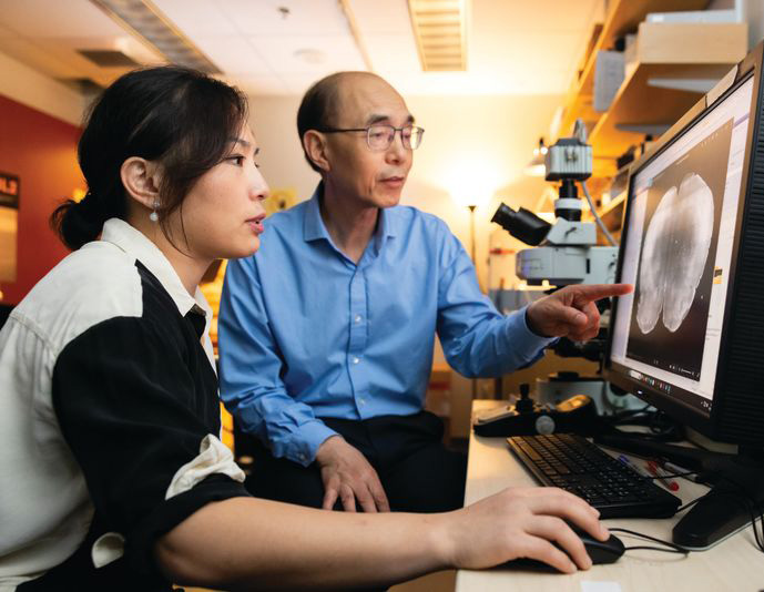
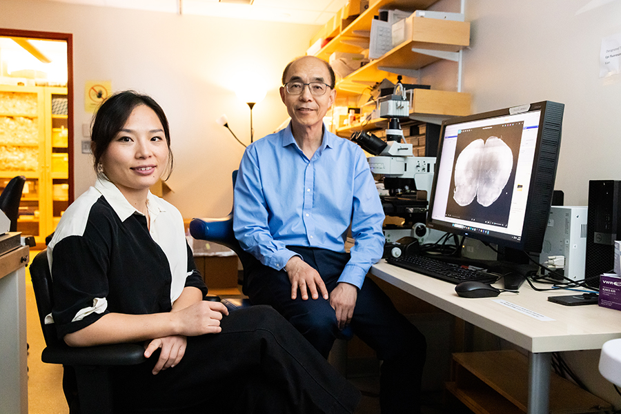
Guoping Feng (right) and his postdoctoral researcher Tinting Zhou (left) examine a mouse brain carrying a genetic mutation associated with schizophrenia. Photo: Steph Stevens
In work supported by the Poitras Center, the Stelling Family Research Fund, and the Yang Tan Collective at MIT, Feng’s lab has engineered three strains of mice that carry ultra-rare schizophrenia-associated mutations. Their first significant findings come from mice with a mutation in a gene called Grin2a. People who inherit a dysfunctional Grin2a gene, which neurons need to detect and respond to a signaling molecule called NMDA, are 20 times more likely to develop schizophrenia than people in whom Grin2a is intact.
Tingting Zhou, a postdoctoral researcher in Feng’s lab, says the team had to think carefully about how to assess mice for schizophrenia-like symptoms. You can’t ask mice about hallucinations or delusions. Instead, Zhou designed an experiment that tested how well mice use new information to update their beliefs about the world—a process that is thought to be impaired in people who experience delusions.
To illustrate how failure to update beliefs can skew someone’s ideas about reality, Zhou describes a situation in which a person watches a stranger reach for something in their pocket, fearing that person intends to harm them. Then, the stranger’s hand emerges with a lollipop. The new information should alleviate concern—but a person with schizophrenia might hold on to their original belief, convinced the lollipop-holding stranger is a threat.
In Zhou’s experiments testing animals’ belief-updating abilities, mice had to keep up with changing information to earn as many treats as possible. Those with the Grin2a mutation were slow to adapt when experimenters adjusted the relative values of their choices. “Once the animal learns something, it’s very hard for them to update the information,” Zhou explains.
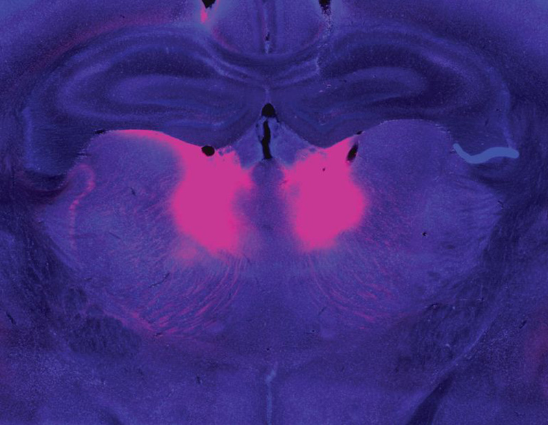
Zhou and Feng linked this behavioral difference to abnormally low activity in a part of the brain called the mediodorsal thalamus. The mediodorsal thalamus acts like a switchboard in the brain, routing and coordinating information between different parts of the cortex to support thinking, decision-making, and flexible behavior. Studies with patients have implicated this region in schizophrenia as well, showing that it has fewer cells and is less active in people with the disorder than those without.
The mediodorsal thalamus (pink) is less active in people with schizophrenia and mouse models of the disease. Image: Guoping Feng, Tingting Zhou
Feng’s lab and others are now looking for belief-updating deficits in other genetic models of schizophrenia. “The goal is to look at whether this is a converging mechanism…then you can start to look at what other [brain] regions are involved,” he says.
In mice with Grin2a mutations, the researchers were able to restore normal belief updating by activating neurons in the mediodorsal thalamus, offering hope that manipulating the same circuitry might benefit patients. “It will not be easy,” Feng says, “but at least you have something you can work on. Previously, it was just very hard to imagine how to develop a new therapeutic for schizophrenia.”
Internal noise
It’s not just the genes associated with schizophrenia that differ across affected individuals. The symptoms of the disorder vary, too. People experience some combination of delusions, hallucinations, disorganized speech, and cognitive problems—but none of these are experienced by everyone with the disorder. This heterogeneity complicates the diagnosis, treatment, and study of schizophrenia. For this reason, some researchers are focusing their efforts on understanding its individual symptoms.
Evelina Fedorenko, a McGovern Investigator and associate professor of brain and cognitive sciences, specializes in understanding how the brain processes speech and language. But recently, her group has teamed up with physician-researcher Ann Shinn at McLean Hospital to begin exploring why some people hear voices when no one is speaking.
About three out of four people with schizophrenia experience auditory hallucinations, which most commonly involve voices.
These hallucinations can be distressing, sometimes involving threatening language or commands to cause harm. Some people with mood disorders or post-traumatic stress disorder also hear them.
Tamar Regev was the 2022–2024 Poitras Center Postdoctoral Fellow in Evelina Fedorenko’s lab. Photo: Steph Stevens
To investigate, Tamar Regev, a research scientist in the Fedorenko lab, asked people who experience auditory hallucinations to listen to different kinds of sounds inside an MRI scanner, then compared how their brains responded versus the brains of people without auditory hallucinations. Her study included participants with schizophrenia and bipolar disorder, both with and without a history of auditory hallucinations, as well as healthy controls.
Inside the scanner, participants listened to three kinds of audio: spoken language, gibberish, and gibberish so scrambled that it barely resembled speech. Regev analyzed how these sounds impacted activity in areas the brain uses to process auditory input at different levels: a part of the auditory cortex that is sensitive to all sounds; a higher-level region within the auditory cortex that usually responds to anything that sounds like speech, even if its content is unclear; and the brain’s language-processing network, which is called on to understand the content of speech, as well as written or signed communications.
Regev found that in people with hallucinations, the part of the brain that usually responds only to language responded to meaningless speech as well. “In this pathway from auditory to speech to language processing, the stimuli that should be filtered out somewhere on the way are now passing to higher stations,” she explains. While auditory hallucinations don’t require external sounds, Fedorenko and Regev propose that the brain’s language areas might be similarly activated by “internal noise” in auditory circuits.
Scrambled language
In people who experience auditory hallucinations, the brain’s language regions respond to sounds that aren’t language–including scrambled meaningless gibberish. Below is a sample gibberish clip used in Fedorenko’s study.
Early identification
McGovern scientists have also used brain imaging to investigate what happens in the brain before people develop clear symptoms of schizophrenia. The disorder is usually diagnosed in adolescence or young adulthood, when patients exhibit the first signs of psychosis—but its origins in the brain likely take root years before that.
“One of the things we’re super interested in is, can you identify people at risk early on, before they have a big problem,” says McGovern Investigator John Gabrieli, whose work is also supported by the Poitras Center and the Stelling Family Research Fund. That might give clinicians an opportunity to intervene and lessen or prevent the disorder’s most devastating effects, he says.
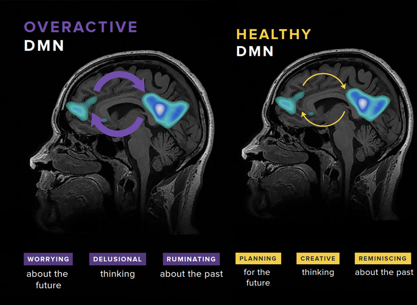
Gabrieli and his colleagues have studied the brains of children who, because they have a parent or sibling with schizophrenia, have an elevated risk of developing the disorder themselves. They found that a system called the default mode network (DMN), which is overactive in adults with schizophrenia, is already working overtime when children in this high-risk group are seven- to 12-years-old.
Gabrieli explains that the DMN is active when people are not actively engaged in an activity or thinking about the external world. “It turns on when you think about your family, your values, your hopes for the future, or important events of your life. It’s almost like a system of who /you are,” he says. Hallucinations and delusions experienced by people with schizophrenia may be associated with overactivity in this network.
The default mode network (DMN) is a large-scale brain network that is active when a person is not focused on the outside world and the brain is at wakeful rest. The DMN is often over-engaged in adolescents with depression and anxiety, as well as teens at risk for these and other disorders like schizophrenia (left). DMN activation and connectivity can be “tuned” to a healthier state through the practice of mindfulness (right).
“They’re kind of living in their internal world of beliefs, as opposed to the reality that most of us occupy,” Gabrieli explains.
He and his colleagues think overactivity in the DMN might make people vulnerable to schizophrenia—and their data show this atypical activity can be detected many years before the core symptoms of schizophrenia appear. With further validation, children with hyperactivity of the DMN might be candidates for early intervention.
With new and better interventions, the ability to identify people who may be on a path toward schizophrenia will be even more impactful—underscoring the need for continued research on multiple fronts. A recent gift of $8 million to the Poitras Center from Patricia and James Poitras is helping accelerate this work in labs at the McGovern Institute and beyond.
One of the symptoms of schizophrenia is difficulty incorporating new information about the world. This can lead patients to struggle with making decisions and, eventually, to lose touch with reality.
MIT neuroscientists have now identified a gene mutation that appears to give rise to this type of difficulty. In a study of mice, the researchers found that the mutated gene impairs the function of a brain circuit that is responsible for updating beliefs based on new input.
This mutation, in a gene called grin2a, was originally identified in a large-scale screen of patients with schizophrenia. The new study suggests that drugs targeting this brain circuit could help with some of the cognitive impairments seen in schizophrenia patients.
“If this circuit doesn’t work well, you cannot quickly integrate information,” says Guoping Feng, the James W. and Patricia T. Poitras Professor in Brain and Cognitive Sciences at MIT, a member of the Broad Institute of Harvard and MIT, and the associate director of the McGovern Institute for Brain Research at MIT. “We are quite confident this circuit is one of the mechanisms that contributes to the cognitive impairment that is a major part of the pathology of schizophrenia.”
Feng and Michael Halassa, an associate professor of psychiatry and neuroscience at Tufts University, are the senior authors of the new study, which appears today in Nature Neuroscience. Tingting Zhou, a research scientist at the McGovern Institute, and Yi-Yun Ho, a former MIT postdoc, are the lead authors of the paper.
McGovern Institute Investigator Guoping Feng (right) and his postdoctoral researcher Tinting Zhou (left) in the lab. Photo: Steph Stevens
Adapting to new information
Schizophrenia is known to have a strong genetic component. For the general population, the risk of developing the disease is about 1 percent, but that goes up to 10 percent for those who have a parent or sibling with the disease, and 50 percent for people who have an identical twin with the disease.
Researchers at the Stanley Center for Psychiatric Research at the Broad Institute have identified more than 100 gene variants linked to schizophrenia, using genome-wide association studies. However, many of those variants are located in non-coding regions of the genome, making it difficult to figure out how they might influence development of the disease.
More recently, researchers at the Stanley Center used a different strategy, known as whole-exome sequencing, to reveal gene mutations linked to schizophrenia. This technique sequences only the protein-coding regions of the genome, so it can reveal mutations that are located in known genes.
Using this approach on about 25,000 sequences from people with schizophrenia and 100,000 sequences from control subjects, the researchers identified 10 genes in which mutations significantly increase the risk of developing schizophrenia.
In the new Nature Neuroscience study, Feng and his students created a mouse model with a mutation in one of those genes, grin2a. This gene encodes a protein that forms part of the NMDA receptor — a receptor that is activated by the neurotransmitter glutamate and is often found on the surface of neurons.
Zhou then investigated whether these mice displayed any of the characteristic behaviors seen in schizophrenia patients. These patients show many complex symptoms, including psychoses such as hallucinations and delusions (loss of contact with reality). Those are difficult to study in mice, but it is possible to study related symptoms such as difficulty in interpreting new sensory input.
Over the past two decades, schizophrenia researchers have hypothesized that psychosis may stem from an impaired ability to update beliefs based on new information.
“Our brain can form a prior belief of reality, and when sensory input comes into the brain, a neurotypical brain can use this new input to update the prior belief. This allows us to generate a new belief that’s close to what the reality is,” Zhou says. “What happens in schizophrenia patients is that they weigh too heavily on the prior belief. They don’t use as much current input to update what they believed before, so the new belief is detached from reality.”
To study this, Zhou designed an experiment that required mice to choose between two levers to press to earn a food reward. One lever was low-reward — mice had to push it six times to get one drop of milk. A high-reward lever dispensed three drops per push.
At the beginning of the study, all of the mice learned to prefer the high-reward lever. However, as the experiment went on, the number of presses required to dispense the higher reward gradually went up, while there were no changes to the low-reward lever.
As the effort required went up, healthy mice start to switch back and forth between the two levers. Once they had to press the high-reward lever around 18 times for three drops of milk, making the effort per drop about the same for each lever, they eventually switched permanently to the low-reward lever. However, mice with a mutation in grin2a showed a different behavior pattern. They spent more time switching back and forth between the two levers, and they made the switch to the low-reward side much later.
“We find that neurotypical animals make adaptive decisions in this changing environment,” Zhou says. “They can switch from the high-reward side to the low-reward side around the equal value point, while for the animals with the mutation, the switch happens much later. Their adaptive decision-making is much slower compared to the wild-type animals.”
An impaired circuit
Using functional ultrasound imaging and electrical recordings, the researchers found that the brain region affected most by the grin2a mutation was the mediodorsal thalamus. This part of the brain connects with the prefrontal cortex to form a thalamocortical circuit that is responsible for regulating cognitive functions such as executive control and decision-making.
The researchers found that neuronal activity in the mediodorsal thalamus appears to keep track of the changes in value of the two reward options. Additionally, the mice showed different patterns of neural activity depending on which state they were — either an exploratory state or committed to one side.
The researchers also showed that they could use optogenetics to reverse the behavioral symptoms of the mice with mutated grin2a. They engineered the neurons of the mediodorsal thalamus so that they could be activated by light, and when these neurons were activated, the mice began behaving similarly to mice without the grin2a mutation.
While only a very small percentage of schizophrenia patients have mutations in the grin2a gene, it’s possible that this circuit dysfunction is a converging mechanism of cognitive impairment for a subset of schizophrenia patients with different causes.
Targeting this circuit could offer a way to overcome some of the cognitive impairments seen in schizophrenia patients, the researchers say. To do that, they are now working on identifying targets within the circuit that could be potentially druggable.
The research was funded by the National Institutes of Mental Health, the Poitras Center for Psychiatric Disorders Research at MIT, the Yang Tan Collective at MIT, the K. Lisa Yang and Hock E. Tan Center for Molecular Therapeutics at MIT, the Stelling Family Research Fund at MIT, the Stanley Center for Psychiatric Research, and the Brain and Behavior Research Foundation.
Today, Stanford University neuroscientist Liqun Luo was announced as the recipient of the 2026 Edward M. Scolnick Prize in Neuroscience by the McGovern Institute for Brain Research at MIT. Luo is the Ann and Bill Swindells Professor in the School of Humanities and Sciences, Professor of Biology, and Professor of Neurobiology by courtesy at Stanford University, and a Howard Hughes Medical Institute Investigator. The McGovern Institute presents the Scolnick Prize annually to recognize outstanding achievements in neuroscience.
“Liqun Luo’s development of first-in-kind genetic tools and detailed, innovative experimentation has succeeded in defining rules that govern how transient cell-cell contacts ultimately establish functional neural circuits in the developing brain,” says McGovern Institute Director Robert Desimone, who is also chair of the selection committee. “Luo’s methodologies for visualizing specific subsets of neurons based on their developmental trajectory or their activity are widely used in the field and have driven the identification of neurons responsible for a range of behaviors, including sleep and social interactions.”
Liqun Luo was born in Shanghai, China and attained his bachelor’s degree in molecular biology from the University of Science and Technology of China in 1986. He moved to the US for graduate studies at Brandeis University with Kalpana White, where he characterized the homolog of the Alzheimer’s amyloid precursor protein in the fruit fly Drosophila. After receiving a PhD in 1992, he moved to the University of California, San Francisco for postdoctoral training with Lily Jan and Yuh-Nung Jan where he published a number of papers about how small GTPase proteins regulate cellular morphology. Luo descends from a line of mentors trained by his scientific hero Seymour Benzer, who is widely known for founding the field of neurogenetics.
In 1996, Luo joined the faculty at Stanford University and established his own research group to focus on the molecular mechanisms of neuronal morphogenesis in the brain. Luo’s laboratory developed groundbreaking techniques—including Mosaic Analysis with a Repressible Cell Marker (MARCM) in fruit flies and Mosaic Analysis with Double Markers (MADM) in mice—that allowed the labeling and genetic manipulation of individual neurons within otherwise normal brains. These innovations gave researchers the ability to image genetically defined and altered neurons as they grow, connect, and change over time. Luo and his colleagues used these tools to reveal how neurons sculpt their branching structures, prune away unnecessary connections, and find the precise partners they need to form functional circuits. His work illuminated the molecular choreography that ensures each neuron wires into the correct network—an essential step in building circuits for sensation, movement, memory, and emotion. Another impactful innovation from Luo’s group, known as TRAP (Targeted Recombination in Active Populations), allows for the genetic tagging of neurons that are active during specific experiences. This technique has helped reveal how neural populations encode thirst, motivation, and long-term memories.
Most recently, Luo and his group have wholly defined the molecular codes that neurons use to recognize their correct partners in the olfactory system of fruit flies. His research demonstrated that a combinatorial pattern of cell-surface proteins precisely guides neurons to connect to one another and form a functional network. His team then succeeded in genetically altering the molecular cues that govern synaptic connections to rewire a neural circuit and produce a predicted change in the fly’s mating behavior.
Colleagues emphasize that Luo’s influence extends far beyond his own discoveries. Many of the molecular principles he has uncovered in simple model organisms have since proven to be conserved across species, underscoring their fundamental importance. His genetic tracing methods have been adopted by laboratories worldwide and applied not only in neuroscience but also in fields such as cancer biology, where tracing cell lineage is critical. He has also trained a generation of neuroscientists who have gone on to lead major research programs of their own, amplifying his impact across the field.
Luo has received numerous honors, including election to the National Academy of Sciences, the NAS Award in the Neurosciences, the Pradel Research Award, and the Society for Neuroscience’s Award for Education in Neuroscience. He has been a Howard Hughes Medical Institute Investigator since 2005. He is also the author of Principles of Neurobiology, a widely used textbook that has been translated into Chinese, Japanese, and Italian.
The Scolnick Prize recognizes discoveries that advance the understanding of the brain and its disorders. Luo’s work exemplifies this mission, providing tools and conceptual frameworks for understanding how neural circuits form and are refined to become functional, and how mutations disrupt these processes. As neuroscience enters an era defined by increasingly precise control over brain circuits, Liqun Luo’s contributions stand as both enabling and visionary.
The McGovern Institute will award the Scolnick Prize to Luo on June 16, 2026. At 4:00 pm he will deliver a lecture titled “Wiring Specificity of Neural Circuits” to be followed by a reception at the McGovern Institute, 43 Vassar Street (building 46, room 3002) in Cambridge. The event is free and open to the public.
As people age, their immune system function declines. T cell populations become smaller and can’t react to pathogens as quickly, making people more susceptible to a variety of infections.
To try to overcome that decline, researchers at MIT and the Broad Institute have found a way to temporarily program cells in the liver to improve T-cell function. This reprogramming can compensate for the age-related decline of the thymus, where T cell maturation normally occurs.
Using mRNA to deliver three key factors that usually promote T-cell survival, the researchers were able to rejuvenate the immune systems of mice. Aged mice that received the treatment showed much larger and more diverse T cell populations in response to vaccination, and they also responded better to cancer immunotherapy treatments. Their findings are published in the December 17 issue of the journal Nature.
If developed for use in patients, this type of treatment could help people lead healthier lives as they age, the researchers say.
“If we can restore something essential like the immune system, hopefully we can help people stay free of disease for a longer span of their life,” says Feng Zhang, the James and Patricia Poitras Professor of Neuroscience at MIT, who has joint appointments in the departments of Brain and Cognitive Sciences and Biological Engineering.
Zhang, who is also an investigator at the McGovern Institute for Brain Research at MIT, a core institute member at the Broad Institute of MIT and Harvard, an investigator in the Howard Hughes Medical Institute, and co-director of the K. Lisa Yang and Hock E. Tan Center for Molecular Therapeutics at MIT, is the senior author of the new study. Former MIT postdoc Mirco Friedrich is the lead author of the paper, which appears today in Nature.
A temporary factory
The thymus, a small organ located in front of the heart, plays a critical role in T-cell development. Within the thymus, immature T cells go through a checkpoint process that ensures a diverse repertoire of T cells. The thymus also secretes cytokines and growth factors that help T cells to survive.
However, starting in early adulthood, the thymus begins to shrink. This process, known as thymic involution, leads to a decline in the production of new T cells. By the age of approximately 75, the thymus is greatly reduced.
“As we get older, the immune system begins to decline. We wanted to think about how can we maintain this kind of immune protection for a longer period of time, and that’s what led us to think about what we can do to boost immunity,” Friedrich says.
Previous work on rejuvenating the immune system has focused on delivering T cell growth factors into the bloodstream, but that can have harmful side effects. Researchers are also exploring the possibility of using transplanted stem cells to help regrow functional tissue in the thymus.
The MIT team took a different approach: They wanted to see if they could create a temporary “factory” in the body that would generate the T-cell-stimulating signals that are normally produced by the thymus.
“Our approach is more of a synthetic approach,” Zhang says. “We’re engineering the body to mimic thymic factor secretion.”
For their factory location, they settled on the liver, for several reasons. First, the liver has a high capacity for producing proteins, even in old age. Also, it’s easier to deliver mRNA to the liver than to most other organs of the body. The liver was also an appealing target because all of the body’s circulating blood has to flow through it, including T cells.
To create their factory, the researchers identified three immune cues that are important for T-cell maturation. They encoded these three factors into mRNA sequences that could be delivered by lipid nanoparticles. When injected into the bloodstream, these particles accumulate in the liver and the mRNA is taken up by hepatocytes, which begin to manufacture the proteins encoded by the mRNA.
The factors that the researchers delivered are DLL1, FLT-3, and IL-7, which help immature progenitor T cells mature into fully differentiated T cells.
Immune rejuvenation
Tests in mice revealed a variety of beneficial effects. First, the researchers injected the mRNA particles into 18-month-old mice, equivalent to humans in their 50s. Because mRNA is short-lived, the researchers gave the mice multiple injections over four weeks to maintain a steady production by the liver.
After this treatment, T cell populations showed significant increases in size and function.
The researchers then tested whether the treatment could enhance the animals’ response to vaccination. They vaccinated the mice with ovalbumin, a protein found in egg whites that is commonly used to study how the immune system responds to a specific antigen. In 18-month-old mice that received the mRNA treatment before vaccination, the researchers found that the population of cytotoxic T-cells specific to ovalbumin doubled, compared to mice of the same age that did not receive the mRNA treatment.
The mRNA treatment can also boost the immune system’s response to cancer immunotherapy, the researchers found. They delivered the mRNA treatment to 18-month-old mice, who were then implanted with tumors and treated with a checkpoint inhibitor drug. This drug, which targets the protein PD-L1, is designed to help take the brakes off the immune system and stimulate T cells to attack tumor cells.
Mice that received the treatment showed much higher survival rates and longer lifespan that those that received the checkpoint inhibitor drug but not the mRNA treatment.
The researchers found that all three factors were necessary to induce this immune enhancement; none could achieve all aspects of it on their own. They now plan to study the treatment in other animal models and to identify additional signaling factors that may further enhance immune system function. They also hope to study how the treatment affects other immune cells, including B cells.
Other authors of the paper include Julie Pham, Jiakun Tian, Hongyu Chen, Jiahao Huang, Niklas Kehl, Sophia Liu, Blake Lash, Fei Chen, Xiao Wang, and Rhiannon Macrae.
The research was funded, in part, by the Howard Hughes Medical Institute, the K. Lisa Yang Brain-Body Center, part of the Yang Tan Collective at MIT, Broad Institute Programmable Therapeutics Gift Donors, the Pershing Square Foundation, J. and P. Poitras, and an EMBO Postdoctoral Fellowship.
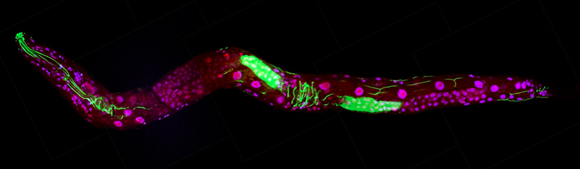
For decades, scientists with big questions about biology have found answers in a tiny worm. That worm–a millimeter-long creature called Caenorhabditis elegans–has helped researchers uncover fundamental features of how cells and organisms work. The impact of that work is enormous: Discoveries made using C. elegans have been recognized with four Nobel prizes and have led to the development of new treatments for human disease.
McGovern Investigator Robert Horvitz shared the 2002 Nobel Prize in Medicine with colleagues Sydney Brenner and John Sulston for discoveries that helped explain how genes regulate programmed cell death and organ development. Photo: AP Images/Aynsley Floyd
In a perspective piece published in the November 2025 issue of the journal PNAS, eleven biologists including Robert Horvitz, the David H. Koch (1962) Professor of Biology at MIT, celebrate Nobel Prize-winning advances made through research in C. elegans. The authors discuss how that work has led to advances for human health and highlight how a uniquely collaborative community among worm researchers has fueled the field.
MIT scientists are well represented in that community: The prominent worm biologists who coauthored the PNAS paper include former MIT graduate students Andy Fire and Paul Sternberg, now at Stanford University and the California Institute of Technology, and two past postdoctoral researchers in Horvitz’s lab, University of Massachusetts Medical School professor Victor Ambros and Massachusetts General Hospital investigator Gary Ruvkun. Ann Rougvie at the University of Minnesota is the paper’s corresponding author.
Early worm discoveries
“This tiny worm is beautiful—elegant both in its appearance and in its many contributions to our understanding of the biological universe in which we live,” says Horvitz, who in 2002 was awarded the Nobel Prize in Medicine along with colleagues Sydney Brenner and John Sulston for discoveries that helped explain how genes regulate programmed cell death and organ development. Horvitz is also a member of MIT’s McGovern Institute for Brain Research and Koch Institute for Integrative Cancer Research as well as an investigator at the Howard Hughes Medical Institute.
Those discoveries were among the early successes in C. elegans research, made by pioneering scientists who recognized the power of the microscopic roundworm. C. elegans offers many advantages for researchers: The worms are easy to grow and maintain in labs; their transparent bodies make cells and internal processes readily visible under a microscope; they are cellularly very simple (e.g., they have only 302 nerve cells, compared with about 100 billion in a human) and their genomes can be readily manipulated to study gene function.
Caenorhabditis elegans, a transparent roundworm only 1mm in length, has provided answers to many fundamental questions in biology. Image: Robert Horvitz
Most importantly, many of the molecules and processes that operate in C. elegans have been retained throughout evolution, meaning discoveries made using the worm can have direct relevance to other organisms, including humans. “Many aspects of biology are ancient and evolutionarily conserved,” Horvitz explains. “Such shared mechanisms can be most readily revealed by analyzing organisms that are highly tractable in the laboratory.”
In the 1960s, Brenner, a molecular biologist who was curious about how animals’ nervous systems develop and function, recognized that C. elegans offered unique opportunities to study these processes. Once he began developing the worm into a model for laboratory studies, it did not take long for other biologists to join him to take advantage of the new system.
In the 1970s, the unique features of the worm allowed Sulston to track the transformation of a fertilized egg into an adult animal, tracing the origins of each of the adult worm’s 959 cells. His studies revealed that in every developing worm, cells divide and mature in predictable ways. He also learned that some of the cells created during development do not survive into adulthood and are instead eliminated by a process termed programmed cell death.
“This tiny worm is beautiful—elegant both in its appearance and in its many contributions to our understanding of the biological universe in which we live,” says Horvitz.
By seeking mutations that perturbed the process of programmed cell death, Horvitz and his colleagues identified key regulators of that process, which is sometimes referred to as apoptosis. These regulators, which both promote and oppose apoptosis, turned out to be vital for programmed cell death across the animal kingdom.
In humans, apoptosis shapes developing organs, refines brain circuits, and optimizes other tissue structures. It also modulates our immune systems and eliminates cells that are in danger of becoming cancerous. The human version of CED-9, the anti-apoptotic regulator that Horvitz’s team discovered in worms, is BCL-2. Researchers have shown that activating apoptotic cell death by blocking BCL-2 is an effective treatment for certain blood cancers. Today, researchers are also exploring new ways of treating immune disorders and neurodegenerative disease by manipulating apoptosis pathways.
Collaborative worm community
Horvitz and his colleagues’ discoveries about apoptosis helped demonstrate that understanding C. elegans biology has direct relevance to human biology and disease. Since then, a vibrant and closely connected community of worm biologists—including many who trained in Horvitz’s lab—has continued to carry out impactful work. In their PNAS article, Horvitz and his coauthors highlight that early work, as well as the Nobel Prize-winning work of:
Andrew Fire and Craig Mello, whose discovery of an RNA-based system of gene silencing led to powerful new tools to manipulate gene activity. The innate process they discovered in worms, known as RNA interference, is now used as the basis of six FDA-approved therapeutics for genetic disorders, silencing faulty genes to stop their harmful effects.
Martin Chalfie, who used a fluorescent protein made by jellyfish to visualize and track specific cells in C. elegans, helping launch the development of a set of tools that transformed biologists’ ability to observe molecules and processes that are important for both health and disease.
Victor Ambros and Gary Ruvkun, who discovered a class of molecules called microRNAs that regulate gene activity not just in worms, but in all multicellular organisms. This prize-winning work was started when Ambros and Ruvkun were postdoctoral researchers in Horvitz’s lab. Humans rely on more than 1,000 microRNAs to ensure our genes are used at the right times and places. Disruptions to microRNAs have been linked to neurological disorders, cancer, cardiovascular disease, and autoimmune disease, and researchers are now exploring how these small molecules might be used for diagnosis or treatment.
Horvitz and his coauthors stress that while the worm itself made these discoveries possible, so too did a host of resources that facilitate collaboration within the worm community and enable its scientists to build upon the work of others. Scientists who study C. elegans have embraced this open, collaborative spirit since the field’s earliest days, Horvitz says, citing the Worm Breeder’s Gazette, an early newsletter where scientists shared their observations, methods, and ideas.
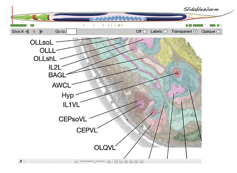
Today, scientists who study C. elegans—whether the organism is the centerpiece of their lab or they are looking to supplement studies of other systems—contribute to and rely on online resources like WormAtlas and WormBase, as well as the Caenorhabditis Genetics Center, to share data and genetic tools. Horvitz says these resources have been crucial to his own lab’s work; his team uses them every day.
WormAtlas provides users with numerous anatomical resources including tools to view electron microscopy slices of the same cell. Image: WormAtlas.org
Just as molecules and processes discovered in C. elegans have pointed researchers toward important pathways in human cells, the worm has also been a vital proving ground for developing methods and approaches later deployed to study more complex organisms. For example, C. elegans, with its 302 neurons, was the first animal for which neuroscientists successfully mapped all of the connections of the nervous system. The resulting wiring diagram, or connectome, has guided countless experiments exploring how neurons work together to process information and control behavior. Informed by both the power and limitations of the C. elegans’ connectome, scientists are now mapping more complex circuitry, such as the 139,000-neuron brain of the fruit fly, whose connectome was completed in 2024.
C. elegans remains a mainstay of biological research, including in neuroscience. Scientists worldwide are using the worm to explore new questions about neural circuits, neurodegeneration, development, and disease. Horvitz’s lab continues to turn to C. elegans to investigate the genes that control animal development and behavior. His team is now using the worm to explore how animals develop a sense of time and transmit that information to their offspring.
Also at MIT, Steven Flavell’s team in the Department of Brain and Cognitive Sciences and the Picower Institute for Learning and Memory is using the worm to investigate how neural connectivity, activity, and modulation integrate internal states, such as hunger, with sensory information, such as the smell of food, to produce sometimes long-lasting behaviors. Flavell is Horvitz’s academic grandson, as Flavell trained with one of Horvitz’s postdoctoral trainees. As new technologies accelerate the pace of scientific discovery, Horvitz and his colleagues are confident that the humble worm will bring more unexpected insights.
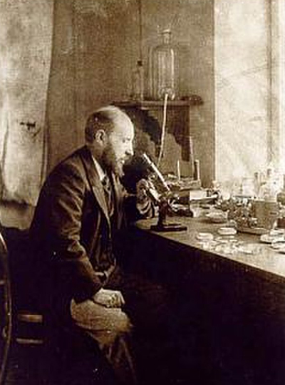
A self-portrait of Santiago Ramón y Cajal looking through a microscope. Image: CC 2.0
On this day, December 10th, nearly 120 years ago, Santiago Ramón y Cajal received a Nobel Prize for capturing and interpreting the very first images of the brain’s most essential components — neurons.
“Many scientists consider Cajal the progenitor of neuroscience because he was the first to really see the brain for what it was: a computational engine made up of individual units,” says Mark Harnett, an investigator at the McGovern Institute and an associate professor in the Department of Brain and Cognitive Sciences. His lab explores how the biophysical features of neurons enable them to perform complex computations that drive thought and behavior.
For Harnett, Cajal is one of the greatest scientific minds to have helped us understand ourselves and our place in the world. Cajal was the first to uncover what neurons look like and propose how they function — equipping the field to solve a slew of the mind’s mysteries. Scientists built on this framework to learn how these remarkable cells relay information — by zapping electrical signals to each other — so we can think, feel, move, communicate, and create.
From art to science and back again
Cajal was born on May 1, 1852, in a small village nestled in the Spanish countryside. It was there Cajal fell deeply and madly in love with … art. But his father was a physician, and urged him to trade his sketches for a scalpel. Begrudgingly, Cajal eventually did. After graduating from medical school in 1873, he worked as an army doctor, but around 1880, he turned his attention to studying the nervous system.
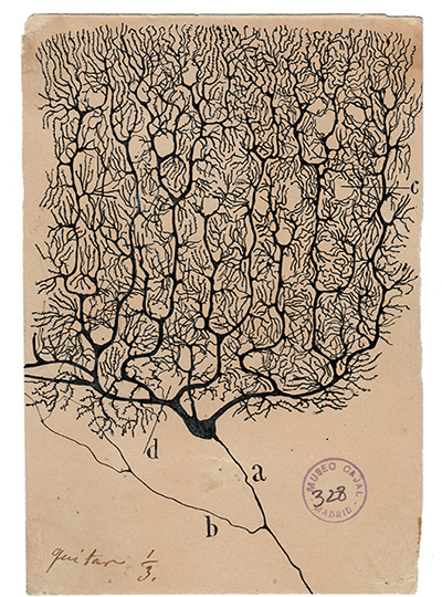
A Purkinje neuron from the human cerebellum. Image: Cajal Institute (CSIC), Madrid
Nineteenth-century scientists didn’t think of the brain as a network of cells but more as plumbing, like the blood vessels in the circulatory system — a series of hollow tubes through which information somehow flowed. Cajal and others were skeptical of this perspective, yet had no way of visualizing the brain at a detailed, cellular level to confirm their suspicions. Scientists at the time stained thin slices of tissue to make cells visible under a microscope, but even the most sophisticated methods stained all cells at once, leaving an indecipherable mass under the microscope’s lens.
This changed in 1887 when Cajal encountered a technique devised by Camillo Golgi that stained only some cells. “Rather than seeing all the cells simultaneously, you saw one at a time,” Harnett explains, making it easier to view a cell’s precise form (Golgi shared the 1906 Nobel Prize with Cajal for this method). If he could refine Golgi’s approach and apply it to neural tissue, Cajal thought, he might finally determine the brain’s architecture.
When he did, a remarkable landscape appeared — black bulbs with sprawling branches, each casting a stringy silhouette. The scene awakened a prior passion. While viewing brain slices under a microscope, Cajal drew what he saw, with surgical precision and an artist’s eye. He had captured — for the first time — the mind’s timberland of cells.
A new theory of the mind
Cajal’s illustrations revealed that brain cells did not form a singular plumbing network, but were distinctly separate, with small gaps between them. “This completely upended what people at the time thought about the brain,” Harnett explains. “It wasn’t made up of connected tubes, but individual cells,” which a few years later in 1891 would be called neurons. Over nearly five decades Cajal created around 2,900 drawings — a collage of neurons from humans and a menagerie of fauna: mice, pigeons, lizards, newts, and fish — spanning a host of cell types, from Purkinje cells to basket and chandelier interneurons.
“Part of Cajal’s genius was that he proposed what the incredible anatomical diversity among neurons meant. He reasoned that maybe one part of the cell could work like an antenna to take in signals, and another might be a cable to send signals out. Cajal was already thinking about input and output at neurons, and synapses as points of contact between them,” Harnett notes. “Each neuron becomes a very complex engine for computation, as opposed to tube-based things that can’t really compute.”
Cajal’s notion that the brain was a network of individual cells would come to be known as the neuron doctrine, a bedrock principle that underlies all of neuroscience today. In his autobiography, Cajal describes neurons as “the mysterious butterflies of the soul, the beating of whose wings may someday – who knows? – clarify the secret of mental life.” And in many ways, they have.
One of thousands of neuron illustrations created by Santiago Ramón y Cajal. Image: CC 2.0
One scientist’s enduring influence
Much of scientists’ current approach to studying the brain is guided by Cajal’s blueprint. This is certainly true for the Harnett lab. “As many in the field do, we share Cajal’s aspiration to apply cutting-edge imaging to reveal hidden aspects of the brain and hypothesize about their function,” Harnett says. “Thankfully, unlike Cajal, we now have the advantage of functional tests to try to validate our hypotheses.”
An ultra high resolution image of a neuron taken by the Harnett lab. Image: Mark Harnett
In a study published in 2022, the Harnett lab used a super-resolution imaging tool to find that filopodia — tiny structures that protrude from dendrites (the signal-receiving “antennas” of neurons) — were far more abundant in the brain than previously thought. Through a battery of tests, they found that these “silent synapses” can become active to facilitate new neural connections. Such pliable sites were believed to only be present very early in life, but the researchers observed filopodia in adult mice, suggesting that they support continuous learning and computational flexibility over the lifespan.
Harnett explains that Cajal’s impact extends beyond neuroscience. “Where does the power of artificial intelligence (AI) come from? It comes, originally, from Cajal.” It’s no wonder, he says, that AI uses neural networks — a mimicry of one of nature’s most powerful designs, first described by Cajal. “The idea that neurons are computational units is really critical to the power and complexity you can achieve within a network. Cajal even hypothesized that changing the strength of signaling between neurons was how learning worked, an idea that was later validated and became one of the critical insights for revolutionizing deep learning in AI.”
By unveiling what’s really happening beneath our skulls, Cajal’s work would both motivate and guide studies of the brain for over a hundred years to come. “Many of his early hypotheses have proven to be true decades and decades later,” Harnett says. “He has and continues to inspire generations of neuroscientists.”
When it comes to brain function, neurons get a lot of the glory. But healthy brains depend on the cooperation of many kinds of cells. The most abundant of the brain’s non-neuronal cells are astrocytes, star-shaped cells with a lot of responsibilities. Astrocytes help shape neural circuits, participate in information processing, and provide nutrient and metabolic support to neurons. Individual cells can take on new roles throughout their lifetimes, and at any given time, the astrocytes in one part of the brain will look and behave differently than the astrocytes somewhere else.
After an extensive analysis by scientists at MIT’s McGovern Institute, neuroscientists now have an atlas detailing astrocytes’ dynamic diversity. Its maps depict the regional specialization of astrocytes across the brains of both mice and marmosets—two powerful models for neuroscience research—and show how their populations shift as brains develop, mature, and age. The study, reported in the November 20 issue of the journal Neuron, was led by Guoping Feng, the James W. (1963) and Patricia T. Poitras Professor of Brain and Cognitive Sciences at MIT. This work was supported by the Hock E. Tan and K. Lisa Yang Center for Autism Research, part of the Yang Tan Collective at MIT, and the National Institutes of Health’s BRAIN Initiative.
Probing the unknown
“It’s really important for us to pay attention to non-neuronal cells’ role in health and disease,” says Feng, who is also the associate director of the McGovern Institute, the director of the Hock E. Tan and K. Lisa Yang Center for Autism Research at MIT, and a member of the Broad Institute of MIT and Harvard. And indeed, these cells—once seen as merely supporting players—have gained more of the spotlight in recent years. Astrocytes are known to play vital roles in the brain’s development and function, and their dysfunction seems to contribute to many psychiatric disorders and neurodegenerative diseases. “But compared to neurons, we know a lot less—especially during development,” Feng adds.
Feng and Margaret Schroeder, a former graduate student in his lab, thought it was important to understand astrocyte diversity across three axes: space, time, and species. They knew from earlier work in the lab, done in collaboration with Steve McCarroll’s lab at Harvard and led by Fenna Krienen in his group, that in adult animals, different parts of the brain have distinctive sets of astrocytes.
“The natural question was, how early in development do we think this regional patterning of astrocytes starts?” Schroeder says.
To find out, she and her colleagues collected brain cells from mice and marmosets at six stages of life, spanning embryonic development to old age. For each animal, they sampled cells from four different brain regions: the prefrontal cortex, the motor cortex, the striatum, and the thalamus.
Then, working with Krienen, who is now an assistant professor at Princeton University, they analyzed the molecular contents of those cells, creating a profile of genetic activity for each one. That profile was based on the mRNA copies of genes found inside the cell, which are known collectively as the cell’s transcriptome. Determining which genes a cell is using and how active those genes are gives researchers insight into a cell’s function and is one way of defining its identity.
Dynamic diversity
After assessing the transcriptomes of about 1.4 million brain cells, the group focused in on the astrocytes, analyzing and comparing their patterns of gene expression. At every life stage, from before birth to old age, the team found regional specialization: Astrocytes from different brain regions had similar patterns of gene expression, which were distinct from those of astrocytes in other brain regions.
This regional specialization was also apparent in the distinct shapes of astrocytes in different parts of the brain, which the team was able to see with expansion microscopy, a high-resolution imaging method developed by McGovern colleague Edward Boyden that reveals fine cellular features.
Notably, the astrocytes in each region changed as animals matured. “When we looked at our late embryonic time point, the astrocytes were already regionally patterned. But when we compare that to the adult profiles, they had completely shifted again,” Schroeder says. “So there’s something happening over postnatal development.” The most dramatic changes the team detected occurred between birth and early adolescence, a period during which brains rapidly rewire as animals begin to interact with the world and learn from their experiences.
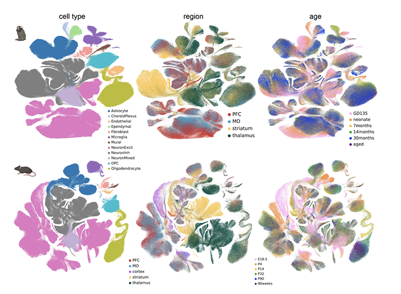
Maps generated by Feng’s team depict the regional specialization of astrocytes across the brains of both mice and marmosets—two powerful models for neuroscience research—and show how their populations shift as brains develop, mature, and age.
Feng and Schroeder suspect that the changes they observed may be driven by the neural circuits that are sculpted and refined as the brain matures. “What we think they’re doing is kind of adapting to their local neuronal niche,” Schroeder says. “The types of genes that they are upregulating and changing during development points to their interaction with neurons.” Feng adds that astrocytes may change their genetic programs in response to nearby neurons, or alternatively, they might help direct the development or function of local circuits as they adopt identities best suited to support particular neurons.
Both mouse and marmoset brains exhibited regional specialization of astrocytes and changes in those populations over time. But when the researchers looked at the specific genes whose activity defined various astrocyte populations, the data from the two species diverged. Schroeder calls this a note of caution for scientists who study astrocytes in animal models, and adds that the new atlas will help researchers assess the potential relevance of findings across species.
Beyond astrocytes
With a new understanding of astrocyte diversity, Feng says his team will pay close attention to how these cells are impacted by the disease-related genes they study and how those effects change during development. He also notes that the gene expression data in the atlas can be used to predict interactions between astrocytes and neurons. “This will really guide future experiments: how these cells’ interactions can shift with changes in the neurons or changes in the astrocytes,” he says.
The Feng lab is eager for other researchers to take advantage of the massive amounts of data they generated as they produced their atlas. Schroeder points out that the team analyzed the transcriptomes of all kinds of cells in the brain regions they studied, not just astrocytes. They are sharing their findings so researchers can use them to understand when and where specific genes are used in the brain, or dig in more deeply to further to explore the brain’s cellular diversity.
More than 300 million people worldwide are living with rare disorders — many of which have a genetic cause and affect the brain and nervous system — yet the vast majority of these conditions lack an approved therapy. Because each rare disorder affects fewer than 65 out of every 100,000 people, studying these disorders and creating new treatments for them is especially challenging.
Thanks to a generous philanthropic gift from Ana Méndez ’91 and Rajeev Jayavant ’86, EE ’88, SM ’88, MIT is now poised to fill the gaps in this research landscape. By establishing the Rare Brain Disorders Nexus — or RareNet — at MIT’s McGovern Institute, the alumni aim to convene leaders in neuroscience research, clinical medicine, patient advocacy, and industry to streamline the lab-to-clinic pipeline for rare brain disorder treatments.
“Ana and Rajeev’s commitment to MIT will form crucial partnerships to propel the translation of scientific discoveries into promising therapeutics and expand the Institute’s impact on the rare brain disorders community,” says MIT President Sally Kornbluth. “We are deeply grateful for their pivotal role in advancing such critical science and bringing attention to conditions that have long been overlooked.”
Building new coalitions
Several hurdles have slowed the lab-to-clinic pipeline for rare brain disorder research. It is difficult to secure a sufficient number of patients per study, and current research efforts are fragmented since each study typically focuses on a single disorder (there are more than 7,000 known rare disorders, according to the World Health Organization). Pharmaceutical companies are often reluctant to invest in emerging treatments due to a limited market size and the high costs associated with preparing drugs for commercialization.
Méndez and Jayavant envision that RareNet will finally break down these barriers. “Our hope is that RareNet will allow leaders in the field to come together under a shared framework and ignite scientific breakthroughs across multiple conditions. A discovery for one rare brain disorder could unlock new insights that are relevant to another,” says Jayavant. “By congregating the best minds in the field, we are confident that MIT will create the right scientific climate to produce drug candidates that may benefit a spectrum of uncommon conditions.”
Guoping Feng, the James W. (1963) and Patricia T. Poitras Professor in Neuroscience and associate director of the McGovern Institute for Brain Research at MIT, will serve as RareNet’s inaugural faculty director. Feng holds a strong record of advancing studies on therapies for neurodevelopmental disorders, including autism spectrum disorders, Williams syndrome, and uncommon forms of epilepsy. His team’s gene therapy for Phelan-McDermid syndrome, a rare and profound autism spectrum disorder, has been licensed to Jaguar Gene Therapy and is currently undergoing clinical trials. “RareNet pioneers a unique model for biomedical research — one that is reimagining the role academia can play in developing therapeutics,” says Feng.
An early version of a gene therapy for SHANK3 mutations — linked to a rare brain disorder called Phelan-McDermid syndrome — correctly finds its way to neurons. Image: Feng lab
RareNet plans to deploy two major initiatives: a global consortium and a therapeutic pipeline accelerator. The consortium will form an international network of researchers, clinicians, and patient groups from the outset. It seeks to connect siloed research efforts, secure more patient samples, promote data sharing, and drive a strong sense of trust and goal alignment across the RareNet community. Partnerships within the consortium will support the aim of the therapeutic pipeline accelerator: to de-risk early lab discoveries and expedite their translation to clinic. By fostering more targeted collaborations — especially between academia and industry — the accelerator will prepare potential treatments for clinical use as efficiently as possible.
MIT labs are focusing on four uncommon conditions in the first wave of RareNet projects: Rett syndrome, prion disease, disorders linked to SYNGAP1 mutations, and Sturge-Weber syndrome. The teams are working to develop novel therapies that can slow, halt, or reverse dysfunctions in the brain and nervous system.
These efforts will build new bridges to connect key stakeholders across the rare brain disorders community and disrupt conventional research approaches. “Rajeev and I are motivated to seed powerful collaborations between MIT researchers, clinicians, patients, and industry,” says Méndez. “Guoping Feng clearly understands our goal to create an environment where foundational studies can thrive and seamlessly move toward clinical impact.”
“Patient and caregiver experiences, and our foreseeable impact on their lives, will guide us and remain at the forefront of our work,” Feng adds. “For far too long the rare brain disorders community has been deprived of life-changing treatments — and, importantly, hope. RareNet gives us the opportunity to transform how we study these conditions and to do so at a moment when it’s needed more than ever.”
The question of how we know ourselves might seem the subject of philosophers, but it is just as much a matter of biology. As modern neuroscientists obtain an increasingly sophisticated understanding of how the brain generates emotions, responds to the external world, and learns from experience, some researchers are returning to a central question: How do we know our experiences, emotions, and physical sensations belong to us?
Curiosity about how the brain generates our sense of self has been a driving force for the research of McGovern Investigator Fan Wang. Following that curiosity has drawn Wang into diverse studies, exploring the origins of pain and the mechanisms we use to control our movements.
“We cannot pinpoint a set of active neurons and say that’s the sense of self. That still remains a mystery,” says Wang, who is also a professor of brain and cognitive sciences and co-director of the K. Lisa Yang and Hock E. Tan Center for Molecular Therapeutics at MIT. But she and other neuroscientists are drilling down into different functions of the brain that together might generate our awareness of ourselves.
McGovern Investigator Fan Wang (right) with research scientist Vincent Prevosto, who studies brain regions implicated in whisker movement. Photo: Steph Stevens
Wang, who teaches the undergraduate course, “Neurobiology of Self,” explains that there are lots of ways to think about our sense of self, which are probably deeply integrated in the brain. Some are mostly about our physical bodies: How do we experience touch? How do we understand
where we are in space, or recognize the boundary between ourselves and rest of the world? Some consider more internal sensations, like how we experience pain or hunger. Emotion is also key to our sense of self: How do we know that anger or joy are our own, and why do these states change the way our bodies feel?
Wang can trace her initial interest in the brain’s sense of self to work she did as a graduate student in Richard Axel’s lab at Columbia University. The lab had identified receptors expressed by sensory neurons in the nose that detect odorous substances. Wang and others discovered the pathways that information about these smells takes to the brain, and how the brain distinguishes one smell from another.
Who is the “knower” of this information? “The answer,” Wang says, “is ‘I’ or ‘me.’ But understanding where I get the sense of self and how that is constructed, is what drives me to do neuroscience.”
Mechanisms of movement
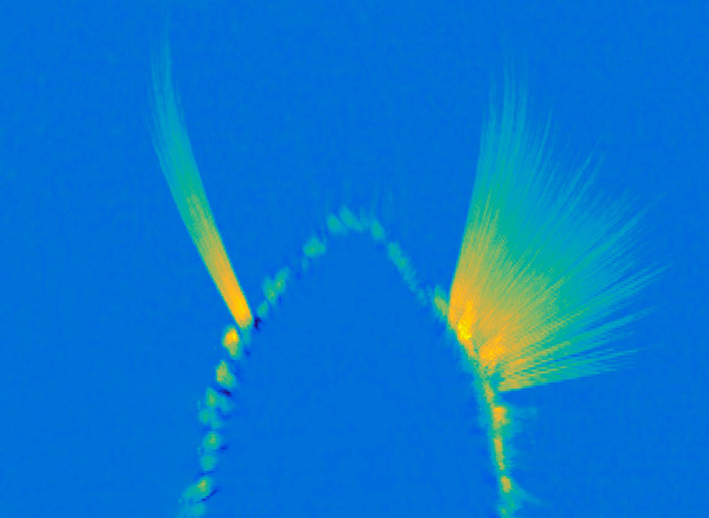
In her lab at the McGovern Institute, Wang is studying how the brain controls the body’s movements, which she sees as closely tied to the awareness of our physical selves. “The reason I think I am in my body is because I can control my movement. I generate the movement. I cannot control your movement,” says Wang. “Volitional movement gives us a sense of agency, and this sense of agency resembles the sense of self.” For the mice that the group studies, one crucial type of movement comes from the whiskers, which the animals depend on as they explore their environments. Wang’s group has traced the neural circuity that controls whiskers’ rhythmic back-and-forth, which is initiated in the brainstem, where many of the body’s most vital functions are controlled. Wang describes the simple circuit as an oscillator, or a self-generated loop.
A maximum projection image showing tracked whiskers on the mouse muzzle. The right (control) side shows the back-and-forth rhythmic sweeping of the whiskers, while the experimental side where the whisking oscillator neurons are silenced, the whiskers move very little. Image: Wang Lab
Once it’s started, “the movement can go on unless some other signals stop it,” she says. The movement the circuit generates is simple but voluntary, and can be fine-tuned based on the sensory feedback the whiskers relay back to the brain. They’ve also been investigating how mice move the larynx to generate the squeaks and calls they use to communicate. These intentional movements must be coordinated with the ongoing cycles of respiration since we produce normal sounds only during expiration. Wang’s team has found neurons in the brainstem that generate vocalization-specific movements, and also discovered how respiration-controlling neural circuits can override them, ensuring that breathing is prioritized.
Wang says understanding the circuitry that controls these simple movements sets the stage for figuring out how the brain modifies activity in those circuits to create more complex, intentional movements. “That brings me closer to understanding where this volition is generated — and closer to this sense of self,” she says.
Emotional pain
Still, she knows that volitional movements — even those generated in response to perceptions of the environment — do not, on their own, define a sense of self. As a counterexample, she looks to self-driving cars: “There’s sensory information coming into the central computer, which then generates a motor output — where to drive, where to turn, where to stop. But none of us think a Waymo taxi has a sense of self.”
Wang says when she pondered the ways in which AI-powered cars lack a sense of self, she began thinking about emotions and pain. “If the self-driving Waymo crashes, it will not feel pain,” she says. “But if we hurt ourselves, we will feel pain. And we will hate that, and then we’ll learn.” So her lab is also exploring how the nervous system generates pain perception, including the emotional response that it evokes.
Ensembles of neurons in the amygdala activated by general anesthesia. Image: Fan Wang
In both humans and mice, pain causes emotional suffering that can be recognized and measured through changes in body functions like heart rate and blood pressure. With funding from the K. Lisa Yang Brain-Body Center at MIT, Wang’s lab is carefully tracking these involuntary, or autonomic, functions to gain a more complete understanding of pain’s emotional impact. This approach has helped clarify the role of pain-suppressing neurons in the brain’s amygdala — an important emotion-processing center — that Wang’s team discovered in 2020. When researchers selectively activate those cells in mice, the animals’ behavior makes it clear that the neurons are suppressing pain. Now, the group has learned that activating these neurons suppresses the autonomic response to pain.
Wang says there’s hope that modulating pain’s emotional response might be a way to treat chronic pain in patients. She explains that some patients with damage to another one of the brain’s emotional centers, the cingulate cortex, feel painful stimuli, but experience them as merely intense sensations. That suggests that it might be possible to modulate the emotional response to pain to eliminate patients’ suffering, without blocking the protective information that pain can provide.
The team has also been focusing on another set of anesthesia-activated neurons, which they have found suppress anxiety. When anxiety-suppressing neurons are activated in mice, the animals’ heart rates slow and they become more willing to explore bright, open spaces. Another anxiety-associated measure — heart rate variability — increases. Wang explains that this change is particularly significant: “If you have persistent low heart rate variability, especially in veterans, that is a very good predictor for anxiety developing into depression in the future,” she says.
The team’s findings, which suggest that changes in autonomic functions may themselves relieve anxiety, point toward potential new targets for anti-anxiety therapies. And by highlighting the connection between emotion and bodily responses, they offer more clues about our sense of self. “These neurons are now changing some high-level concept about anxiety,” Wang points out.
That link between emotion and body seems to Wang to be key to the sense of self. The big questions remain unanswered, but that simply stokes her curiosity. “I can be aware of my bodily responses: I am aware of ‘I am anxious’ or ‘I am in pain.’ I can see the pathways from which stimuli go into these nervous systems and come back down to the body and control the response. But I still don’t know who is the person — the knower,” she says. “I haven’t found it, so I’m going to keep looking.”