Modeling the social mind

Typically, it would take two graduate students to do the research that Setayesh Radkani is doing.

Driven by an insatiable curiosity about the human mind, she is working on two PhD thesis projects in two different cognitive neuroscience labs at MIT. For one, she is studying punishment as a social tool to influence others. For the other, she is uncovering the neural processes underlying social learning — that is, learning from others. By piecing together these two research programs, Radkani is hoping to gain a better understanding of the mechanisms underpinning social influence in the mind and brain.

Radkani lived in Iran for most of her life, growing up alongside her younger brother in Tehran. The two spent a lot of time together and have long been each other’s best friends. Her father is a civil engineer, and her mother is a midwife. Her parents always encouraged her to explore new things and follow her own path, even if it wasn’t quite what they imagined for her. And her uncle helped cultivate her sense of curiosity, teaching her to “always ask why” as a way to understand how the world works.

Growing up, Radkani most loved learning about human psychology and using math to model the world around her. But she thought it was impossible to combine her two interests. Prioritizing math, she pursued a bachelor’s degree in electrical engineering at the Sharif University of Technology in Iran.

Then, late in her undergraduate studies, Radkani took a psychology course and discovered the field of cognitive neuroscience, in which scientists mathematically model the human mind and brain. She also spent a summer working in a computational neuroscience lab at the Swiss Federal Institute of Technology in Lausanne. Seeing a way to combine her interests, she decided to pivot and pursue the subject in graduate school.

An experience leading a project in her engineering ethics course during her final year of undergrad further helped her discover some of the questions that would eventually form the basis of her PhD. The project investigated why some students cheat and how to change this.

“Through this project I learned how complicated it is to understand the reasons that people engage in immoral behavior, and even more complicated than that is how to devise policies and react in these situations in order to change people’s attitudes,” Radkani says. “It was this experience that made me realize that I’m interested in studying the human social and moral mind.”

She began looking into social cognitive neuroscience research and stumbled upon a relevant TED talk by Rebecca Saxe, the John W. Jarve Professor in Brain and Cognitive Sciences at MIT, who would eventually become one of Radkani’s research advisors. Radkani knew immediately that she wanted to work with Saxe. But she needed to first get into the BCS PhD program at MIT, a challenging obstacle given her minimal background in the field.

After two application cycles and a year’s worth of graduate courses in cognitive neuroscience, Radkani was accepted into the program. But to come to MIT, she had to leave her family behind. Coming from Iran, Radkani has a single-entry visa, making it difficult for her to travel outside the U.S. She hasn’t been able to visit her family since starting her PhD and won’t be able to until at least after she graduates. Her visa also limits her research contributions, restricting her from attending conferences outside the U.S. “That is definitely a huge burden on my education and on my mental health,” she says.

Still, Radkani is grateful to be at MIT, indulging her curiosity in the human social mind. And she’s thankful for her supportive family, who she calls over FaceTime every day.

Modeling how people think about punishment

In Saxe’s lab, Radkani is researching how people approach and react to punishment, through behavioral studies and neuroimaging. By synthesizing these findings, she’s developing a computational model of the mind that characterizes how people make decisions in situations involving punishment, such as when a parent disciplines a child, when someone punishes their romantic partner, or when the criminal justice system sentences a defendant. With this model, Radkani says she hopes to better understand “when and why punishment works in changing behavior and influencing beliefs about right and wrong, and why sometimes it fails.”

Punishment isn’t a new research topic in cognitive neuroscience, Radkani says, but in previous studies, scientists have often only focused on people’s behavior in punitive situations and haven’t considered the thought processes that underlie those behaviors. Characterizing these thought processes, though, is key to understanding whether punishment in a situation can be effective in changing people’s attitudes.

People bring their prior beliefs into a punitive situation. Apart from moral beliefs about the appropriateness of different behaviors, “you have beliefs about the characteristics of the people involved, and you have theories about their intentions and motivations,” Radkani says. “All those come together to determine what you do or how you are influenced by punishment,” given the circumstances. Punishers decide a suitable punishment based on their interpretation of the situation, in light of their beliefs. Targets of punishment then decide whether they’ll change their attitude as a result of the punishment, depending on their own beliefs. Even outside observers make decisions, choosing whether to keep or change their moral beliefs based on what they see.

To capture these decision-making processes, Radkani is developing a computational model of the mind for punitive situations. The model mathematically represents people’s beliefs and how they interact with certain features of the situation to shape their decisions. The model then predicts a punisher’s decisions, and how punishment will influence the target and observers. Through this model, Radkani will provide a foundational understanding of how people think in various punitive situations.

Researching the neural mechanisms of social learning

In parallel, working in the lab of Professor Mehrdad Jazayeri, Radkani is studying social learning, uncovering its underlying neural processes. Through social learning, people learn from other people’s experiences and decisions, and incorporate this socially acquired knowledge into their own decisions or beliefs.

Humans are extraordinary in their social learning abilities, however our primary form of learning, shared by all other animals, is learning from self-experience. To investigate how learning from others is similar to or different from learning from our own experiences, Radkani has designed a two-player video game that involves both types of learning. During the game, she and her collaborators in Jazayeri’s lab record neural activity in the brain. By analyzing these neural measurements, they plan to uncover the computations carried out by neural circuits during social learning, and compare those to learning from self-experience.

Radkani first became curious about this comparison as a way to understand why people sometimes draw contrasting conclusions from very similar situations. “For example, if I get Covid from going to a restaurant, I’ll blame the restaurant and say it was not clean,” Radkani says. “But if I hear the same thing happen to my friend, I’ll say it’s because they were not careful.” Radkani wanted to know the root causes of this mismatch in how other people’s experiences affect our beliefs and judgements differently from our own similar experiences, particularly because it can lead to “errors that color the way that we judge other people,” she says.

By combining her two research projects, Radkani hopes to better understand how social influence works, particularly in moral situations. From there, she has a slew of research questions that she’s eager to investigate, including: How do people choose who to trust? And which types of people tend to be the most influential? As Radkani’s research grows, so does her curiosity.

Studies of autism tend to exclude women, researchers find

In recent years, researchers who study autism have made an effort to include more women and girls in their studies. However, despite these efforts, most studies of autism consistently enroll small numbers of female subjects or exclude them altogether, according to a new study from MIT.

The researchers found that a screening test commonly used to determine eligibility for studies of autism consistently winnows out a much higher percentage of women than men, creating a “leaky pipeline” that results in severe underrepresentation of women in studies of autism.

This lack of representation makes it more difficult to develop useful interventions or provide accurate diagnoses for girls and women, the researchers say.

“I think the findings favor having a more inclusive approach and widening the lens to end up being less biased in terms of who participates in research,” says John Gabrieli, the Grover Hermann Professor of Health Sciences and Technology and a professor of brain and cognitive sciences at MIT. “The more we understand autism in men and women and nonbinary individuals, the better services and more accurate diagnoses we can provide.”

Gabrieli, who is also a member of MIT’s McGovern Institute for Brain Research, is the senior author of the study, which appears in the journal Autism Research. Anila D’Mello, a former MIT postdoc who is now an assistant professor at the University of Texas Southwestern, is the lead author of the paper. MIT Technical Associate Isabelle Frosch, Research Coordinator Cindy Li, and Research Specialist Annie Cardinaux are also authors of the paper.

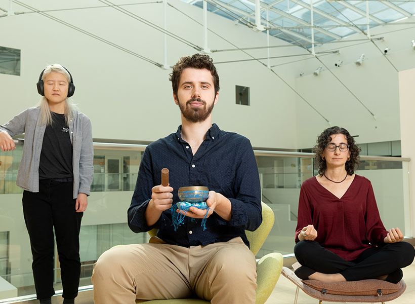
Gabrieli lab researchers Annie Cardinaux (left), Anila D’Mello (center), Cindy Li (right), and Isabelle Frosch (not pictured) have
uncovered sex biases in ASD research. Photo: Steph Stevens

Screening out females

Autism spectrum disorders are diagnosed based on observation of traits such as repetitive behaviors and difficulty with language and social interaction. Doctors may use a variety of screening tests to help them make a diagnosis, but these screens are not required.

For research studies of autism, it is routine to use a screening test called the Autism Diagnostic Observation Schedule (ADOS) to determine eligibility for the study. This test, which assesses social interaction, communication, play, and repetitive behaviors, provides a quantitative score in each category, and only participants who reach certain scores qualify for inclusion in studies.

While doing a study exploring how quickly the brains of autistic adults adapt to novel events in the environment, scientists in Gabrieli’s lab began to notice that the ADOS appeared to have unequal effects on male and female participation in research. As the study progressed, D’Mello noticed some significant brain differences between the male and female subjects in the study.

To investigate these differences further, D’Mello tried to find more female participants using an MIT database of autistic adults who have expressed interest in participating in research studies. However, when she sorted through the subjects, she found that only about half of the women in the database had met the ADOS cutoff scores typically required for inclusion in autism studies, compared to 80 percent of the males.

“We realized then that there’s a discrepancy and that the ADOS is essentially screening out who eventually participated in research,” D’Mello says. “We were really surprised at how many males we retained and how many females we lost to the ADOS.”

To see if this phenomenon was more widespread, the researchers looked at six publicly available datasets, which include more than 40,000 adults who have been diagnosed as autistic. For some of these datasets, participants were screened with ADOS to determine their eligibility to participate in studies, while for others, a “community diagnosis” — diagnosis from a doctor or other health care provider — was sufficient.

The researchers found that in datasets that required ADOS screening for eligibility, the ratio of male to female participants ended up being around 8:1, while in those that required only a community diagnosis the ratios ranged from about 2:1 to 1:1.

Previous studies have found differences between behavioral patterns in autistic men and women, but the ADOS test was originally developed using a largely male sample, which may explain why it often excludes women from research studies, D’Mello says.

“There were few females in the sample that was used to create this assessment, so it might be that it’s not great at picking up the female phenotype, which may differ in certain ways — primarily in domains like social communication,” she says.

Effects of exclusion

Failure to include more women and girls in studies of autism may contribute to shortcomings in the definitions of the disorder, the researchers say.

“The way we think about it is that the field evolved perhaps an implicit bias in how autism is defined, and it was driven disproportionately by analysis of males, and recruitment of males, and so on,” Gabrieli says. “So, the definition doesn’t fit as well, on average, with the different expression of autism that seems to be more common in females.”

This implicit bias has led to documented difficulties in receiving a diagnosis for girls and women, even when their symptoms are the same as those presented by autistic boys and men.

“Many females might be missed altogether in terms of diagnoses, and then our study shows that in the research setting, what is already a small pool gets whittled down at a much larger rate than that of males,” D’Mello says.

Excluding girls and women from this kind of research study can lead to treatments that don’t work as well for them, and it contributes to the perception that autism doesn’t affect women as much as men.

“The goal is that research should directly inform treatment, therapies, and public perception,” D’Mello says. “If the research is saying that there aren’t females with autism, or that the brain basis of autism only looks like the patterns established in males, then you’re not really helping females as much as you could be, and you’re not really getting at the truth of what the disorder might be.”

The researchers now plan to further explore some of the gender and sex-based differences that appear in autism, and how they arise. They also plan to expand the gender categories that they include. In the current study, the surveys that each participant filled out asked them to choose male or female, but the researchers have updated their questionnaire to include nonbinary and transgender options.

The research was funded by the Hock E. Tan and K. Lisa Yang Center for Autism Research, the Simons Center for the Social Brain at MIT, and the National Institutes of Mental Health.

These neurons have food on the brain

A gooey slice of pizza. A pile of crispy French fries. Ice cream dripping down a cone on a hot summer day. When you look at any of these foods, a specialized part of your visual cortex lights up, according to a new study from MIT neuroscientists.

This newly discovered population of food-responsive neurons is located in the ventral visual stream, alongside populations that respond specifically to faces, bodies, places, and words. The unexpected finding may reflect the special significance of food in human culture, the researchers say.

“Food is central to human social interactions and cultural practices. It’s not just sustenance,” says Nancy Kanwisher, the Walter A. Rosenblith Professor of Cognitive Neuroscience and a member of MIT’s McGovern Institute for Brain Research and Center for Brains, Minds, and Machines. “Food is core to so many elements of our cultural identity, religious practice, and social interactions, and many other things that humans do.”

The findings, based on an analysis of a large public database of human brain responses to a set of 10,000 images, raise many additional questions about how and why this neural population develops. In future studies, the researchers hope to explore how people’s responses to certain foods might differ depending on their likes and dislikes, or their familiarity with certain types of food.

MIT postdoc Meenakshi Khosla is the lead author of the paper, along with MIT research scientist N. Apurva Ratan Murty. The study appears today in the journal Current Biology.

Visual categories

More than 20 years ago, while studying the ventral visual stream, the part of the brain that recognizes objects, Kanwisher discovered cortical regions that respond selectively to faces. Later, she and other scientists discovered other regions that respond selectively to places, bodies, or words. Most of those areas were discovered when researchers specifically set out to look for them. However, that hypothesis-driven approach can limit what you end up finding, Kanwisher says.

“There could be other things that we might not think to look for,” she says. “And even when we find something, how do we know that that’s actually part of the basic dominant structure of that pathway, and not something we found just because we were looking for it?”

To try to uncover the fundamental structure of the ventral visual stream, Kanwisher and Khosla decided to analyze a large, publicly available dataset of full-brain functional magnetic resonance imaging (fMRI) responses from eight human subjects as they viewed thousands of images.

“We wanted to see when we apply a data-driven, hypothesis-free strategy, what kinds of selectivities pop up, and whether those are consistent with what had been discovered before. A second goal was to see if we could discover novel selectivities that either haven’t been hypothesized before, or that have remained hidden due to the lower spatial resolution of fMRI data,” Khosla says.

To do that, the researchers applied a mathematical method that allows them to discover neural populations that can’t be identified from traditional fMRI data. An fMRI image is made up of many voxels — three-dimensional units that represent a cube of brain tissue. Each voxel contains hundreds of thousands of neurons, and if some of those neurons belong to smaller populations that respond to one type of visual input, their responses may be drowned out by other populations within the same voxel.

The new analytical method, which Kanwisher’s lab has previously used on fMRI data from the auditory cortex, can tease out responses of neural populations within each voxel of fMRI data.

Using this approach, the researchers found four populations that corresponded to previously identified clusters that respond to faces, places, bodies, and words. “That tells us that this method works, and it tells us that the things that we found before are not just obscure properties of that pathway, but major, dominant properties,” Kanwisher says.

Intriguingly, a fifth population also emerged, and this one appeared to be selective for images of food.

“We were first quite puzzled by this because food is not a visually homogenous category,” Khosla says. “Things like apples and corn and pasta all look so unlike each other, yet we found a single population that responds similarly to all these diverse food items.”

The food-specific population, which the researchers call the ventral food component (VFC), appears to be spread across two clusters of neurons, located on either side of the FFA. The fact that the food-specific populations are spread out between other category-specific populations may help explain why they have not been seen before, the researchers say.

“We think that food selectivity had been harder to characterize before because the populations that are selective for food are intermingled with other nearby populations that have distinct responses to other stimulus attributes. The low spatial resolution of fMRI prevents us from seeing this selectivity because the responses of different neural population get mixed in a voxel,” Khosla says.

“The technique which the researchers used to identify category-sensitive cells or areas is impressive, and it recovered known category-sensitive systems, making the food category findings most impressive,” says Paul Rozin, a professor of psychology at the University of Pennsylvania, who was not involved in the study. “I can’t imagine a way for the brain to reliably identify the diversity of foods based on sensory features. That makes this all the more fascinating, and likely to clue us in about something really new.”

Food vs non-food

The researchers also used the data to train a computational model of the VFC, based on previous models Murty had developed for the brain’s face and place recognition areas. This allowed the researchers to run additional experiments and predict the responses of the VFC. In one experiment, they fed the model matched images of food and non-food items that looked very similar — for example, a banana and a yellow crescent moon.

“Those matched stimuli have very similar visual properties, but the main attribute in which they differ is edible versus inedible,” Khosla says. “We could feed those arbitrary stimuli through the predictive model and see whether it would still respond more to food than non-food, without having to collect the fMRI data.”

They could also use the computational model to analyze much larger datasets, consisting of millions of images. Those simulations helped to confirm that the VFC is highly selective for images of food.

From their analysis of the human fMRI data, the researchers found that in some subjects, the VFC responded slightly more to processed foods such as pizza than unprocessed foods like apples. In the future they hope to explore how factors such as familiarity and like or dislike of a particular food might affect individuals’ responses to that food.

They also hope to study when and how this region becomes specialized during early childhood, and what other parts of the brain it communicates with. Another question is whether this food-selective population will be seen in other animals such as monkeys, who do not attach the cultural significance to food that humans do.

The research was funded by the National Institutes of Health, the National Eye Institute, and the National Science Foundation through the MIT Center for Brains, Minds, and Machines.

Why do we dream?

As part of our Ask the Brain series, science writer Shafaq Zia answers the question, “Why do we dream?”

_____

One night, Albert Einstein dreamt that he was walking through a farm where he found a herd of cows against an electric fence. When the farmer switched on the fence, the cows suddenly jumped back, all at the same time. But to the farmer’s eyes, who was standing at the other end of the field, they seemed to have jumped one after another, in a wave formation. Einstein woke up and the Theory of Relativity was born.

Dreaming is one of the oldest biological phenomena; for as long as humans have slept, they’ve dreamt. But through most of our history, dreams have remained mystified, leaving scientists, philosophers, and artists alike searching for meaning.

In many aboriginal cultures, such as the Esa Eja community in Peruvian Amazon, dreaming is a sacred practice for gaining knowledge, or solving a problem, through the dream narrative. But in the last century or so, technological advancements have allowed neuroscientists to take up dreams as a matter of scientific inquiry in order to answer a much-pondered question — what is the purpose of dreaming?

Falling asleep

The human brain is a fascinating place. It is composed of approximately 80 billion neurons and it is their combined electrical chatter that generates oscillations known as brain waves. There are five types of brain waves —  alpha, beta, theta, delta, and gamma — that each indicate a different state between sleep and wakefulness.

Using EEG, a test that records electrical activity in the brain, scientists have identified that when we’re awake, our brain emits beta and gamma waves. These tend to have a stimulating effect and help us remain actively engaged in mental activities.

The differently named frequency bands of neural oscillations, or brainwaves: delta, theta, alpha, beta, and gamma.

But during the transition to sleep, the number of beta waves lowers significantly and the brain produces high levels of alpha waves. These waves regulate attention and help filter out distractions. A recent study led by McGovern Institute Director Robert Desimone, showed that people can actually enhance their attention by controlling their own alpha brain waves using neurofeedback training. It’s unknown how long these effects might last and whether this kind of control could be achieved with other types of brain waves, but the researchers are now planning additional studies to explore these questions.

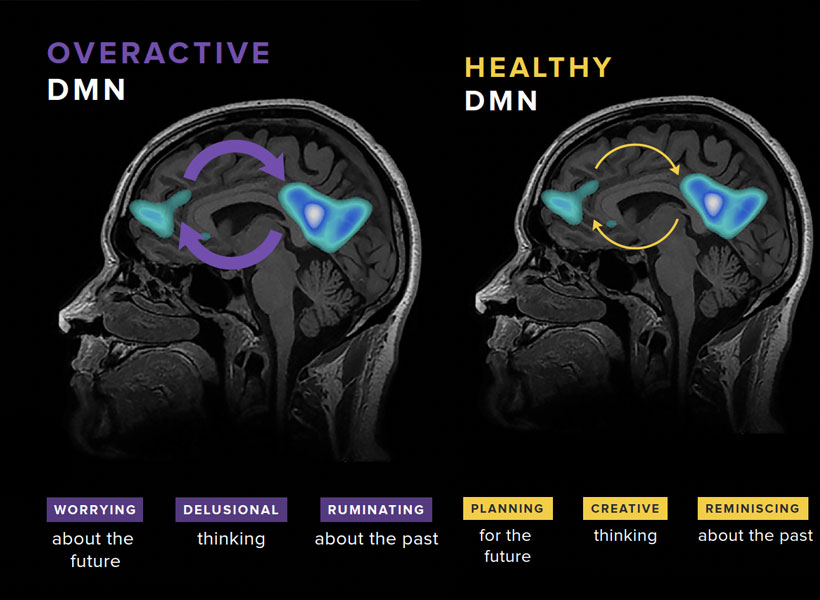
Alpha waves are also produced when we daydream, meditate, or listen to the sound of rain. As our minds wander, many parts of the brain are engaged, including a specialized system called the “default mode network.” Disturbances in this network, explains Susan Whitfield-Gabrieli, a professor of psychology at Northeastern University and a McGovern Institute research affiliate, have been linked to various brain disorders including schizophrenia, depression and ADHD. By identifying the brain circuits associated with mind wandering, she says, we can begin to develop better treatment options for people suffering from these disorders.

Finally, as we enter a dreamlike state, the prefrontal cortex of the brain, responsible for keeping impulses in check, slowly grows less active. This is when there’s a spur in theta waves that leads to an unconstrained window of consciousness; there is little censorship from the mind, allowing for visceral dreams and creative thoughts.

The dreaming brain

“Every time you learn something, it happens so quickly,” said Dheeraj Roy, postdoctoral fellow in Guoping Feng’s lab at the McGovern Institute. “The brain is continuously recording information, but how do you take a break and then make sense of it all?”

This is where dreams come in, says Roy. During sleep, newly-formed memories are gradually stabilized into a more permanent form of long-term storage in the brain. Dreaming, he says, is influenced by the consolidation of these memories during sleep. Most dreams are made up of experiences, thoughts, emotion, places, and people we have already encountered in our lives. But, during dreaming, bits and pieces of these memories seem to be reorganized to create a particularly bizarre scenario: you’re talking to your sister when it suddenly begins to rain roses and you’re dancing at a New Year’s party.

This re-organization may not be so random; as the brain is processing memories, it pulls together the ones that are seemingly related to each other. Perhaps you dreamt of your sister because you were at a store recently where a candle smelt like her rose-scented perfume, which reminded you of the time you made a new year resolution to spend less money on flowers.

Some brain disorders, like Parkinson’s disease, have been associated with vivid, unpleasant dreams and erratic brain wave patterns. Researchers at the McGovern Institute hope that a better understanding of mechanics of the brain – including neural circuits and brain waves – will help people with Parkinson’s and other brain disorders.

So perhaps dreams aren’t instilled with meaning, symbolism, and wisdom in the way we’ve always imagined, and they simply reflect important biological processes taking place in our brain. But with all that science has uncovered about dreaming and the ways in which it links to creativity and memory, the magical essence of this universal human experience remains untainted.

_____

Do you have a question for The Brain? Ask it here.

Whether speaking Turkish or Norwegian, the brain’s language network looks the same

Over several decades, neuroscientists have created a well-defined map of the brain’s “language network,” or the regions of the brain that are specialized for processing language. Found primarily in the left hemisphere, this network includes regions within Broca’s area, as well as in other parts of the frontal and temporal lobes.

However, the vast majority of those mapping studies have been done in English speakers as they listened to or read English texts. MIT neuroscientists have now performed brain imaging studies of speakers of 45 different languages. The results show that the speakers’ language networks appear to be essentially the same as those of native English speakers.

The findings, while not surprising, establish that the location and key properties of the language network appear to be universal. The work also lays the groundwork for future studies of linguistic elements that would be difficult or impossible to study in English speakers because English doesn’t have those features.

“This study is very foundational, extending some findings from English to a broad range of languages,” says Evelina Fedorenko, the Frederick A. and Carole J. Middleton Career Development Associate Professor of Neuroscience at MIT and a member of MIT’s McGovern Institute for Brain Research. “The hope is that now that we see that the basic properties seem to be general across languages, we can ask about potential differences between languages and language families in how they are implemented in the brain, and we can study phenomena that don’t really exist in English.”

Fedorenko is the senior author of the study, which appears today in Nature Neuroscience. Saima Malik-Moraleda, a PhD student in the Speech and Hearing Bioscience and Technology program at Harvard University, and Dima Ayyash, a former research assistant, are the lead authors of the paper.

Mapping language networks

The precise locations and shapes of language areas differ across individuals, so to find the language network, researchers ask each person to perform a language task while scanning their brains with functional magnetic resonance imaging (fMRI). Listening to or reading sentences in one’s native language should activate the language network. To distinguish this network from other brain regions, researchers also ask participants to perform tasks that should not activate it, such as listening to an unfamiliar language or solving math problems.

Several years ago, Fedorenko began designing these “localizer” tasks for speakers of languages other than English. While most studies of the language network have used English speakers as subjects, English does not include many features commonly seen in other languages. For example, in English, word order tends to be fixed, while in other languages there is more flexibility in how words are ordered. Many of those languages instead use the addition of morphemes, or segments of words, to convey additional meaning and relationships between words.

“There has been growing awareness for many years of the need to look at more languages, if you want make claims about how language works, as opposed to how English works,” Fedorenko says. “We thought it would be useful to develop tools to allow people to rigorously study language processing in the brain in other parts of the world. There’s now access to brain imaging technologies in many countries, but the basic paradigms that you would need to find the language-responsive areas in a person are just not there.”

For the new study, the researchers performed brain imaging of two speakers of 45 different languages, representing 12 different language families. Their goal was to see if key properties of the language network, such as location, left lateralization, and selectivity, were the same in those participants as in people whose native language is English.

The researchers decided to use “Alice in Wonderland” as the text that everyone would listen to, because it is one of the most widely translated works of fiction in the world. They selected 24 short passages and three long passages, each of which was recorded by a native speaker of the language. Each participant also heard nonsensical passages, which should not activate the language network, and was asked to do a variety of other cognitive tasks that should not activate it.

The team found that the language networks of participants in this study were found in approximately the same brain regions, and had the same selectivity, as those of native speakers of English.

“Language areas are selective,” Malik-Moraleda says. “They shouldn’t be responding during other tasks such as a spatial working memory task, and that was what we found across the speakers of 45 languages that we tested.”

Additionally, language regions that are typically activated together in English speakers, such as the frontal language areas and temporal language areas, were similarly synchronized in speakers of other languages.

The researchers also showed that among all of the subjects, the small amount of variation they saw between individuals who speak different languages was the same as the amount of variation that would typically be seen between native English speakers.

Similarities and differences

While the findings suggest that the overall architecture of the language network is similar across speakers of different languages, that doesn’t mean that there are no differences at all, Fedorenko says. As one example, researchers could now look for differences in speakers of languages that predominantly use morphemes, rather than word order, to help determine the meaning of a sentence.

“There are all sorts of interesting questions you can ask about morphological processing that don’t really make sense to ask in English, because it has much less morphology,” Fedorenko says.

Another possibility is studying whether speakers of languages that use differences in tone to convey different word meanings would have a language network with stronger links to auditory brain regions that encode pitch.

Right now, Fedorenko’s lab is working on a study in which they are comparing the ‘temporal receptive fields’ of speakers of six typologically different languages, including Turkish, Mandarin, and Finnish. The temporal receptive field is a measure of how many words the language processing system can handle at a time, and for English, it has been shown to be six to eight words long.

“The language system seems to be working on chunks of just a few words long, and we’re trying to see if this constraint is universal across these other languages that we’re testing,” Fedorenko says.

The researchers are also working on creating language localizer tasks and finding study participants representing additional languages beyond the 45 from this study.

The research was funded by the National Institutes of Health and research funds from MIT’s Department of Brain and Cognitive Sciences, the McGovern Institute, and the Simons Center for the Social Brain. Malik-Moraleda was funded by a la Caixa Fellowship and a Friends of McGovern fellowship.

A voice for change — in Spanish

Jessica Chomik-Morales had a bicultural childhood. She was born in Boca Raton, Florida, where her parents had come seeking a better education for their daughter than she would have access to in Paraguay. But when she wasn’t in school, Chomik-Morales was back in that small, South American country with her family. One of the consequences of growing up in two cultures was an early interest in human behavior. “I was always in observer mode,” Chomik-Morales says, recalling how she would tune in to the nuances of social interactions in order to adapt and fit in.

Today, that fascination with human behavior is driving Chomik-Morales as she works with MIT professor of cognitive science Laura Schulz and Walter A. Rosenblith Professor of Cognitive Neuroscience and McGovern Institute for Brain Research investigator Nancy Kanwisher as a post-baccalaureate research scholar, using functional brain imaging to investigate how the brain recognizes and understands causal relationships. Since arriving at MIT last fall, she’s worked with study volunteers to collect functional MRI (fMRI) scans and used computational approaches to interpret the images. She’s also refined her own goals for the future.

Jessica Chomik-Morales (right) with postdoctoral associate Héctor De Jesús-Cortés. Photo: Steph Stevens

She plans to pursue a career in clinical neuropsychology, which will merge her curiosity about the biological basis of behavior with a strong desire to work directly with people. “I’d love to see what kind of questions I could answer about the neural mechanisms driving outlier behavior using fMRI coupled with cognitive assessment,” she says. And she’s confident that her experience in MIT’s two-year post-baccalaureate program will help her get there. “It’s given me the tools I need, and the techniques and methods and good scientific practice,” she says. “I’m learning that all here. And I think it’s going to make me a more successful scientist in grad school.”

The road to MIT

Chomik-Morales’s path to MIT was not a straightforward trajectory through the U.S. school system. When her mom, and later her dad, were unable to return to the U.S., she started eight grade in the capital city of Asunción. It did not go well. She spent nearly every afternoon in the principal’s office, and soon her father was encouraging her to return to the United States. “You are an American,” he told her. “You have a right to the educational system there.”

Back in Florida, Chomik-Morales became a dedicated student, even while she worked assorted jobs and shuffled between the homes of families who were willing to host her. “I had to grow up,” she says. “My parents are sacrificing everything just so I can have a chance to be somebody. People don’t get out of Paraguay often, because there aren’t opportunities and it’s a very poor country. I was given an opportunity, and if I waste that, then that is disrespect not only to my parents, but to my lineage, to my country.”

As she graduated from high school and went on to earn a degree in cognitive neuroscience at Florida Atlantic University, Chomik-Morales found herself experiencing things that were completely foreign to her family. Though she spoke daily with her mom via WhatsApp, it was hard to share what she was learning in school or what she was doing in the lab. And while they celebrated her academic achievements, Chomik-Morales knew they didn’t really understand them. “Neither of my parents went to college,” she says. “My mom told me that she never thought twice about learning about neuroscience. She had this misconception that it was something that she would never be able to digest.”

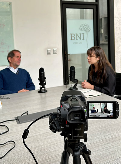
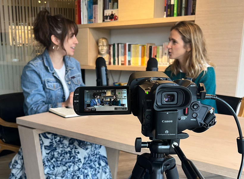
Chomik-Morales believes that the wonders of neuroscience are for everybody. But she also knows that Spanish speakers like her mom have few opportunities to hear the kinds of accessible, engaging stories that might draw them in. So she’s working to change that. With support from the McGovern Institute, the National Science Foundation funded Science and Technology Center for Brains, Minds, and Machines, Chomik-Morales is hosting and producing a weekly podcast called “Mi Última Neurona” (“My Last Neuron”), which brings conversations with neuroscientists to Spanish speakers around the world.

Listeners hear how researchers at MIT and other institutions are exploring big concepts like consciousness and neurodegeneration, and learn about the approaches they use to study the brain in humans, animals, and computational models. Chomik-Morales wants listeners to get to know neuroscientists on a personal level too, so she talks with her guests about their career paths, their lives outside the lab, and often, their experiences as immigrants in the United States.

After recording an interview with Chomik-Morales that delved into science, art, and the educational system in his home country of Peru, postdoc Arturo Deza thinks “Mi Última Neurona” has the potential to inspire Spanish speakers in Latin America, as well immigrants in other countries. “Even if you’re not a scientist, it’s really going to captivate you and you’re going to get something out of it,” he says. To that point, Chomik-Morales’s mother has quickly become an enthusiastic listener, and even begun seeking out resources to learn more about the brain on her own.

Chomik-Morales hopes the stories her guests share on “Mi Última Neurona” will inspire a future generation of Hispanic neuroscientists. She also wants listeners to know that a career in science doesn’t have to mean leaving their country behind. “Gain whatever you need to gain from outside, and then, if it’s what you desire, you’re able to go back and help your own community,” she says. With “Mi Última Neurona,” she adds, she feels she is giving back to her roots.

How do illusions trick the brain?

As part of our Ask the Brain series, Jarrod Hicks, a graduate student in Josh McDermott‘s lab and Dana Boebinger, a postdoctoral researcher at the University of Rochester (and former graduate student in Josh McDermott’s lab), answer the question, “How do illusions trick the brain?”

_____

Graduate student Jarrod Hicks studies how the brain processes sound. Photo: M.E. Megan Hicks

Imagine you’re a detective. Your job is to visit a crime scene, observe some evidence, and figure out what happened. However, there are often multiple stories that could have produced the evidence you observe. Thus, to solve the crime, you can’t just rely on the evidence in front of you – you have to use your knowledge about the world to make your best guess about the most likely sequence of events. For example, if you discover cat hair at the crime scene, your prior knowledge about the world tells you it’s unlikely that a cat is the culprit. Instead, a more likely explanation is that the culprit might have a pet cat.

Although it might not seem like it, this kind of detective work is what your brain is doing all the time. As your senses send information to your brain about the world around you, your brain plays the role of detective, piecing together each bit of information to figure out what is happening in the world. The information from your senses usually paints a pretty good picture of things, but sometimes when this information is incomplete or unclear, your brain is left to fill in the missing pieces with its best guess of what should be there. This means that what you experience isn’t actually what’s out there in the world, but rather what your brain thinks is out there. The consequence of this is that your perception of the world can depend on your experience and assumptions.

Optical illusions

Optical illusions are a great way of showing how our expectations and assumptions affect what we perceive. For example, look at the squares labeled “A” and “B” in the image below.

Checkershadow illusion. Image: Edward H. Adelson

Is one of them lighter than the other? Although most people would agree that the square labeled “B” is much lighter than the one labeled “A,” the two squares are actually the exact same color. You perceive the squares differently because your brain knows, from experience, that shadows tend to make things appear darker than what they actually are. So, despite the squares being physically identical, your brain thinks “B” should be lighter.

Auditory illusions

Tricks of perception are not limited to optical illusions. There are also several dramatic examples of how our expectations influence what we hear. For example, listen to the mystery sound below. What do you hear?

Mystery sound

Because you’ve probably never heard a sound quite like this before, your brain has very little idea about what to expect. So, although you clearly hear something, it might be very difficult to make out exactly what that something is. This mystery sound is something called sine-wave speech, and what you’re hearing is essentially a very degraded sound of someone speaking.

Now listen to a “clean” version of this speech in the audio clip below:

Clean speech

You probably hear a person saying, “the floor was quite slippery.” Now listen to the mystery sound above again. After listening to the original audio, your brain has a strong expectation about what you should hear when you listen to the mystery sound again. Even though you’re hearing the exact same mystery sound as before, you experience it completely differently. (Audio clips courtesy of University of Sussex).


Dana Boebinger describes the science of illusions in this McGovern Minute.

Subjective perceptions

These illusions have been specifically designed by scientists to fool your brain and reveal principles of perception. However, there are plenty of real-life situations in which your perceptions strongly depend on expectations and assumptions. For example, imagine you’re watching TV when someone begins to speak to you from another room. Because the noise from the TV makes it difficult to hear the person speaking, your brain might have to fill in the gaps to understand what’s being said. In this case, different expectations about what is being said could cause you to hear completely different things.

Which phrase do you hear?

Listen to the clip below to hear a repeating loop of speech. As the sound plays, try to listen for one of the phrases listed in teal below.

Because the audio is somewhat ambiguous, the phrase you perceive depends on which phrase you listen for. So even though it’s the exact same audio each time, you can perceive something totally different! (Note: the original audio recording is from a football game in which the fans were chanting, “that is embarrassing!”)

Illusions like the ones above are great reminders of how subjective our perceptions can be. In order to make sense of the messy information coming in from our senses, our brains are constantly trying to fill in the blanks and with its best guess of what’s out there. Because of this guesswork, our perceptions depend on our experiences, leading each of us to perceive and interact with the world in a way that’s uniquely ours.

Jarrod Hicks is a PhD candidate in the Department of Brain and Cognitive Sciences at MIT working with Josh McDermott in the Laboratory for Computational Audition. He studies sound segregation, a key aspect of real-world hearing in which a sound source of interest is estimated amid a mixture of competing sources. He is broadly interested in teaching/outreach, psychophysics, computational approaches to represent stimulus spaces, and neural coding of high-level sensory representations.

_____

Do you have a question for The Brain? Ask it here.

What words can convey

From search engines to voice assistants, computers are getting better at understanding what we mean. That’s thanks to language processing programs that make sense of a staggering number of words, without ever being told explicitly what those words mean. Such programs infer meaning instead through statistics—and a new study reveals that this computational approach can assign many kinds of information to a single word, just like the human brain.

The study, published April 14, 2022, in the journal Nature Human Behavior, was co-led by Gabriel Grand, a graduate student at MIT’s Computer Science and Artificial Intelligence Laboratory, and Idan Blank, an assistant professor at the University of California, Los Angeles, and supervised by McGovern Investigator Ev Fedorenko, a cognitive neuroscientist who studies how the human brain uses and understands language, and Francisco Pereira at the National Institute of Mental Health. Fedorenko says the rich knowledge her team was able to find within computational language models demonstrates just how much can be learned about the world through language alone.

Early language models

The research team began its analysis of statistics-based language processing models in 2015, when the approach was new. Such models derive meaning by analyzing how often pairs of words co-occur in texts and using those relationships to assess the similarities of words’ meanings. For example, such a program might conclude that “bread” and “apple” are more similar to one another than they are to “notebook,” because “bread” and “apple” are often found in proximity to words like “eat” or “snack,” whereas “notebook” is not.

The models were clearly good at measuring words’ overall similarity to one another. But most words carry many kinds of information, and their similarities depend on which qualities are being evaluated. “Humans can come up with all these different mental scales to help organize their understanding of words,” explains Grand, a former undergraduate researcher in the Fedorenko lab. For examples, he says, “dolphins and alligators might be similar in size, but one is much more dangerous than the other.”

Grand and Idan Blank, who was then a graduate student at the McGovern Institute, wanted to know whether the models captured that same nuance. And if they did, how was the information organized?

To learn how the information in such a model stacked up to humans’ understanding of words, the team first asked human volunteers to score words along many different scales: Were the concepts those words conveyed big or small, safe or dangerous, wet or dry? Then, having mapped where people position different words along these scales, they looked to see whether language processing models did the same.

Grand explains that distributional semantic models use co-occurrence statistics to organize words into a huge, multidimensional matrix. The more similar words are to one another, the closer they are within that space. The dimensions of the space are vast, and there is no inherent meaning built into its structure. “In these word embeddings, there are hundreds of dimensions, and we have no idea what any dimension means,” he says. “We’re really trying to peer into this black box and say, ‘is there structure in here?’”

Word-vectors in the category ‘animals’ (blue circles) are orthogonally projected (light-blue lines) onto the feature subspace for ‘size’ (red line), defined as the vector difference between large−→−− and small−→−− (red circles). The three dimensions in this figure are arbitrary and were chosen via principal component analysis to enhance visualization (the original GloVe word embedding has 300 dimensions, and projection happens in that space). Image: Fedorenko lab

Specifically, they asked whether the semantic scales they had asked their volunteers use were represented in the model. So they looked to see where words in the space lined up along vectors defined by the extremes of those scales. Where did dolphins and tigers fall on line from “big” to “small,” for example? And were they closer together along that line than they were on a line representing danger (“safe” to “dangerous”)?

Across more than 50 sets of world categories and semantic scales, they found that the model had organized words very much like the human volunteers. Dolphins and tigers were judged to be similar in terms of size, but far apart on scales measuring danger or wetness. The model had organized the words in a way that represented many kinds of meaning—and it had done so based entirely on the words’ co-occurrences.

That, Fedorenko says, tells us something about the power of language. “The fact that we can recover so much of this rich semantic information from just these simple word co-occurrence statistics suggests that this is one very powerful source of learning about things that you may not even have direct perceptual experience with.”

Unexpected synergy

This story originally appeared in the Spring 2022 issue of BrainScan.

***

Recent results from cognitive neuroscientist Nancy Kanwisher’s lab have left her pondering the role of music in human evolution. “Music is this big mystery,” she says. “Every human society that’s been studied has music. No other animals have music in the way that humans do. And nobody knows why humans have music at all. This has been a puzzle for centuries.”

MIT neuroscientist and McGovern Investigator Nancy Kanwisher. Photo: Jussi Puikkonen/KNAW

Some biologists and anthropologists have reasoned that since there’s no clear evolutionary advantage for humans’ unique ability to create and respond to music, these abilities must have emerged when humans began to repurpose other brain functions. To appreciate song, they’ve proposed, we draw on parts of the brain dedicated to speech and language. It makes sense, Kanwisher says: music and language are both complex, uniquely human ways of communicating. “It’s very sensible to think that there might be common machinery,” she says. “But there isn’t.”

That conclusion is based on her team’s 2015 discovery of neurons in the human brain that respond only to music. They first became clued in to these music-sensitive cells when they asked volunteers to listen to a diverse panel of sounds inside an MRI scanner. Functional brain imaging picked up signals suggesting that some neurons were specialized to detect only music but the broad map of brain activity generated by an fMRI couldn’t pinpoint those cells.

Singing in the brain

Kanwisher’s team wanted to know more but neuroscientists who study the human brain can’t always probe its circuitry with the exactitude of their colleagues who study the brains of mice or rats. They can’t insert electrodes into human brains to monitor the neurons they’re interested in. Neurosurgeons, however, sometimes do — and thus, collaborating with neurosurgeons has created unique opportunities for Kanwisher and other McGovern investigators to learn about the human brain.

Kanwisher’s team collaborated with clinicians at Albany Medical Center to work with patients who are undergoing monitoring prior to surgical treatment for epilepsy. Before operating, a neurosurgeon must identify the spot in their patient’s brain that is triggering seizures. This means inserting electrodes into the brain to monitor specific areas over a few days or weeks. The electrodes they implant pinpoint activity far more precisely, both spatially and temporally, than an MRI. And with patients’ permission, researchers like Kanwisher can take advantage of the information they collect.

“The intracranial recording from human brains that’s possible from collaboration with neurosurgeons is extremely precious to us,” Kanwisher says. “All of the research is kind of opportunistic, on whatever the surgeons are doing for clinical reasons. But sometimes we get really lucky and the electrodes are right in an area where we have long-standing scientific questions that those data can answer.”

Song-selective neural population (yellow) in the “inflated” human brain. Image: Sam Norman-Haignere

The unexpected discovery of song-specific neurons, led by postdoctoral researcher Sam Norman-Haignere, who is now an assistant professor at the University of Rochester Medical Center, emerged from such a collaboration. The team worked with patients at Albany Medical Center whose presurgical monitoring encompassed the auditory-processing part of the brain that they were curious about. Sure enough, certain electrodes picked up activity only when patients were listening to music. The data indicated that in some of those locations, it didn’t matter what kind of music was playing: the cells fired in response to a range of sounds that included flute solos, heavy metal, and rap. But other locations became active exclusively in response to vocal music. “We did not have that hypothesis at all, Kanwisher says. “It reallytook our breath away,” she says.

When that discovery is considered along with findings from McGovern colleague Ev Fedorenko, who has shown that the brain’s language-processing regions do not respond to music, Kanwisher says it’s now clear that music and language are segregated in the human brain. The origins of our unique appreciation for music, however, remain a mystery.

Clinical advantage

Clinical collaborations are also important to researchers in Ann Graybiels lab, who rely largely on model organisms like mice and rats to investigate the fine details of neural circuits. Working with clinicians helps keep them focused on answering questions that matter to patients.

In studying how the brain makes decisions, the Graybiel lab has zeroed in on connections that are vital for making choices that carry both positive and negative consequences. This is the kind of decision-making that you might call on when considering whether to accept a job that pays more but will be more demanding than your current position, for example. In experiments with rats, mice, and monkeys, they’ve identified different neurons dedicated to triggering opposing actions “approach” or “avoid” in these complex decision-making tasks. They’ve also found evidence that both age and stress change how the brain deals with these kinds of decisions.

In work led by former Graybiel lab research scientist Ken-ichi Amemori, they have worked with psychiatrist Diego Pizzagalli at McLean Hospital to learn what happens in the human brain when people make these complex decisions.

By monitoring brain activity as people made decisions inside an MRI scanner, the team identified regions that lit up when people chose to “approach” or “avoid.” They also found parallel activity patterns in monkeys that performed the same task, supporting the relevance of animal studies to understanding this circuitry.

In people diagnosed with major depression, however, the brain responded to approach-avoidance conflict somewhat differently. Certain areas were not activated as strongly as they were in people without depression, regardless of whether subjects ultimately chose to “approach” or “avoid.” The team suspects that some of these differences might reflect a stronger tendency toward avoidance, in which potential rewards are less influential for decision-making, while an individual is experiencing major depression.

The brain activity associated with approach-avoidance conflict in humans appears to align with what Graybiel’s team has seen in mice, although clinical imaging cannot reveal nearly as much detail about the involved circuits. Graybiel says that gives her confidence that what they are learning in the lab, where they can manipulate and study neural circuits with precision, is important. “I think there’s no doubt that this is relevant to humans,” she says. “I want to get as far into the mechanisms as possible, because maybe we’ll hit something that’s therapeutically valuable, or maybe we will really get an intuition about how parts of the brain work. I think that will help people.”

Singing in the brain

Press Mentions

For the first time, MIT neuroscientists have identified a population of neurons in the human brain that lights up when we hear singing, but not other types of music.

These neurons, found in the auditory cortex, appear to respond to the specific combination of voice and music, but not to either regular speech or instrumental music. Exactly what they are doing is unknown and will require more work to uncover, the researchers say.

“The work provides evidence for relatively fine-grained segregation of function within the auditory cortex, in a way that aligns with an intuitive distinction within music,” says Sam Norman-Haignere, a former MIT postdoc who is now an assistant professor of neuroscience at the University of Rochester Medical Center.

The work builds on a 2015 study in which the same research team used functional magnetic resonance imaging (fMRI) to identify a population of neurons in the brain’s auditory cortex that responds specifically to music. In the new work, the researchers used recordings of electrical activity taken at the surface of the brain, which gave them much more precise information than fMRI.

“There’s one population of neurons that responds to singing, and then very nearby is another population of neurons that responds broadly to lots of music. At the scale of fMRI, they’re so close that you can’t disentangle them, but with intracranial recordings, we get additional resolution, and that’s what we believe allowed us to pick them apart,” says Norman-Haignere.

Norman-Haignere is the lead author of the study, which appears today in the journal Current Biology. Josh McDermott, an associate professor of brain and cognitive sciences, and Nancy Kanwisher, the Walter A. Rosenblith Professor of Cognitive Neuroscience, both members of MIT’s McGovern Institute for Brain Research and Center for Brains, Minds and Machines (CBMM), are the senior authors of the study.

Neural recordings

In their 2015 study, the researchers used fMRI to scan the brains of participants as they listened to a collection of 165 sounds, including different types of speech and music, as well as everyday sounds such as finger tapping or a dog barking. For that study, the researchers devised a novel method of analyzing the fMRI data, which allowed them to identify six neural populations with different response patterns, including the music-selective population and another population that responds selectively to speech.

In the new study, the researchers hoped to obtain higher-resolution data using a technique known as electrocorticography (ECoG), which allows electrical activity to be recorded by electrodes placed inside the skull. This offers a much more precise picture of electrical activity in the brain compared to fMRI, which measures blood flow in the brain as a proxy of neuron activity.

“With most of the methods in human cognitive neuroscience, you can’t see the neural representations,” Kanwisher says. “Most of the kind of data we can collect can tell us that here’s a piece of brain that does something, but that’s pretty limited. We want to know what’s represented in there.”

Electrocorticography cannot be typically be performed in humans because it is an invasive procedure, but it is often used to monitor patients with epilepsy who are about to undergo surgery to treat their seizures. Patients are monitored over several days so that doctors can determine where their seizures are originating before operating. During that time, if patients agree, they can participate in studies that involve measuring their brain activity while performing certain tasks. For this study, the MIT team was able to gather data from 15 participants over several years.

For those participants, the researchers played the same set of 165 sounds that they used in the earlier fMRI study. The location of each patient’s electrodes was determined by their surgeons, so some did not pick up any responses to auditory input, but many did. Using a novel statistical analysis that they developed, the researchers were able to infer the types of neural populations that produced the data that were recorded by each electrode.

“When we applied this method to this data set, this neural response pattern popped out that only responded to singing,” Norman-Haignere says. “This was a finding we really didn’t expect, so it very much justifies the whole point of the approach, which is to reveal potentially novel things you might not think to look for.”

That song-specific population of neurons had very weak responses to either speech or instrumental music, and therefore is distinct from the music- and speech-selective populations identified in their 2015 study.

Music in the brain

In the second part of their study, the researchers devised a mathematical method to combine the data from the intracranial recordings with the fMRI data from their 2015 study. Because fMRI can cover a much larger portion of the brain, this allowed them to determine more precisely the locations of the neural populations that respond to singing.

“This way of combining ECoG and fMRI is a significant methodological advance,” McDermott says. “A lot of people have been doing ECoG over the past 10 or 15 years, but it’s always been limited by this issue of the sparsity of the recordings. Sam is really the first person who figured out how to combine the improved resolution of the electrode recordings with fMRI data to get better localization of the overall responses.”

The song-specific hotspot that they found is located at the top of the temporal lobe, near regions that are selective for language and music. That location suggests that the song-specific population may be responding to features such as the perceived pitch, or the interaction between words and perceived pitch, before sending information to other parts of the brain for further processing, the researchers say.

The researchers now hope to learn more about what aspects of singing drive the responses of these neurons. They are also working with MIT Professor Rebecca Saxe’s lab to study whether infants have music-selective areas, in hopes of learning more about when and how these brain regions develop.

The research was funded by the National Institutes of Health, the U.S. Army Research Office, the National Science Foundation, the NSF Science and Technology Center for Brains, Minds, and Machines, the Fondazione Neurone, the Howard Hughes Medical Institute, and the Kristin R. Pressman and Jessica J. Pourian ’13 Fund at MIT.