Singing in the brain

Press Mentions

For the first time, MIT neuroscientists have identified a population of neurons in the human brain that lights up when we hear singing, but not other types of music.

These neurons, found in the auditory cortex, appear to respond to the specific combination of voice and music, but not to either regular speech or instrumental music. Exactly what they are doing is unknown and will require more work to uncover, the researchers say.

“The work provides evidence for relatively fine-grained segregation of function within the auditory cortex, in a way that aligns with an intuitive distinction within music,” says Sam Norman-Haignere, a former MIT postdoc who is now an assistant professor of neuroscience at the University of Rochester Medical Center.

The work builds on a 2015 study in which the same research team used functional magnetic resonance imaging (fMRI) to identify a population of neurons in the brain’s auditory cortex that responds specifically to music. In the new work, the researchers used recordings of electrical activity taken at the surface of the brain, which gave them much more precise information than fMRI.

“There’s one population of neurons that responds to singing, and then very nearby is another population of neurons that responds broadly to lots of music. At the scale of fMRI, they’re so close that you can’t disentangle them, but with intracranial recordings, we get additional resolution, and that’s what we believe allowed us to pick them apart,” says Norman-Haignere.

Norman-Haignere is the lead author of the study, which appears today in the journal Current Biology. Josh McDermott, an associate professor of brain and cognitive sciences, and Nancy Kanwisher, the Walter A. Rosenblith Professor of Cognitive Neuroscience, both members of MIT’s McGovern Institute for Brain Research and Center for Brains, Minds and Machines (CBMM), are the senior authors of the study.

Neural recordings

In their 2015 study, the researchers used fMRI to scan the brains of participants as they listened to a collection of 165 sounds, including different types of speech and music, as well as everyday sounds such as finger tapping or a dog barking. For that study, the researchers devised a novel method of analyzing the fMRI data, which allowed them to identify six neural populations with different response patterns, including the music-selective population and another population that responds selectively to speech.

In the new study, the researchers hoped to obtain higher-resolution data using a technique known as electrocorticography (ECoG), which allows electrical activity to be recorded by electrodes placed inside the skull. This offers a much more precise picture of electrical activity in the brain compared to fMRI, which measures blood flow in the brain as a proxy of neuron activity.

“With most of the methods in human cognitive neuroscience, you can’t see the neural representations,” Kanwisher says. “Most of the kind of data we can collect can tell us that here’s a piece of brain that does something, but that’s pretty limited. We want to know what’s represented in there.”

Electrocorticography cannot be typically be performed in humans because it is an invasive procedure, but it is often used to monitor patients with epilepsy who are about to undergo surgery to treat their seizures. Patients are monitored over several days so that doctors can determine where their seizures are originating before operating. During that time, if patients agree, they can participate in studies that involve measuring their brain activity while performing certain tasks. For this study, the MIT team was able to gather data from 15 participants over several years.

For those participants, the researchers played the same set of 165 sounds that they used in the earlier fMRI study. The location of each patient’s electrodes was determined by their surgeons, so some did not pick up any responses to auditory input, but many did. Using a novel statistical analysis that they developed, the researchers were able to infer the types of neural populations that produced the data that were recorded by each electrode.

“When we applied this method to this data set, this neural response pattern popped out that only responded to singing,” Norman-Haignere says. “This was a finding we really didn’t expect, so it very much justifies the whole point of the approach, which is to reveal potentially novel things you might not think to look for.”

That song-specific population of neurons had very weak responses to either speech or instrumental music, and therefore is distinct from the music- and speech-selective populations identified in their 2015 study.

Music in the brain

In the second part of their study, the researchers devised a mathematical method to combine the data from the intracranial recordings with the fMRI data from their 2015 study. Because fMRI can cover a much larger portion of the brain, this allowed them to determine more precisely the locations of the neural populations that respond to singing.

“This way of combining ECoG and fMRI is a significant methodological advance,” McDermott says. “A lot of people have been doing ECoG over the past 10 or 15 years, but it’s always been limited by this issue of the sparsity of the recordings. Sam is really the first person who figured out how to combine the improved resolution of the electrode recordings with fMRI data to get better localization of the overall responses.”

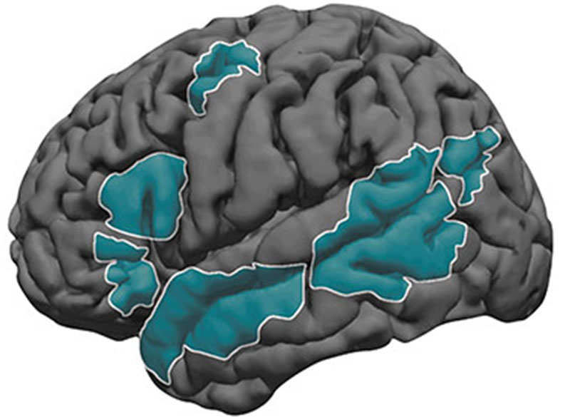
The song-specific hotspot that they found is located at the top of the temporal lobe, near regions that are selective for language and music. That location suggests that the song-specific population may be responding to features such as the perceived pitch, or the interaction between words and perceived pitch, before sending information to other parts of the brain for further processing, the researchers say.

The researchers now hope to learn more about what aspects of singing drive the responses of these neurons. They are also working with MIT Professor Rebecca Saxe’s lab to study whether infants have music-selective areas, in hopes of learning more about when and how these brain regions develop.

The research was funded by the National Institutes of Health, the U.S. Army Research Office, the National Science Foundation, the NSF Science and Technology Center for Brains, Minds, and Machines, the Fondazione Neurone, the Howard Hughes Medical Institute, and the Kristin R. Pressman and Jessica J. Pourian ’13 Fund at MIT.

National Academy of Sciences honors cognitive neuroscientist Nancy Kanwisher

MIT neuroscientist and McGovern Investigator Nancy Kanwisher. Photo: Jussi Puikkonen/KNAW

The National Academy of Sciences (NAS) has announced today that Nancy Kanwisher, the Walter A. Rosenblith Professor of Cognitive Neuroscience in MIT’s Department of Brain and Cognitive Sciences, has received the 2022 NAS Award in the Neurosciences for her “pioneering research into the functional organization of the human brain.” The $25,000 prize, established by the Fidia Research Foundation, is presented every three years to recognize “extraordinary contributions to the neuroscience fields.”

“I am deeply honored to receive this award from the NAS,” says Kanwisher, who is also an investigator in MIT’s McGovern Institute and a member of the Center for Brains, Minds and Machines. “It has been a profound privilege, and a total blast, to watch the human brain in action as these data began to reveal an initial picture of the organization of the human mind. But the biggest joy has been the opportunity to work with the incredible group of talented young scientists who actually did the work that this award recognizes.”

A window into the mind

Kanwisher is best known for her landmark insights into how humans recognize and process faces. Psychology had long-suggested that recognizing a face might be distinct from general object recognition. But Kanwisher galvanized the field in 1997 with her seminal discovery that the human brain contains a small region specialized to respond only to faces. The region, which Kanwisher termed the fusiform face area (FFA), became activated when subjects viewed images of faces in an MRI scanner, but not when they looked at scrambled faces or control stimuli.

Since her 1997 discovery (now the most highly cited manuscript in its area), Kanwisher and her students have applied similar methods to find brain specializations for the recognition of scenes, the mental states of others, language, and music. Taken together, her research provides a compelling glimpse into the architecture of the brain, and, ultimately, what makes us human.

“Nancy’s work over the past two decades has argued that many aspects of human cognition are supported by specialized neural circuitry, a conclusion that stands in contrast to our subjective sense of a singular mental experience,” says McGovern Institute Director Robert Desimone. “She has made profound contributions to the psychological and cognitive sciences and I am delighted that the National Academy of Sciences has recognized her outstanding achievements.”

One-in-a-million mentor

Beyond the lab, Kanwisher has a reputation as a tireless communicator and mentor who is actively engaged in the policy implications of brain research. The statistics speak for themselves: her 2014 TED talk, “A Neural portrait of the human mind” has been viewed over a million times online and her introductory MIT OCW course on the human brain has generated more than nine million views on YouTube.

Nancy Kanwisher works with researchers from her lab in MIT’s Martinos Imaging Center. Photo: Kris Brewer

Kanwisher also has an exceptional track record in training women scientists who have gone on to successful independent research careers, in many cases becoming prominent figures in their own right.

“Nancy is the one-in-a-million mentor, who is always skeptical of your ideas and your arguments, but immensely confident of your worth,” says Rebecca Saxe, John W. Jarve (1978) Professor of Brain and Cognitive Sciences, investigator at the McGovern Institute, and associate dean of MIT’s School of Science. Saxe was a graduate student in Kanwisher’s lab where she earned her PhD in cognitive neuroscience in 2003. “She has such authentic curiosity,” Saxe adds. “It’s infectious and sustaining. Working with Nancy was a constant reminder of why I wanted to be a scientist.”

The NAS will present Kanwisher with the award during its annual meeting on May 1, 2022 in Washington, DC. The event will be webcast live. Kanwisher plans to direct her prize funds to the non-profit organization Malengo, established by a former student and which provides quality undergraduate education to individuals who would otherwise not be able to afford it.

Babies can tell who has close relationships based on one clue: saliva

Learning to navigate social relationships is a skill that is critical for surviving in human societies. For babies and young children, that means learning who they can count on to take care of them.

MIT neuroscientists have now identified a specific signal that young children and even babies use to determine whether two people have a strong relationship and a mutual obligation to help each other: whether those two people kiss, share food, or have other interactions that involve sharing saliva.

In a new study, the researchers showed that babies expect people who share saliva to come to one another’s aid when one person is in distress, much more so than when people share toys or interact in other ways that do not involve saliva exchange. The findings suggest that babies can use these cues to try to figure out who around them is most likely to offer help, the researchers say.

“Babies don’t know in advance which relationships are the close and morally obligating ones, so they have to have some way of learning this by looking at what happens around them,” says Rebecca Saxe, the John W. Jarve Professor of Brain and Cognitive Sciences, a member of MIT’s McGovern Institute for Brain Research, and the senior author of the new study.

MIT postdoc Ashley Thomas is the lead author of the study, which appears today in Science. Brandon Woo, a Harvard University graduate student; Daniel Nettle, a professor of behavioral science at Newcastle University; and Elizabeth Spelke, a professor of psychology at Harvard, are also authors of the paper.

Sharing saliva

In human societies, people typically distinguish between “thick” and “thin” relationships. Thick relationships, usually found between family members, feature strong levels of attachment, obligation, and mutual responsiveness. Anthropologists have also observed that people in thick relationships are more willing to share bodily fluids such as saliva.

“That inspired both the question of whether infants distinguish between those types of relationships, and whether saliva sharing might be a really good cue they could use to recognize them,” Thomas says.

To study those questions, the researchers observed toddlers (16.5 to 18.5 months) and babies (8.5 to 10 months) as they watched interactions between human actors and puppets. In the first set of experiments, a puppet shared an orange with one actor, then tossed a ball back and forth with a different actor.

After the children watched these initial interactions, the researchers observed the children’s reactions when the puppet showed distress while sitting between the two actors. Based on an earlier study of nonhuman primates, the researchers hypothesized that babies would look first at the person whom they expected to help. That study showed that when baby monkeys cry, other members of the troop look to the baby’s parents, as if expecting them to step in.

The MIT team found that the children were more likely to look toward the actor who had shared food with the puppet, not the one who had shared a toy, when the puppet was in distress.

In a second set of experiments, designed to focus more specifically on saliva, the actor either placed her finger in her mouth and then into the mouth of the puppet, or placed her finger on her forehead and then onto the forehead of the puppet. Later, when the actor expressed distress while standing between the two puppets, children watching the video were more likely to look toward the puppet with whom she had shared saliva.

Social cues

The findings suggest that saliva sharing is likely an important cue that helps infants to learn about their own social relationships and those of people around them, the researchers say.

“The general skill of learning about social relationships is very useful,” Thomas says. “One reason why this distinction between thick and thin might be important for infants in particular, especially human infants, who depend on adults for longer than many other species, is that it might be a good way to figure out who else can provide the support that they depend on to survive.”

The researchers did their first set of studies shortly before Covid-19 lockdowns began, with babies who came to the lab with their families. Later experiments were done over Zoom. The results that the researchers saw were similar before and after the pandemic, confirming that pandemic-related hygiene concerns did not affect the outcome.

“We actually know the results would have been similar if it hadn’t been for the pandemic,” Saxe says. “You might wonder, did kids start to think very differently about sharing saliva when suddenly everybody was talking about hygiene all the time? So, for that question, it’s very useful that we had an initial data set collected before the pandemic.”

Doing the second set of studies on Zoom also allowed the researchers to recruit a much more diverse group of children because the subjects were not limited to families who could come to the lab in Cambridge during normal working hours.

In future work, the researchers hope to perform similar studies with infants in cultures that have different types of family structures. In adult subjects, they plan to use functional magnetic resonance imaging (fMRI) to study what parts of the brain are involved in making saliva-based assessments about social relationships.

The research was funded by the National Institutes of Health; the Patrick J. McGovern Foundation; the Guggenheim Foundation; a Social Sciences and Humanities Research Council Doctoral Fellowship; MIT’s Center for Brains, Minds, and Machines; and the Siegel Foundation.

The craving state

This story originally appeared in the Winter 2022 issue of BrainScan.

***

For people struggling with substance use disorders — and there are about 35 million of them worldwide — treatment options are limited. Even among those who seek help, relapse is common. In the United States, an epidemic of opioid addiction has been declared a public health emergency.

A 2019 survey found that 1.6 million people nationwide had an opioid use disorder, and the crisis has surged since the start of the COVID-19 pandemic. The Centers for Disease Control and Prevention estimates that more than 100,000 people died of drug overdose between April 2020 and April 2021 — nearly 30 percent more overdose deaths than occurred during the same period the previous year.

In the United States, an epidemic of opioid addiction has been declared a public health emergency.

A deeper understanding of what addiction does to the brain and body is urgently needed to pave the way to interventions that reliably release affected individuals from its grip. At the McGovern Institute, researchers are turning their attention to addiction’s driving force: the deep, recurring craving that makes people prioritize drug use over all other wants and needs.

McGovern Institute co-founder, Lore Harp McGovern.

“When you are in that state, then it seems nothing else matters,” says McGovern Investigator Fan Wang. “At that moment, you can discard everything: your relationship, your house, your job, everything. You only want the drug.”

With a new addiction initiative catalyzed by generous gifts from Institute co-founder Lore Harp McGovern and others, McGovern scientists with diverse expertise have come together to begin clarifying the neurobiology that underlies the craving state. They plan to dissect the neural transformations associated with craving at every level — from the drug-induced chemical changes that alter neuronal connections and activity to how these modifications impact signaling brain-wide. Ultimately, the McGovern team hopes not just to understand the craving state, but to find a way to relieve it — for good.

“If we can understand the craving state and correct it, or at least relieve a little bit of the pressure,” explains Wang, who will help lead the addiction initiative, “then maybe we can at least give people a chance to use their top-down control to not take the drug.”

The craving cycle

For individuals suffering from substance use disorders, craving fuels a cyclical pattern of escalating drug use. Following the euphoria induced by a drug like heroin or cocaine, depression sets in, accompanied by a drug craving motivated by the desire to relieve that suffering. And as addiction progresses, the peaks and valleys of this cycle dip lower: the pleasant feelings evoked by the drug become weaker, while the negative effects a person experiences in its absence worsen. The craving remains, and increasing use of the drug are required to relieve it.

By the time addiction sets in, the brain has been altered in ways that go beyond a drug’s immediate effects on neural signaling.

These insidious changes leave individuals susceptible to craving — and the vulnerable state endures. Long after the physical effects of withdrawal have subsided, people with substance use disorders can find their craving returns, triggered by exposure to a small amount of the drug, physical or social cues associated with previous drug use, or stress. So researchers will need to determine not only how different parts of the brain interact with one another during craving and how individual cells and the molecules within them are affected by the craving state — but also how things change as addiction develops and progresses.

Circuits, chemistry and connectivity

One clear starting point is the circuitry the brain uses to control motivation. Thanks in part to decades of research in the lab of McGovern Investigator Ann Graybiel, neuroscientists know a great deal about how these circuits learn which actions lead to pleasure and which lead to pain, and how they use that information to establish habits and evaluate the costs and benefits of complex decisions.

Graybiel’s work has shown that drugs of abuse strongly activate dopamine-responsive neurons in a part of the brain called the striatum, whose signals promote habit formation. By increasing the amount of dopamine that neurons release, these drugs motivate users to prioritize repeated drug use over other kinds of rewards, and to choose the drug in spite of pain or other negative effects. Her group continues to investigate the naturally occurring molecules that control these circuits, as well as how they are hijacked by drugs of abuse.

Distribution of opioid receptors targeted by morphine (shown in blue) in two regions in the dorsal striatum and nucleus accumbens of the mouse brain. Image: Ann Graybiel

In Fan Wang’s lab, work investigating the neural circuits that mediate the perception of physical pain has led her team to question the role of emotional pain in craving. As they investigated the source of pain sensations in the brain, they identified neurons in an emotion-regulating center called the central amygdala that appear to suppress physical pain in animals. Now, Wang wants to know whether it might be possible to modulate neurons involved in emotional pain to ameliorate the negative state that provokes drug craving.

These animal studies will be key to identifying the cellular and molecular changes that set the brain up for recurring cravings. And as McGovern scientists begin to investigate what happens in the brains of rodents that have been trained to self-administer addictive drugs like fentanyl or cocaine, they expect to encounter tremendous complexity.

McGovern Associate Investigator Polina Anikeeva, whose lab has pioneered new technologies that will help the team investigate the full spectrum of changes that underlie craving, says it will be important to consider impacts on the brain’s chemistry, firing patterns, and connectivity. To that end, multifunctional research probes developed in her lab will be critical to monitoring and manipulating neural circuits in animal models.

Imaging technology developed by investigator Ed Boyden will also enable nanoscale protein visualization brain-wide. An important goal will be to identify a neural signature of the craving state. With such a signal, researchers can begin to explore how to shut off that craving — possibly by directly modulating neural signaling.

Targeted treatments

“One of the reasons to study craving is because it’s a natural treatment point,” says McGovern Associate Investigator Alan Jasanoff. “And the dominant kind of approaches that people in our team think about are approaches that relate to neural circuits — to the specific connections between brain regions and how those could be changed.” The hope, he explains, is that it might be possible to identify a brain region whose activity is disrupted during the craving state, then use clinical brain stimulation methods to restore normal signaling — within that region, as well as in other connected parts of the brain.

To identify the right targets for such a treatment, it will be crucial to understand how the biology uncovered in laboratory animals reflects what’s happens in people with substance use disorders. Functional imaging in John Gabrieli’s lab can help bridge the gap between clinical and animal research by revealing patterns of brain activity associated with the craving state in both humans and rodents. A new technique developed in Jasanoff’s lab makes it possible to focus on the activity between specific regions of an animal’s brain. “By doing that, we hope to build up integrated models of how information passes around the brain in craving states, and of course also in control states where we’re not experiencing craving,” he explains.

In delving into the biology of the craving state, McGovern scientists are embarking on largely unexplored territory — and they do so with both optimism and urgency. “It’s hard to not appreciate just the size of the problem, and just how devastating addiction is,” says Anikeeva. “At this point, it just seems almost irresponsible to not work on it, especially when we do have the tools and we are interested in the general brain regions that are important for that problem. I would say that there’s almost a civic duty.”

Jacqueline Lees and Rebecca Saxe named associate deans of science

Jaqueline Lees and Rebecca Saxe have been named associate deans serving in the MIT School of Science. Lees is the Virginia and D.K. Ludwig Professor for Cancer Research and is currently the associate director of the Koch Institute for Integrative Cancer Research, as well as an associate department head and professor in the Department of Biology at MIT. Saxe is the John W. Jarve (1978) Professor in Brain and Cognitive Sciences and the associate head of the Department of Brain and Cognitive Sciences (BCS); she is also an associate investigator in the McGovern Institute for Brain Research.

Lees and Saxe will both contribute to the school’s diversity, equity, inclusion, and justice (DEIJ) activities, as well as develop and implement mentoring and other career-development programs to support the community. From their home departments, Saxe and Lees bring years of DEIJ and mentorship experience to bear on the expansion of school-level initiatives.

Lees currently serves on the dean’s science council in her capacity as associate director of the Koch Institute. In this new role as associate dean for the School of Science, she will bring her broad administrative and programmatic experiences to bear on the next phase for DEIJ and mentoring activities.

Lees joined MIT in 1994 as a faculty member in MIT’s Koch Institute (then the Center for Cancer Research) and Department of Biology. Her research focuses on regulators that control cellular proliferation, terminal differentiation, and stemness — functions that are frequently deregulated in tumor cells. She dissects the role of these proteins in normal cell biology and development, and establish how their deregulation contributes to tumor development and metastasis.

Since 2000, she has served on the Department of Biology’s graduate program committee, and played a major role in expanding the diversity of the graduate student population. Lees also serves on DEIJ committees in her home department, as well as at the Koch Institute.

With co-chair with Boleslaw Wyslouch, director of the Laboratory for Nuclear Science, Lees led the ReseArch Scientist CAreer LadderS (RASCALS) committee tasked to evaluate career trajectories for research staff in the School of Science and make recommendations to recruit and retain talented staff, rewarding them for their contributions to the school’s research enterprise.

“Jackie is a powerhouse in translational research, demonstrating how fundamental work at the lab bench is critical for making progress at the patient bedside,” says Nergis Mavalvala, dean of the School of Science. “With Jackie’s dedicated and thoughtful partnership, we can continue to lead in basic research and develop the recruitment, retention, and mentoring and necessary to support our community.”

Saxe will join Lees in supporting and developing programming across the school that could also provide direction more broadly at the Institute.

“Rebecca is an outstanding researcher in social cognition and a dedicated educator — someone who wants our students not only to learn, but to thrive,” says Mavalvala. “I am grateful that Rebecca will join the dean’s leadership team and bring her mentorship and leadership skills to enhance the school.”

For example, in collaboration with former department head James DiCarlo, the BCS department has focused on faculty mentorship of graduate students; and, in collaboration with Professor Mark Bear, the department developed postdoc salary and benefit standards. Both initiatives have become models at MIT.

With colleague Laura Schulz, Saxe also served as co-chair of the Committee on Medical Leave and Hospitalizations (CMLH), which outlined ways to enhance MIT’s current leave and hospitalization procedures and policies for undergraduate and graduate students. Saxe was also awarded MIT’s Committed to Caring award for excellence in graduate student mentorship, as well as the School of Science’s award for excellence in undergraduate teaching.

In her research, Saxe studies human social cognition, using a combination of behavioral testing and brain imaging technologies. She is best known for her work on brain regions specialized for abstract concepts, such as “theory of mind” tasks that involve understanding the mental states of other people. Her TED Talk, “How we read each other’s minds” has been viewed more than 3 million times. She also studies the development of the human brain during early infancy.

She obtained her PhD from MIT and was a Harvard University junior fellow before joining the MIT faculty in 2006. In 2014, the National Academy of Sciences named her one of two recipients of the Troland Award for investigators age 40 or younger “to recognize unusual achievement and further empirical research in psychology regarding the relationships of consciousness and the physical world.” In 2020, Saxe was named a John Simon Guggenheim Foundation Fellow.

Saxe and Lees will also work closely with Kuheli Dutt, newly hired assistant dean for diversity, equity, and inclusion, and other members of the dean’s science council on school-level initiatives and strategy.

“I’m so grateful that Rebecca and Jackie have agreed to take on these new roles,” Mavalvala says. “And I’m super excited to work with these outstanding thought partners as we tackle the many puzzles that I come across as dean.”

Investigating the embattled brain

Omar Rutledge served as a US Army infantryman in the 1st Armored and 25th Infantry Divisions. He was deployed in support of Operation Iraqi Freedom from March 2003 to July 2004. Photo: Omar Rutledge

As an Iraq war veteran, Omar Rutledge is deeply familiar with post-traumatic stress – recurring thoughts and memories that persist long after a danger has passed – and he knows that a brain altered by trauma is not easily fixed. But as a graduate student in the Department of Brain and Cognitive Sciences, Rutledge is determined to change that. He wants to understand exactly how trauma alters the brain – and whether the tools of neuroscience can be used to help fellow veterans with post-traumatic stress disorder (PTSD) heal from their experiences.

“In the world of PTSD research, I look to my left and to my right, and I don’t see other veterans, certainly not former infantrymen,” says Rutledge, who served in the US Army and was deployed to Iraq from March 2003 to July 2004. “If there are so few of us in this space, I feel like I have an obligation to make a difference for all who suffer from the traumatic experiences of war.”

Rutledge is uniquely positioned to make such a difference in the lab of McGovern Investigator John Gabrieli, where researchers use technologies like magnetic resonance imaging (MRI), electroencephalography (EEG), and magnetoencephalography (MEG) to peer into the human brain and explore how it powers our thoughts, memories, and emotions. Rutledge is studying how PTSD weakens the connection between the amygdala, which is responsible for emotions like fear, and the prefrontal cortex, which regulates or controls these emotional responses. He hopes these studies will eventually lead to the development of wearable technologies that can retrain the brain to be less responsive to triggering events.

“I feel like it has been a mission of mine to do this kind of work.”

Though Covid-19 has unexpectedly paused some aspects of his research, Rutledge is pursuing another line of research inspired both by the mandatory social distancing protocols imposed during the lockdown and his own experiences with social isolation. Does chronic social isolation cause physical or chemical changes in the brain similar to those seen in PTSD? And does loneliness exacerbate symptoms of PTSD?

“There’s this hypervigilance that occurs in loneliness, and there’s also something very similar that occurs in PTSD — a heightened awareness of potential threats,” says Rutledge, who is the recipient of Michael Ferrara Graduate Fellowship provided by the Poitras Center, a fellowship made possible by the many friends and family of Michael Ferrara. “The combination of the two may lead to more adverse reactions in people with PTSD.”

In the future, Rutledge hopes to explore whether chronic loneliness impairs reasoning and logic skills and has a deeper impact on veterans who have PTSD.

Although his research tends to resurface painful memories of his own combat experiences, Rutledge says if it can help other veterans heal, it’s worth it.  “In the process, it makes me a little bit stronger as well,” he adds.

Individual neurons responsible for complex social reasoning in humans identified

This story is adapted from a January 27, 2021 press release from Massachusetts General Hospital.

The ability to understand others’ hidden thoughts and beliefs is an essential component of human social behavior. Now, neuroscientists have for the first time identified specific neurons critical for social reasoning, a cognitive process that requires individuals to acknowledge and predict others’ hidden beliefs and thoughts.

The findings, published in Nature, open new avenues of study into disorders that affect social behavior, according to the authors.

In the study, a team of Harvard Medical School investigators based at Massachusetts General Hospital and colleagues from MIT took a rare look at how individual neurons represent the beliefs of others. They did so by recording neuron activity in patients undergoing neurosurgery to alleviate symptoms of motor disorders such as Parkinson’s disease.

Theory of mind

The researcher team, which included McGovern scientists Ev Fedorenko and Rebecca Saxe, focused on a complex social cognitive process called “theory of mind.” To illustrate this, let’s say a friend appears to be sad on her birthday. One may infer she is sad because she didn’t get a present or she is upset at growing older.

“When we interact, we must be able to form predictions about another person’s unstated intentions and thoughts,” said senior author Ziv Williams, HMS associate professor of neurosurgery at Mass General. “This ability requires us to paint a mental picture of someone’s beliefs, which involves acknowledging that those beliefs may be different from our own and assessing whether they are true or false.”

This social reasoning process develops during early childhood and is fundamental to successful social behavior. Individuals with autism, schizophrenia, bipolar affective disorder, and traumatic brain injuries are believed to have a deficit of theory-of-mind ability.

For the study, 15 patients agreed to perform brief behavioral tasks before undergoing neurosurgery for placement of deep-brain stimulation for motor disorders. Microelectrodes inserted into the dorsomedial prefrontal cortex recorded the behavior of individual neurons as patients listened to short narratives and answered questions about them.

For example, participants were presented with the following scenario to evaluate how they considered another’s belief of reality: “You and Tom see a jar on the table. After Tom leaves, you move the jar to a cabinet. Where does Tom believe the jar to be?”

Social computation

The participants had to make inferences about another’s beliefs after hearing each story. The experiment did not change the planned surgical approach or alter clinical care.

“Our study provides evidence to support theory of mind by individual neurons,” said study first author Mohsen Jamali, HMS instructor in neurosurgery at Mass General. “Until now, it wasn’t clear whether or how neurons were able to perform these social cognitive computations.”

The investigators found that some neurons are specialized and respond only when assessing another’s belief as false, for example. Other neurons encode information to distinguish one person’s beliefs from another’s. Still other neurons create a representation of a specific item, such as a cup or food item, mentioned in the story. Some neurons may multitask and aren’t dedicated solely to social reasoning.

“Each neuron is encoding different bits of information,” Jamali said. “By combining the computations of all the neurons, you get a very detailed representation of the contents of another’s beliefs and an accurate prediction of whether they are true or false.”

Now that scientists understand the basic cellular mechanism that underlies human theory of mind, they have an operational framework to begin investigating disorders in which social behavior is affected, according to Williams.

“Understanding social reasoning is also important to many different fields, such as child development, economics, and sociology, and could help in the development of more effective treatments for conditions such as autism spectrum disorder,” Williams said.

Previous research on the cognitive processes that underlie theory of mind has involved functional MRI studies, where scientists watch which parts of the brain are active as volunteers perform cognitive tasks.

But the imaging studies capture the activity of many thousands of neurons all at once. In contrast, Williams and colleagues recorded the computations of individual neurons. This provided a detailed picture of how neurons encode social information.

“Individual neurons, even within a small area of the brain, are doing very different things, not all of which are involved in social reasoning,” Williams said. “Without delving into the computations of single cells, it’s very hard to build an understanding of the complex cognitive processes underlying human social behavior and how they go awry in mental disorders.”

Adapted from a Mass General news release.

The pursuit of reward

View the interactive version of this story in our Spring 2021 issue of BrainScan.

The brain circuits that influence our decisions, cognitive functions, and ultimately, our actions are intimately connected with the circuits that give rise to our motivations. By exploring these relationships, scientists at McGovern are seeking knowledge that might suggest new strategies for changing our habits or treating motivation-disrupting conditions such as depression and addiction.

Risky decisions

MIT Institute Professor Ann Graybiel. Photo: Justin Knight

In Ann Graybiel’s lab, researchers have been examining how the brain makes choices that carry both positive and negative consequences — deciding to take on a higher-paying but more demanding job, for example. Psychologists call these dilemmas approach-avoidance conflicts, and resolving them not only requires weighing the good versus the bad, but also motivation to engage with the decision.

Emily Hueske, a research scientist in the Graybiel lab, explains that everyone has their own risk tolerance when it comes to such decisions, and certain psychiatric conditions, including depression and anxiety disorders, can shift the tipping point at which a person chooses to “approach” or “avoid.”

Studies have shown that neurons in the striatum (see image below), a region deep in the brain involved in both motivation and movement, activate as we grapple with these decisions. Graybiel traced this activity even further, to tiny compartments within the striatum called striosomes.

(She discovered striosomes many years ago and has been studying their function for decades.)

A motivational switch

In 2015, Graybiel’s team manipulated striosome signaling within genetically engineered mice and changed the way animals behave in approach-avoidance conflict situations. Taking cues from an assessment used to evaluate approach-avoidance behavior in patients, they presented mice with opportunities to obtain chocolate while experiencing unwelcome exposure in a brightly lit area.

Experimentally activating neurons in striosomes had a dramatic effect, causing mice to venture into brightly lit areas that they would normally avoid. With striosomal circuits switched on, “this animal all of a sudden is like a different creature,” Graybiel says.

Two years later, they found that chronic stress and other factors can also disrupt this signaling and change the choices animals make.

An image of the mouse striatum showing clusters of striosomes (red and yellow). Image: Graybiel lab

Age of ennui

This November, Alexander Friedman, who worked as a research scientist in the Graybiel lab, and Hueske reported in Cell that they found an age-related decline in motivation-modulated learning in mice and rats. Neurons within striosomes became more active than the cells that surround them as animals learned to assign positive and negative values to potential choices. And older mice were less engaged than their younger counterparts in the type of learning required to make these cost-benefit analyses. A similar lack of motivation was observed in a mouse model of Huntington’s disease, a neurodegenerative disorder that is often associated with mood
disturbances in patients.

“This coincides with our previous findings that striosomes are critically important for decisions that involve a conflict.”

“This coincides with our previous findings that striosomes are critically important for decisions that involve a conflict,” says Friedman, who is now an assistant professor at the University of Texas at El Paso.

Graybiel’s team is continuing to investigate these uniquely positioned compartments in the brain, expecting to shed light on the mechanisms that underlie both learning and motivation.

“There’s no learning without motivation, and in fact, motivation can be influenced by learning,” Hueske says. “The more you learn, the more excited you might be to engage in the task. So the two are intertwined.”

The aging brain

Researchers in John Gabrieli’s lab are also seeking to understand the circuits that link motivation to learning, and recently, his team reported that they, too, had found an age-related decline in motivation-modulated learning.

Studies in young adults have shown that memory improves when the brain circuits that process motivation and memory interact. Gabrieli and neurologist Maiya Geddes, who worked in Gabrieli’s lab as a postdoctoral fellow, wondered whether this holds true in older adults, particularly as memory declines.

To find out, the team recruited 40 people to participate in a brain imaging study. About half of the participants were between the ages of 18 and 30, while the others were between the ages of 49 and 84. While inside an fMRI scanner, each participant was asked to commit certain words to memory and told their success would determine how much money they received for participating in the experiment.

Diminished drive

MRI scan
Younger adults show greater activation in the reward-related regions of the brain during incentivized memory tasks compared to older adults. Image: Maiya Geddes

Not surprisingly, when participants were asked 24 hours later to recall the words, the younger group performed better overall than the older group. In young people, incentivized memory tasks triggered activity in parts of the brain involved in both memory and motivation. But in older adults, while these two parts of the brain could be activated independently, they did not seem to be communicating with one another.

“It seemed that the older adults, at least in terms of their brain response, did care about the kind of incentives that we were offering,” says Geddes, who is now an assistant professor at McGill University. “But for whatever reason, that wasn’t allowing them to benefit in terms of improved memory performance.”

Since the study indicates the brain still can anticipate potential rewards, Geddes is now exploring whether other sources of motivation, such as social rewards, might more effectively increase healthful decisions and behaviors in older adults.

Circuit control

Understanding how the brain generates and responds to motivation is not only important for improving learning strategies. Lifestyle choices such as exercise and social engagement can help people preserve cognitive function and improve their quality of life as they age, and Gabrieli says activating the right motivational circuits could help encourage people to implement healthy changes.

By pinpointing these motivational circuits in mice, Graybiel hopes that her research will lead to better treatment strategies for people struggling with motivational challenges, including Parkinson’s disease. Her team is now exploring whether striosomes serve as part of a value-sensitive switch, linking our intentions to dopamine-containing neurons in the midbrain that can modulate our actions.

“Perhaps this motivation is critical for the conflict resolution, and striosomes combine two worlds, dopaminergic motivation and cortical knowledge, resulting in motivation to learn,” Friedman says.

“Now we know that these challenges have a biological basis, and that there are neural circuits that can promote or reduce our feeling of motivational energy,” explains Graybiel. “This realization in itself is a major step toward learning how we can control these circuits both behaviorally and by highly selective therapeutic targeting.”

Stars, brains, and enzymes: a celebration of MIT science

“Our topic tonight, science and discovery, lives at the heart of MIT.” In his welcoming remarks for the first virtual MIT Better World gathering, W. Eric L. Grimson, MIT chancellor for academic advancement, detailed some of the ways MIT excels as a hub of scientific research and innovation. “Institute researchers are plumbing the secrets of the universe; modeling climate at a local, regional, and global scale; striving to understand how brains and bodies give rise to cognition and mind; and racing to find treatments and cures for diseases ranging from the acute, like Covid-19, to the chronic, like cancers and maladies of the aging brain,” said Grimson, who is also the Bernard M. Gordon Professor of Medical Engineering.

Members of the MIT community from around the globe were invited to attend the MIT Better World (Science) event, held online in November, to hear from Institute leaders, faculty, students, and alumni about the pursuit of scientific knowledge. Alumni in more than 80 countries registered to attend, and the evening put a special emphasis on Canada, which is home to a group of alumni and friends who served as virtual hosts, and to which Grimson and all of the opening session speakers captured in the video above have personal ties.

Grimson’s remarks were followed by presentations from the new dean of the MIT School of Science, Nergis Mavalvala; as well as Rebecca Saxe, the John W. Jarve (1978) Professor in Brain and Cognitive Sciences and associate investigator at the McGovern Institute for Brain Research; and microbiology PhD student Linda Zhong-Johnson.

Mavalvala, the Curtis (1963) and Kathleen Marble Professor of Astrophysics, described how she and colleagues have worked to improve the sensitivity of instruments used to detect gravitational waves through LIGO—the landmark research endeavor that has revealed, among other recent discoveries, that colliding neutron stars are the “factories” in which heavy elements like gold and platinum are manufactured. Having begun the role of School of Science dean this fall, Mavalvala now takes joy in enabling discoveries across the MIT community, including those focused on our own corner of the universe. “It’s a vast world out there, and for us to make a better world, we must first understand that world. At MIT, that’s just what we do.”

Saxe, who uses brain imaging to study human social cognition, described prescient experiments on social isolation conducted by her lab between 2017 and 2019. “Sometimes we do science just out of curiosity,” said Saxe as she explained why she, former postdoc Livia Tomova, and fellow researchers pursued a project with uncertain applications — only to find themselves writing what Saxe now calls “the most timely and relevant paper in my life” in March, just as the Covid-19 pandemic triggered widespread isolation measures.

The third speaker, Linda Zhong-Johnson, discussed her PhD research in the labs of Anthony J. Sinskey, professor of biology, and Christopher A. Voigt, the Daniel I.C. Wang Professor of Advanced Biotechnology. Her goal is to reduce the amount of plastic in landfills and oceans by studying enzymes that could digest polyethylene terephthalate, or PET, the plastic used to make most water bottles. “We’re getting closer to the answer,” she said. “I’m grateful to be at MIT, where we have the mandate and resources to keep exploring.”

More virtual MIT Better World events on the topics of health and sustainability are planned for this coming February and March. Meanwhile, watch the full session (above) and a range of breakout sessions on topics such as the politics of molecular medicine and the Mars 2020 mission, and learn more about the MIT Campaign for a Better World at betterworld.mit.edu.

A hunger for social contact

Since the coronavirus pandemic began in the spring, many people have only seen their close friends and loved ones during video calls, if at all. A new study from MIT finds that the longings we feel during this kind of social isolation share a neural basis with the food cravings we feel when hungry.

The researchers found that after one day of total isolation, the sight of people having fun together activates the same brain region that lights up when someone who hasn’t eaten all day sees a picture of a plate of cheesy pasta.

“People who are forced to be isolated crave social interactions similarly to the way a hungry person craves food.”

“Our finding fits the intuitive idea that positive social interactions are a basic human need, and acute loneliness is an aversive state that motivates people to repair what is lacking, similar to hunger,” says Rebecca Saxe, the John W. Jarve Professor of Brain and Cognitive Sciences at MIT, a member of MIT’s McGovern Institute for Brain Research, and the senior author of the study.

The research team collected the data for this study in 2018 and 2019, long before the coronavirus pandemic and resulting lockdowns. Their new findings, described today in Nature Neuroscience, are part of a larger research program focusing on how social stress affects people’s behavior and motivation.

Former MIT postdoc Livia Tomova, who is now a research associate at Cambridge University, is the lead author of the paper. Other authors include Kimberly Wang, a McGovern Institute research associate; Todd Thompson, a McGovern Institute scientist; Atsushi Takahashi, assistant director of the Martinos Imaging Center; Gillian Matthews, a research scientist at the Salk Institute for Biological Studies; and Kay Tye, a professor at the Salk Institute.

Social craving

The new study was partly inspired by a recent paper from Tye, a former member of MIT’s Picower Institute for Learning and Memory. In that 2016 study, she and Matthews, then an MIT postdoc, identified a cluster of neurons in the brains of mice that represent feelings of loneliness and generate a drive for social interaction following isolation. Studies in humans have shown that being deprived of social contact can lead to emotional distress, but the neurological basis of these feelings is not well-known.

“We wanted to see if we could experimentally induce a certain kind of social stress, where we would have control over what the social stress was,” Saxe says. “It’s a stronger intervention of social isolation than anyone had tried before.”

To create that isolation environment, the researchers enlisted healthy volunteers, who were mainly college students, and confined them to a windowless room on MIT’s campus for 10 hours. They were not allowed to use their phones, but the room did have a computer that they could use to contact the researchers if necessary.

“There were a whole bunch of interventions we used to make sure that it would really feel strange and different and isolated,” Saxe says. “They had to let us know when they were going to the bathroom so we could make sure it was empty. We delivered food to the door and then texted them when it was there so they could go get it. They really were not allowed to see people.”

After the 10-hour isolation ended, each participant was scanned in an MRI machine. This posed additional challenges, as the researchers wanted to avoid any social contact during the scanning. Before the isolation period began, each subject was trained on how to get into the machine, so that they could do it by themselves, without any help from the researcher.

“Normally, getting somebody into an MRI machine is actually a really social process. We engage in all kinds of social interactions to make sure people understand what we’re asking them, that they feel safe, that they know we’re there,” Saxe says. “In this case, the subjects had to do it all by themselves, while the researcher, who was gowned and masked, just stood silently by and watched.”

Each of the 40 participants also underwent 10 hours of fasting, on a different day. After the 10-hour period of isolation or fasting, the participants were scanned while looking at images of food, images of people interacting, and neutral images such as flowers. The researchers focused on a part of the brain called the substantia nigra, a tiny structure located in the midbrain, which has previously been linked with hunger cravings and drug cravings. The substantia nigra is also believed to share evolutionary origins with a brain region in mice called the dorsal raphe nucleus, which is the area that Tye’s lab showed was active following social isolation in their 2016 study.

The researchers hypothesized that when socially isolated subjects saw photos of people enjoying social interactions, the “craving signal” in their substantia nigra would be similar to the signal produced when they saw pictures of food after fasting. This was indeed the case. Furthermore, the amount of activation in the substantia nigra was correlated with how strongly the patients rated their feelings of craving either food or social interaction.

Degrees of loneliness

The researchers also found that people’s responses to isolation varied depending on their normal levels of loneliness. People who reported feeling chronically isolated months before the study was done showed weaker cravings for social interaction after the 10-hour isolation period than people who reported a richer social life.

“For people who reported that their lives were really full of satisfying social interactions, this intervention had a bigger effect on their brains and on their self-reports,” Saxe says.

The researchers also looked at activation patterns in other parts of the brain, including the striatum and the cortex, and found that hunger and isolation each activated distinct areas of those regions. That suggests that those areas are more specialized to respond to different types of longings, while the substantia nigra produces a more general signal representing a variety of cravings.

Now that the researchers have established that they can observe the effects of social isolation on brain activity, Saxe says they can now try to answer many additional questions. Those questions include how social isolation affect people’s behavior, whether virtual social contacts such as video calls help to alleviate cravings for social interaction, and how isolation affects different age groups.

The researchers also hope to study whether the brain responses that they saw in this study could be used to predict how the same participants responded to being isolated during the lockdowns imposed during the early stages of the coronavirus pandemic.

The research was funded by a SFARI Explorer Grant from the Simons Foundation, a MINT grant from the McGovern Institute, the National Institutes of Health, including an NIH Pioneer Award, a Max Kade Foundation Fellowship, and an Erwin Schroedinger Fellowship from the Austrian Science Fund.