Face-specific brain area responds to faces even in people born blind

More than 20 years ago, neuroscientist Nancy Kanwisher and others discovered that a small section of the brain located near the base of the skull responds much more strongly to faces than to other objects we see. This area, known as the fusiform face area, is believed to be specialized for identifying faces.

Now, in a surprising new finding, Kanwisher and her colleagues have shown that this same region also becomes active in people who have been blind since birth, when they touch a three-dimensional model of a face with their hands. The finding suggests that this area does not require visual experience to develop a preference for faces.

“That doesn’t mean that visual input doesn’t play a role in sighted subjects — it probably does,” she says. “What we showed here is that visual input is not necessary to develop this particular patch, in the same location, with the same selectivity for faces. That was pretty astonishing.”

Kanwisher, the Walter A. Rosenblith Professor of Cognitive Neuroscience and a member of MIT’s McGovern Institute for Brain Research, is the senior author of the study. N. Apurva Ratan Murty, an MIT postdoc, is the lead author of the study, which appears this week in the Proceedings of the National Academy of Sciences. Other authors of the paper include Santani Teng, a former MIT postdoc; Aude Oliva, a senior research scientist, co-director of the MIT Quest for Intelligence, and MIT director of the MIT-IBM Watson AI Lab; and David Beeler and Anna Mynick, both former lab technicians.

Selective for faces

Studying people who were born blind allowed the researchers to tackle longstanding questions regarding how specialization arises in the brain. In this case, they were specifically investigating face perception, but the same unanswered questions apply to many other aspects of human cognition, Kanwisher says.

“This is part of a broader question that scientists and philosophers have been asking themselves for hundreds of years, about where the structure of the mind and brain comes from,” she says. “To what extent are we products of experience, and to what extent do we have built-in structure? This is a version of that question asking about the particular role of visual experience in constructing the face area.”

The new work builds on a 2017 study from researchers in Belgium. In that study, congenitally blind subjects were scanned with functional magnetic resonance imaging (fMRI) as they listened to a variety of sounds, some related to faces (such as laughing or chewing), and others not. That study found higher responses in the vicinity of the FFA to face-related sounds than to sounds such as a ball bouncing or hands clapping.

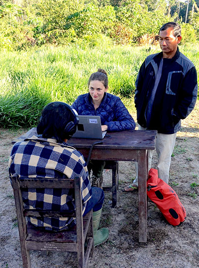
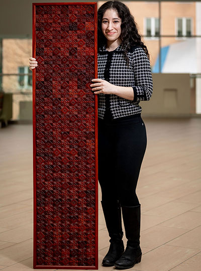
In the new study, the MIT team wanted to use tactile experience to measure more directly how the brains of blind people respond to faces. They created a ring of 3D-printed objects that included faces, hands, chairs, and mazes, and rotated them so that the subject could handle each one while in the fMRI scanner.

They began with normally sighted subjects and found that when they handled the 3D objects, a small area that corresponded to the location of the FFA was preferentially active when the subjects touched the faces, compared to when they touched other objects. This activity, which was weaker than the signal produced when sighted subjects looked at faces, was not surprising to see, Kanwisher says.

“We know that people engage in visual imagery, and we know from prior studies that visual imagery can activate the FFA. So the fact that you see the response with touch in a sighted person is not shocking because they’re visually imagining what they’re feeling,” she says.

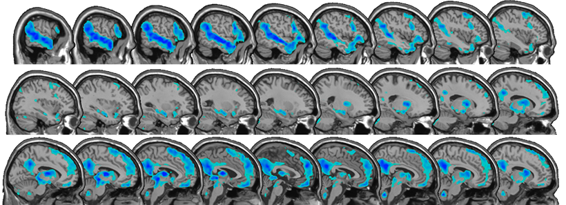
The researchers then performed the same experiments, using tactile input only, with 15 subjects who reported being blind since birth. To their surprise, they found that the brain showed face-specific activity in the same area as the sighted subjects, at levels similar to when sighted people handled the 3D-printed faces.

“When we saw it in the first few subjects, it was really shocking, because no one had seen individual face-specific activations in the fusiform gyrus in blind subjects previously,” Murty says.

Patterns of connection

The researchers also explored several hypotheses that have been put forward to explain why face-selectivity always seems to develop in the same region of the brain. One prominent hypothesis suggests that the FFA develops face-selectivity because it receives visual input from the fovea (the center of the retina), and we tend to focus on faces at the center of our visual field. However, since this region developed in blind people with no foveal input, the new findings do not support this idea.

Another hypothesis is that the FFA has a natural preference for curved shapes. To test that idea, the researchers performed another set of experiments in which they asked the blind subjects to handle a variety of 3D-printed shapes, including cubes, spheres, and eggs. They found that the FFA did not show any preference for the curved objects over the cube-shaped objects.

The researchers did find evidence for a third hypothesis, which is that face selectivity arises in the FFA because of its connections to other parts of the brain. They were able to measure the FFA’s “connectivity fingerprint” — a measure of the correlation between activity in the FFA and activity in other parts of the brain — in both blind and sighted subjects.

They then used the data from each group to train a computer model to predict the exact location of the brain’s selective response to faces based on the FFA connectivity fingerprint. They found that when the model was trained on data from sighted patients, it could accurately predict the results in blind subjects, and vice versa. They also found evidence that connections to the frontal and parietal lobes of the brain, which are involved in high-level processing of sensory information, may be the most important in determining the role of the FFA.

“It’s suggestive of this very interesting story that the brain wires itself up in development not just by taking perceptual information and doing statistics on the input and allocating patches of brain, according to some kind of broadly agnostic statistical procedure,” Kanwisher says. “Rather, there are endogenous constraints in the brain present at birth, in this case, in the form of connections to higher-level brain regions, and these connections are perhaps playing a causal role in its development.”

The research was funded by the National Institutes of Health Shared Instrumentation Grant to the Athinoula Martinos Center at MIT, a National Eye Institute Training Grant, the Smith-Kettlewell Eye Research Institute’s Rehabilitation Engineering Research Center, an Office of Naval Research Vannevar Bush Faculty Fellowship, an NIH Pioneer Award, and a National Science Foundation Science and Technology Center Grant.

Full paper at PNAS

Nine MIT School of Science professors receive tenure for 2020

Beginning July 1, nine faculty members in the MIT School of Science have been granted tenure by MIT. They are appointed in the departments of Brain and Cognitive Sciences, Chemistry, Mathematics, and Physics.

Physicist Ibrahim Cisse investigates living cells to reveal and study collective behaviors and biomolecular phase transitions at the resolution of single molecules. The results of his work help determine how disruptions in genes can cause diseases like cancer. Cisse joined the Department of Physics in 2014 and now holds a joint appointment with the Department of Biology. His education includes a bachelor’s degree in physics from North Carolina Central University, concluded in 2004, and a doctoral degree in physics from the University of Illinois at Urbana-Champaign, achieved in 2009. He followed his PhD with a postdoc at the École Normale Supérieure of Paris and a research specialist appointment at the Howard Hughes Medical Institute’s Janelia Research Campus.

Jörn Dunkel is a physical applied mathematician. His research focuses on the mathematical description of complex nonlinear phenomena in a variety of fields, especially biophysics. The models he develops help predict dynamical behaviors and structure formation processes in developmental biology, fluid dynamics, and even knot strengths for sailing, rock climbing and construction. He joined the Department of Mathematics in 2013 after completing postdoctoral appointments at Oxford University and Cambridge University. He received diplomas in physics and mathematics from Humboldt University of Berlin in 2004 and 2005, respectively. The University of Augsburg awarded Dunkel a PhD in statistical physics in 2008.

A cognitive neuroscientist, Mehrdad Jazayeri studies the neurobiological underpinnings of mental functions such as planning, inference, and learning by analyzing brain signals in the lab and using theoretical and computational models, including artificial neural networks. He joined the Department of Brain and Cognitive Sciences in 2013. He achieved a BS in electrical engineering from the Sharif University of Technology in 1994, an MS in physiology at the University of Toronto in 2001, and a PhD in neuroscience from New York University in 2007. Prior to joining MIT, he was a postdoc at the University of Washington. Jazayeri is also an investigator at the McGovern Institute for Brain Research.

Yen-Jie Lee is an experimental particle physicist in the field of proton-proton and heavy-ion physics. Utilizing the Large Hadron Colliders, Lee explores matter in extreme conditions, providing new insight into strong interactions and what might have existed and occurred at the beginning of the universe and in distant star cores. His work on jets and heavy flavor particle production in nuclei collisions improves understanding of the quark-gluon plasma, predicted by quantum chromodynamics (QCD) calculations, and the structure of heavy nuclei. He also pioneered studies of high-density QCD with electron-position annihilation data. Lee joined the Department of Physics in 2013 after a fellowship at CERN and postdoc research at the Laboratory for Nuclear Science at MIT. His bachelor’s and master’s degrees were awarded by the National Taiwan University in 2002 and 2004, respectively, and his doctoral degree by MIT in 2011. Lee is a member of the Laboratory for Nuclear Science.

Josh McDermott investigates the sense of hearing. His research addresses both human and machine audition using tools from experimental psychology, engineering, and neuroscience. McDermott hopes to better understand the neural computation underlying human hearing, to improve devices to assist hearing impaired, and to enhance machine interpretation of sounds. Prior to joining MIT’s Department of Brain and Cognitive Sciences, he was awarded a BA in 1998 in brain and cognitive sciences by Harvard University, a master’s degree in computational neuroscience in 2000 by University College London, and a PhD in brain and cognitive sciences in 2006 by MIT. Between his doctoral time at MIT and returning as a faculty member, he was a postdoc at the University of Minnesota and New York University, and a visiting scientist at Oxford University. McDermott is also an associate investigator at the McGovern Institute for Brain Research and an investigator in the Center for Brains, Minds and Machines.

Solving environmental challenges by studying and manipulating chemical reactions is the focus of Yogesh Surendranath’s research. Using chemistry, he works at the molecular level to understand how to efficiently interconvert chemical and electrical energy. His fundamental studies aim to improve energy storage technologies, such as batteries, fuel cells, and electrolyzers, that can be used to meet future energy demand with reduced carbon emissions. Surendranath joined the Department of Chemistry in 2013 after a postdoc at the University of California at Berkeley. His PhD was completed in 2011 at MIT, and BS in 2006 at the University of Virginia. Suendranath is also a collaborator in the MIT Energy Initiative.

A theoretical astrophysicist, Mark Vogelsberger is interested in large-scale structures of the universe, such as galaxy formation. He combines observational data, theoretical models, and simulations that require high-performance supercomputers to improve and develop detailed models that simulate galaxy diversity, clustering, and their properties, including a plethora of physical effects like magnetic fields, cosmic dust, and thermal conduction. Vogelsberger also uses simulations to generate scenarios involving alternative forms of dark matter. He joined the Department of Physics in 2014 after a postdoc at the Harvard-Smithsonian Center for Astrophysics. Vogelsberger is a 2006 graduate of the University of Mainz undergraduate program in physics, and a 2010 doctoral graduate of the University of Munich and the Max Plank Institute for Astrophysics. He is also a principal investigator in the MIT Kavli Institute for Astrophysics and Space Research.

Adam Willard is a theoretical chemist with research interests that fall across molecular biology, renewable energy, and material science. He uses theory, modeling, and molecular simulation to study the disorder that is inherent to systems over nanometer-length scales. His recent work has highlighted the fundamental and unexpected role that such disorder plays in phenomena such as microscopic energy transport in semiconducting plastics, ion transport in batteries, and protein hydration. Joining the Department of Chemistry in 2013, Willard was formerly a postdoc at Lawrence Berkeley National Laboratory and then the University of Texas at Austin. He holds a PhD in chemistry from the University of California at Berkeley, achieved in 2009, and a BS in chemistry and mathematics from the University of Puget Sound, granted in 2003.

Lindley Winslow seeks to understand the fundamental particles shaped the evolution of our universe. As an experimental particle and nuclear physicist, she develops novel detection technology to search for axion dark matter and a proposed nuclear decay that makes more matter than antimatter. She started her faculty position in the Department of Physics in 2015 following a postdoc at MIT and a subsequent faculty position at the University of California at Los Angeles. Winslow achieved her BA in physics and astronomy in 2001 and PhD in physics in 2008, both at the University of California at Berkeley. She is also a member of the Laboratory for Nuclear Science.

Universal musical harmony

Many forms of Western music make use of harmony, or the sound created by certain pairs of notes. A longstanding question is why some combinations of notes are perceived as pleasant while others sound jarring to the ear. Are the combinations we favor a universal phenomenon? Or are they specific to Western culture?

Through intrepid research trips to the remote Bolivian rainforest, the McDermott lab at the McGovern Institute has found that aspects of the perception of note combinations may be universal, even though the aesthetic evaluation of note combination as pleasant or unpleasant is culture-specific.

“Our work has suggested some universal features of perception that may shape musical behavior around the world,” says McGovern Associate Investigator Josh McDermott, senior author of the Nature Communications study. “But it also indicates the rich interplay with cultural influences that give rise to the experience of music.”

Remote learning

Questions about the universality of musical perception are difficult to answer, in part because of the challenge in finding people with little exposure to Western music. McDermott, who is also an associate professor in MIT’s Department of Brain and Cognitive Sciences and an investigator in the Center for Brains Minds and Machines, has found a way to address this problem. His lab has performed a series of studies with the participation of an indigenous population, the Tsimane’, who live in relative isolation from Western culture and have had little exposure to Western music. Accessing the Tsimane’ villages is challenging, as they are scattered throughout the rainforest and only reachable during the dry part of the year.

Left to right Josh McDermott (in vehicle), Alex Durango, Sophie Dolan and Malinda McPherson experiencing a travel delay en route to a Tsimane’ village after a heavy rainfall. Photo: Malinda McPherson

“When we enter a village there is always a crowd of curious children to greet us,” says Malinda McPherson, a graduate student in the lab and lead author of the study. “Tsimane’ are friendly and welcoming, and we have visited some villages several times, so now many people recognize us.”

In a study published in 2019, McDermott’s team found evidence that the brain’s ability to detect musical octaves is not universal, but is gained through cultural experience. And in 2016 they published findings suggesting that the preference for consonance over dissonance is culture-specific. In their new study, the team decided to explore whether aspects of the perception of consonance and dissonance might nonetheless be universally present across cultures.

Music lessons

In Western music, harmony is the sound of two or more notes heard simultaneously. Think of the Leonard Cohen song, Hallelujah, where he sings about harmony (“the fourth, the fifth, the minor fall and the major lift”). A combination of two notes is called an interval, and intervals that are perceived to be the most pleasant (or consonant, like the fourth and the fifth, for example) to the Western ear are generally represented by smaller integer ratios.

Intervals that are related by low integer ratios have fascinated scientists for centuries.

“Such intervals are central to Western music, but are also believed to be a common feature of many musical systems around the world,” McPherson explains. “So intervals are a natural target for cross-cultural research, which can help identify aspects of perception that are and aren’t independent of cultural experience.”

Scientists have been drawn to low integer ratios in music in part because they relate to the frequencies in voices and many instruments, known as ‘overtones’. Overtones from sounds like voices form a particular pattern known as the harmonic series. As it happens, the combination of two concurrent notes related by a low integer ratio partially reproduces this pattern. Because the brain presumably evolved to represent natural sounds, such as voices, it has seemed plausible that intervals with low integer ratios might have special perceptual status across cultures.

Since the Tsimane’ do not generally sing or play music together, meaning they have not been trained to hear or sing in harmony, McPherson and her colleagues were presented with a unique opportunity to explore whether there is anything universal about the perception of musical intervals.

Taking notes

In order to probe the perception of musical intervals, McDermott and colleagues took advantage of the fact that ears accustomed to Western musical harmony often have difficulty picking apart two “consonant” notes when they are played at the same time. This auditory confusion is known as “fusion” in the field. By contrast, two “dissonant” notes are easier to hear as separate.

The tendency of “consonant” notes to be heard by Westerners as fused could reflect their common occurrence in Western music. But it could also be driven by the resemblance of low-integer-ratio note combinations to the harmonic series. This similarity of consonant intervals to the acoustic structure of typical natural sounds raises the possibility that the human brain is biologically tuned to “fuse” consonant notes.

Graduate student and lead author, Malinda McPherson, works with a participant and translator in the field. Photo: Malinda McPherson

To explore this question, the team ran identical sets of experiments on two participant groups: US non-musicians residing in the Boston metropolitan area and Tsimane’ residing in villages in the Amazon rain forest. Listeners heard two concurrent notes separated by a particular musical interval (consonant or dissonant), and were asked to judge whether they heard one or two sounds. The experiment was performed with both synthetic and natural sounds.

They found that like the Boston cohort, the Tsimane’ were more likely to mistake two notes as a single sound if they were consonant than if they were dissonant.

“I was surprised by how similar some of the results in Tsimane’ participants were to those in US participants,” says McPherson, “particularly given the striking differences that we consistently see in preferences for musical intervals.”

When it came to whether consonant intervals were more pleasant than dissonant intervals, the results told a very different story. While the US study participants found consonant intervals more pleasant than dissonant intervals, the Tsimane’ showed no preference, implying that our sense of what is pleasant is shaped by culture.

“The fusion results provide an example of a perceptual effect that could influence musical systems, for instance by creating a natural perceptual contrast to exploit,” explains McDermott. “Hopefully our work helps to show how one can conduct rigorous perceptual experiments in the field and learn things that would be hidden if we didn’t consider populations in other parts of the world.”

Saxe Lab examines social impact of COVID-19

After being forced to relocate from their MIT dorms during the COVID19 crisis, two members of the Saxe lab are now applying their psychology skills to study the impact of mandatory relocation and social isolation on mental health.

“When ‘social distancing’ measures hit MIT, we tried to process how the implementation of these policies would impact the landscape of our social lives,” explains graduate student Heather Kosakowski, who conceived of the study late one evening with undergraduate Michelle Hung.  This landscape is broad, examining the effects of being uprooted and physically relocated from a place, but also changes in social connections, including friendships and even dating life.

MIT undergrad Michelle Hung in the Saxe lab. Photo: Michelle Hung

“I started speculating about how my life and the lives of other MIT students would change,” says Hung. “I was overwhelmed, sad, and scared. But then we realized that we were actually equipped to find the answers to our questions by conducting a study.”

Together, Kosakowski and Hung developed a survey to measure how the social behavior of MIT students, postdocs, and staff is changing over the course of the pandemic. Survey questions were designed to measure loneliness and other aspects of mental health. The survey was sent to members of the MIT community and shared on social media in mid-March, when the pandemic hit the US, and MIT made the unprecedented decision to send students home, shift to online instruction, and dramatically ramp down operations on campus.

More than 500 people responded to the initial survey, ranging in age from 18 to 60, living in cities and countries around the world. Many but not all of those who responded were affiliated with MIT. Kosakowski and Hung are sending follow-up surveys to participants every two weeks and the team plans to collect data for the duration of the pandemic.

“Throwing myself into creating the survey was a way to cope with feeling sad about leaving a community I love,” explains Hung, who flew home to California in March and admits that she struggles with feelings of loneliness now that she’s off campus.

Although it is too soon to form any conclusions about their research, Hung predicts that feelings of loneliness may actually diminish over the course of the pandemic.

“Humans have an impressive ability to adapt to change,” she says. “And I think in this virtual world, people will find novel ways to stay connected that we couldn’t have predicted.”

Whether we find ourselves feeling more or less lonely as this COVID-19 crisis comes to an end, both Kosakowski and Hung agree that it will fundamentally change life as we know it.

The Saxe lab is looking for more survey participants. To learn more about this study or to participate in the survey, click here.


Three from MIT awarded 2020 Guggenheim Fellowships

MIT faculty members Sabine Iatridou, Jonathan Gruber, and Rebecca Saxe are among 175 scientists, artists, and scholars awarded 2020 fellowships from the John Simon Guggenheim Foundation. Appointed on the basis of prior achievement and exceptional promise, the 2020 Guggenheim Fellows were selected from almost 3,000 applicants.

“It’s exceptionally encouraging to be able to share such positive news at this terribly challenging time” says Edward Hirsch, president of the foundation. “A Guggenheim Fellowship has always offered practical assistance, helping fellows do their work, but for many of the new fellows, it may be a lifeline at a time of hardship, a survival tool as well as a creative one.”

Since 1925, the foundation has granted more the $375 million in fellowships to over 18,000 individuals, including Nobel laureates, Fields medalists, poets laureate, and winners of the Pulitzer Prize, among other internationally recognized honors. This year’s MIT recipients include a linguist, an economist, and a cognitive neuroscientist.

Rebecca Saxe is an associate investigator of the McGovern Institute and the John W. Jarve (1978) Professor in Brain and Cognitive Sciences. She studies human social cognition, using a combination of behavioral testing and brain imaging technologies. She is best known for her work on brain regions specialized for abstract concepts such as “theory of mind” tasks that involve understanding the mental states of other people. She also studies the development of the human brain during early infancy. She obtained her PhD from MIT and was a Harvard University junior fellow before joining the MIT faculty in 2006. Saxe was chosen in 2012 as a Young Global Leader by the World Economic Forum, and she received the 2014 Troland Award from the National Academy of Sciences. Her TED Talk, “How we read each other’s minds” has been viewed over 3 million times.

Jonathan Gruber is the Ford Professor of Economics at MIT, the director of the Health Care Program at the National Bureau of Economic Research, and the former president of the American Society of Health Economists. He has published more than 175 research articles, has edited six research volumes, and is the author of “Public Finance and Public Policy,” a leading undergraduate text; “Health Care Reform,” a graphic novel; and “Jump-Starting America: How Breakthrough Science Can Revive Economic Growth and the American Dream.” In 2006 he received the American Society of Health Economists Inaugural Medal for the best health economist in the nation aged 40 and under. He served as deputy sssistant secretary for economic policy at the U.S. Department of the Treasury. He was a key architect of Massachusetts’ ambitious health reform effort, and became an inaugural member of the Health Connector Board, the main implementing body for that effort. He served as a technical consultant to the Obama administration and worked with both the administration and Congress to help craft the Affordable Care Act. In 2011, he was named “One of the Top 25 Most Innovative and Practical Thinkers of Our Time” by Slate magazine.

Sabine Iatridou is professor of linguistics in MIT’s Department of Linguistics and Philosophy. Her work focuses on syntax and the syntax-semantics interface, as well as comparative linguistics. She is the author and coauthor of a series of innovative papers about tense and modality that opened up whole new domains of research for the field. Since those publications, she has made foundational contributions to many branches of linguistics that connect form with meaning. She is the recipient of the National Young Investigator Award (USA), of an honorary doctorate from the University of Crete in Greece, and of an award from the Royal Dutch Academy of Sciences. She was elected fellow of the Linguistic Society of America. She is co-founder and co-director of the CreteLing Summer School of Linguistics.

“As we grapple with the difficulties of the moment, it is also important to look to the future,” says Hirsch. “The artists, writers, scholars, and scientific researchers supported by the fellowship will help us understand and learn from what we are enduring individually and collectively, and it is an honor for the foundation to help them do their essential work.”

Uncovering the functional architecture of a historic brain area

In 1840 a patient named Leborgne was admitted to a hospital near Paris: he was only able repeat the word “Tan.” This loss of speech drew the attention of Paul Broca who, after Leborgne’s death, identified lesions in his frontal lobe in the left hemisphere. These results echoed earlier findings from French neurologist Marc Dax. Now known as “Broca’s area,” the roles of this brain region have been extended to mental functions far beyond speech articulation. So much so, that the underlying functional organization of Broca’s area has become a source of discussion and some confusion.

McGovern Investigator Ev Fedorenko is now calling, in a paper at Trends in Cognitive Sciences, for recognition that Broca’s area consists of functionally distinct, specialized regions, with one sub-region very much dedicated to language processing.

“Broca’s area is one of the first regions you learn about in introductory psychology and neuroscience classes, and arguably laid the foundation for human cognitive neuroscience,” explains Ev Fedorenko, who is also an assistant professor in MIT’s Department of Brain and Cognitive Sciences. “This patch of cortex and its connections with other brain areas and networks provides a microcosm for probing some core questions about the human brain.”

Broca’s area, shown in red. Image: Wikimedia

Language is a uniquely human capability, and thus the discovery of Broca’s area immediately captured the attention of researchers.

“Because language is universal across cultures, but unique to the human species, studying Broca’s area and constraining theories of language accordingly promises to provide a window into one of the central abilities that make humans so special,” explains co-author Idan Blank, a former postdoc at the McGovern Institute who is now an assistant professor of psychology at UCLA.

Function over form

Broca’s area is found in the posterior portion of the left inferior frontal gyrus (LIFG). Arguments and theories abound as to its function. Some consider the region as dedicated to language or syntactic processing, others argue that it processes multiple types of inputs, and still others argue it is working at a high level, implementing working memory and cognitive control. Is Broca’s area a highly specialized circuit, dedicated to the human-specific capacity for language and largely independent from the rest high-level cognition, or is it a CPU-like region, overseeing diverse aspects of the mind and orchestrating their operations?

“Patient investigations and neuroimaging studies have now associated Broca’s region with many processes,” explains Blank. “On the one hand, its language-related functions have expanded far beyond articulation, on the other, non-linguistic functions within Broca’s area—fluid intelligence and problem solving, working memory, goal-directed behavior, inhibition, etc.—are fundamental to ‘all of cognition.’”

While brain anatomy is a common path to defining subregions in Broca’s area, Fedorenko and Blank argue that instead this approach can muddy the water. In fact, the anatomy of the brain, in terms of cortical folds and visible landmarks that originally stuck out to anatomists, vary from individual to individual in terms of their alignment with the underlying functions of brain regions. While these variations might seem small, they potentially have a huge impact on conclusions about functional regions based on traditional analysis methods. This means that the same bit of anatomy (like, say, the posterior portion of a gyrus) could be doing different things in different brains.

“In both investigations of patients with brain damage and much of brain imaging work, a lot of confusion has stemmed from the use of macroanatomical areas (like the inferior frontal gyrus (IFG)) as ‘units of analysis’,” explains Fedorenko. “When some researchers found IFG activation for a syntactic manipulation, and others for a working memory manipulation, the field jumped to the conclusion that syntactic processing relies on working memory. But these effects might actually be arising in totally distinct parts of the IFG.”

The only way to circumvent this problem is to turn to functional data and aggregate information from functionally defined areas across individuals. Using this approach, across four lines of evidence from the last decade, Fedorenko and Blank came to a clear conclusion: Broca’s area is not a monolithic region with a single function, but contains distinct areas, one dedicated to language processing, and another that supports domain-general functions like working memory.

“We just have to stop referring to macroanatomical brain regions (like gyri and sulci, or their parts) when talking about the functional architecture of the brain,” explains Fedorenko. “I am delighted to see that more and more labs across the world are recognizing the inter-individual variability that characterizes the human brain– this shift is putting us on the right path to making fundamental discoveries about how our brain works.”

Indeed, accounting for distinct functional regions, within Broca’s area and elsewhere, seems essential going forward if we are to truly understand the complexity of the human brain.

Study explores brain basis of special interests

Did you know that 88% of children on the autism spectrum have an affinity — or special interest that they are particularly passionate about?

We are curious about this.

The Gabrieli lab is exploring the brain basis of these special interests in kids with and without autism. The PAL (Project on Affinities and Language) study uses noninvasive and child-friendly fMRI methods to study whether affinities can activate language regions of the brain. The lab is currently looking for 7–12-year-old children with and without autism who have a special interest or passion.

Interested in participating?

Sign up here

Embracing neurodiversity to better understand autism

Researchers often approach autism spectrum disorder (ASD) through the lens of what might “break down.” While this approach has value, autism is an extremely heterogeneous condition, and diagnosed individuals have a broad range of abilities.

The Gabrieli lab is embracing this diversity and leveraging the strengths of diagnosed individuals by researching their specific “affinities.”

Affinities involve a strong passion for specific topics, ranging from insects to video game characters, and can include impressive feats of knowledge and focus.

The biological basis of these affinities and associated abilities remains unclear, which is intriguing to John Gabrieli and his lab.

“A striking aspect of autism is the great variation from individual to individual,” explains McGovern Investigator John Gabrieli. “Understanding what motivates an individual child may inform how to best help that child reach his or her communicative potential.”

Doug Tan is an artist on the autism spectrum who has a particular interest in Herbie, the fictional Volkswagen Beetle. Nearly all of Tan’s works include a visual reference to his “affinity” (shown here in black). Image: Doug Tan

Affinities have traditionally been seen as a distraction “interfering” with conventional teaching and learning. This mindset was upended by the 2014 book Life Animated by Ron Suskind, whose autistic son Owen seemingly lost his ability to speak around age three. Despite this setback, Owen maintained a deep affinity for Disney movies and characters. Rather than extinguishing this passion, the Suskinds embraced it as a path to connection.

Reframing such affinities as a strength not a frustration, and a path to communication rather than a roadblock, caught the attention of Kristy Johnson, a PhD student at the MIT Media Lab, who also has a non-verbal child with autism.

“My interest is in empowering and understanding populations that have traditionally been hard to study, including those with non-verbal and minimally verbal autism,” explains Johnson. “One way to do that is through affinities.”

But even identifying affinities is difficult. An interest in “trains” might mean 18th-century smokestacks to one child, and the purple line of the MBTA commuter rail to another. Serendipitously, she mentioned her interest to Gabrieli one day. He slammed his hands on the table, jumped up, and ran to find lab members Anila D’Mello and Halie Olson, who were gearing up to pursue the neural basis of affinities in autism. A collaboration was born.

Scientific challenge

What followed was six months of intense discussion. How can an affinity be accurately defined? How can individually tailored experiments be adequately controlled? What makes a robust comparison group? How can task-related performance differences between individuals with autism be accounted for?

The handful of studies that had used fMRI neuroimaging to examine affinities in autism had focused on the brain’s reward circuitry. D’Mello and Olson wanted to examine the language network of the brain — a well-defined network of brain regions whose activation can be measured by fMRI. Affinities trigger communication in some individuals with autism (Suskind’s family were using Disney characters to engage and communicate, not simply as a reward). Was the language network being engaged by affinities? Could these results point to a way of tailoring learning for all types of development?

“The language network involves lots of regions across the brain, including temporal, parietal, frontal, and subcortical areas, which play specific roles in different aspects of language processing” explains Olson. “We were interested in a task that used affinities to tap the language network.”

fMRI reveals regions of the brain that show increased activity for stories related to affinities versus neutral stories; these include regions important for language processing. Image: Anila D’Mello

By studying this network, the team is testing whether affinities can elicit “typical” activation in regions of the brain that are sometimes assumed to not be engaged in autism. The approach may help develop better paradigms for studying other tasks with individuals with autism. Regardless of whether there are differences between the group diagnosed with autism and typically developing children, insight will likely be gained into how personalized special interests influence engagement of the language network.

The resulting study is task-free, removing the variable of differing motor or cognitive skill sets. Kids watch videos of their individual affinity in the fMRI scanner, and then listen to stories based on that affinity. They also watch and listen to “neutral” videos and stories about nature that are consistent across all children. Identifying affinities robustly so that the right stimulus can be presented is critical. Rather than an interest in bugs, affinities are often very specific (bugs that eat other bugs). But identifying and cross-checking affinities is something the group is becoming adept at. The results are emerging, but the effects that the team are seeing are significant, and preliminary data suggest that affinities engage networks beyond reward circuits.

“We have a small sample right now, but across the sample, there seems to be a difference in activation in the brain’s language network when listening to affinity stories compared to neutral stories,” explains D’Mello. “The biggest surprise is that the differences are evident in single subjects.”

Future forward

The work is already raising exciting new questions. Are there other brain regions engaged by affinities? How would such information inform education and intervention paradigms? In addition, the team is showing it’s possible to derive information from individualized, naturalistic experimental paradigms, a message for brain imaging and behavioral studies in general. The researchers also hope the results inspire parents, teachers, and psychologists to perceive and engage with an individual’s affinities in new ways.

“This could really help teach us to communicate with and motivate very young and non-verbal kids on the spectrum in a way that is interesting and meaningful to them,” D’Mello explains.

By studying the strengths of individuals with autism, these researchers are showing that, through embracing neurodiversity, we can enhance science, our understanding of the brain, and perhaps even our understanding of ourselves.

Learn about autism studies at MIT

McGovern lab manager creates art inspired by science

Michal De-Medonsa, technical associate and manager of the Jazayeri lab, created a large wood mosaic for her lab. We asked Michal to tell us a bit about the mosaic, her inspiration, and how in the world she found the time to create such an exquisitely detailed piece of art.

______

Jazayeri lab manager Michal De-Medonsa holds her wood mosaic entitled “JazLab.” Photo: Caitlin Cunningham

Describe this piece of art for us.

To make a piece this big (63″ x 15″), I needed several boards of padauk wood. I could have just etched each board as a whole unit and glued the 13 or so boards to each other, but I didn’t like the aesthetic. The grain and color within each board would look beautiful, but the line between each board would become obvious, segmented, and jarring when contrasted with the uniformity within each board. Instead, I cut out about 18 separate squares out of each board, shuffled all 217 pieces around, and glued them to one another in a mosaic style with a larger pattern (inspired by my grandfather’s work in granite mosaics).

What does this mosaic mean to you?

Once every piece was shuffled, the lines between single squares were certainly visible, but as a feature, were far less salient than had the full boards been glued to one another. As I was working on the piece, I was thinking about how the same concept holds true in society. Even if there is diversity within a larger piece (an institution, for example), there is a tendency for groups to form within the larger piece (like a full board), diversity becomes separated. This isn’t a criticism of any institution, it is human nature to form in-groups. It’s subconscious (so perhaps the criticism is that we, as a society, don’t give that behavior enough thought and try to ameliorate our reflex to group with those who are “like us”). The grain of the wood is uniform, oriented in the same direction, the two different cutting patterns create a larger pattern within the piece, and there are smaller patterns between and within single pieces. I love creating and finding patterns in my art (and life). Alfred North Whitehead wrote that “understanding is the apperception of pattern as such.” True, I believe, in science, art, and the humanities. What a great goal – to understand.​

Tell us about the name of this piece.

Every large piece I make is inspired by the people I make it for, and is therefore named after them. This piece is called JazLab. Having lived around the world, and being a descendant of a nomadic people, I don’t consider any one place home, but am inspired by every place I’ve lived. In all of my work, you can see elements of my Jewish heritage, antiquity, the Middle East, Africa, and now MIT.

How has MIT influenced your art?

MIT has influenced me in the most obvious way MIT could influence anyone – technology. Before this series, I made very small versions of this type of work, designing everything on a piece of paper with a pencil and a ruler, and making every cut by hand. Each of those small squares would take ~2 hours (depending on the design), and I was limited to softer woods.

Since coming to MIT, I learned that I had access to the Hobby Shop with a huge array of power tools and software. I began designing my patterns on the computer and used power tools to make the cuts. I actually struggled a lot with using the tech – not because it was hard (which, it really is when you just start out), but rather because it felt like I was somehow “cheating.” How is this still art? And although this is something I still think about often, I’ve tried to look at it in this way: every generation, in their time, used the most advanced technology. The beauty and value of the piece doesn’t come from how many bruises, cuts, and blisters your machinery gave you, or whether you scraped the wood out with your nails, but rather, once you were given a tool, what did you decide to do with it? My pieces still have a huge hand-on-material work, but I am working on accepting that using technology in no way devalues the work.

Given your busy schedule with the Jazayeri lab, how did you find the time to create this piece of art?

I took advantage of any free hour I could. Two days out of the week, the hobby shop is open until 9pm, and I would additionally go every Saturday. For the parts that didn’t require the shop (adjusting each piece individually with a carving knife, assembling them, even most of the glueing) I would just work  at home – often very late into the night.

______

JazLab is on display in the Jazayeri lab in MIT Bldg 46.

Brain biomarkers predict mood and attention symptoms

Mood and attentional disorders amongst teens are an increasing concern, for parents, society, and for peers. A recent Pew research center survey found conditions such as depression and anxiety to be the number one concern that young students had about their friends, ranking above drugs or bullying.

“We’re seeing an epidemic in teen anxiety and depression,” explains McGovern Research Affiliate Susan Whitfield-Gabrieli.

“Scientists are finding a huge increase in suicide ideation and attempts, something that hit home for me as a mother of teens. Emergency rooms in hospitals now have guards posted outside doors of these teenagers that attempted suicide—this is a pressing issue,” explains Whitfield-Gabrieli who is also director of the Northeastern University Biomedical Imaging Center and a member of the Poitras Center for Psychiatric Disorders Research.

Finding new methods for discovering early biomarkers for risk of psychiatric disorders would allow early interventions and avoid reaching points of crisis such as suicide ideation or attempts. In research published recently in JAMA Psychiatry, Whitfield-Gabrieli and colleagues found that signatures predicting future development of depression and attentional symptoms can be detected in children as young as seven years old.

Long-term view

While previous work had suggested that there may be biomarkers that predict development of mood and attentional disorders, identifying early biomarkers prior to an onset of illness requires following a cohort of pre-teens from a young age, and monitoring them across years. This effort to have a proactive, rather than reactive, approach to the development of symptoms associated with mental disorders is exactly the route Whitfield-Gabrieli and colleagues took.

“One of the exciting aspects of this study is that the cohort is not pre-selected for already having symptoms of psychiatric disorders themselves or even in their family,” explained Whitfield-Gabrieli. “It’s an unbiased cohort that we followed over time.”

McGovern research affiliate Susan Whitfield-Gabrieli has discovered early brain biomarkers linked to psychiatric disorders.

In some past studies, children were pre-selected, for example a major depressive disorder diagnosis in the parents, but Whitfield-Gabrieli and colleagues, Silvia Bunge from Berkeley and Laurie Cutting from Vanderbilt, recruited a range of children without preconditions, and examined them at age 7, then again 4 years later. The researchers examined resting state functional connectivity, and compared this to scores on the child behavioral checklist (CBCL), allowing them to relate differences in the brain to a standardized analysis of behavior that can be linked to psychiatric disorders. The CBCL is used both in research and in the clinic and his highly predictive of disorders including ADHD, so that changes in the brain could be related to changes in a widely used clinical scoring system.

“Over the four years, some people got worse, some got better, and some stayed the same according the CBCL. We could relate this directly to differences in brain networks, and could identify at age 7 who would get worse,” explained Whitfield-Gabrieli.

Brain network changes

The authors analyzed differences in resting state network connectivity, regions across the brain that rise and fall in activity level together, as visualized using fMRI. Reduced connectivity between these regions may allow us to get a handle on reduced “top-down” control of neural circuits. The dorsolateral prefrontal region is linked to executive function, external attention, and emotional control. Increased connection with the medial prefrontal cortex is known to be present in attention deficit hyperactivity disorder (ADHD), while a reduced connection to a different brain region, the sgACC, is seen in major depressive disorder. The question remained as to whether these changes can be seen prior to the onset of diagnosable attentional or mood disorders.

Whitfield-Gabrieli and colleagues found that these resting state networks varied in the brains of children that would later develop anxiety/depression and ADHD symptoms. Weaker scores in connectivity between the dorsolateral and medial prefrontal cortical regions tended to be seen in children whose attention scores went on to improve. Analysis of the resting state networks above could differentiate those who would have typical attentional behavior by age 11 versus those that went on to develop ADHD.

Whitfield-Gabrieli has replicated this finding in an independent sample of children and she is continuing to expand the analysis and check the results, as well as follow this cohort into the future. Should changes in resting state networks be a consistent biomarker, the next step is to initiate interventions prior to the point of crisis.

“We’ve recently been able to use mindfulness interventions, and show these reduce self-perceived stress and amygdala activation in response to fear, and we are also testing the effect of exercise interventions,” explained Whitfield-Gabrieli. “The hope is that by using predictive biomarkers we can augment children’s lifestyles with healthy interventions that can prevent risk converting to a psychiatric disorder.”